-
PDF
- Split View
-
Views
-
Cite
Cite
D. A. Pérennou, G. Mazibrada, V. Chauvineau, R. Greenwood, J. Rothwell, M. A. Gresty, A. M. Bronstein, Lateropulsion, pushing and verticality perception in hemisphere stroke: a causal relationship?, Brain, Volume 131, Issue 9, September 2008, Pages 2401–2413, https://doi.org/10.1093/brain/awn170
Close - Share Icon Share
Abstract
The relationships between perception of verticality by different sensory modalities, lateropulsion and pushing behaviour and lesion location were investigated in 86 patients with a first stroke. Participants sat restrained in a drum-like framework facing along the axis of rotation. They gave estimates of their subjective postural vertical by signalling the point of feeling upright during slow drum rotation which tilted them rightwards–leftwards. The subjective visual vertical was indicated by setting a line to upright on a computer screen. The haptic vertical was assessed in darkness by manually setting a rod to the upright. Normal estimates ranged from −2.5° to 2.5° for visual vertical and postural vertical, and from −4.5° to 4.5° for haptic vertical. Of six patients with brainstem stroke and ipsilesional lateropulsion only one had an abnormal ipsilesional postural vertical tilt (6°); six had an ipsilesional visual vertical tilt (13 ±.4°); two had ipsilesional haptic vertical tilts of 6°. In 80 patients with a hemisphere stroke (35 with contralesional lateropulsion including 6 ‘pushers’), 34 had an abnormal contralesional postural vertical tilt (average −8.5 ± 4.7°), 44 had contralesional visual vertical tilts (average −7 ± 3.2°) and 26 patients had contralesional haptic vertical tilts (−7.8 ± 2.8°); none had ipsilesional haptic vertical or postural vertical tilts. Twenty-one (26%) showed no tilt of any modality, 41 (52%) one or two abnormal modality(ies) and 18 (22%) a transmodal contralesional tilt (i.e. PV + VV + HV). Postural vertical was more tilted in right than in left hemisphere strokes and specifically biased by damage to neural circuits centred around the primary somatosensory cortex and thalamus. This shows that thalamo-parietal projections have a functional role in the processing of the somaesthetic graviceptive information. Tilts of the postural vertical were more closely related to postural disorders than tilts of the visual vertical. All patients with a transmodal tilt showed a severe lateropulsion and 17/18 a right hemisphere stroke. This indicates that the right hemisphere plays a key role in the elaboration of an internal model of verticality, and in the control of body orientation with respect to gravity. Patients with a ‘pushing’ behaviour showed a transmodal tilt of verticality perception and a severe postural vertical tilt. We suggest that pushing is a postural behaviour that leads patients to align their erect posture with an erroneous reference of verticality.
Introduction
Although it is established that patients’ perceptions of verticality can be altered after stroke the consequences of this altered perception for postural disability and recovery are unclear. A clinical feature in stroke is that erect sitting and/or standing posture can be compromised by ‘lateropulsion’; that is, an active lateral tilt of the body which is usually ipsilesional in caudal brainstem strokes (Bjerver and Silfverskiold, 1968; Dieterich and Brandt, 1992; Brandt and Dieterich, 1994) and contralesional in rostral brainstem strokes (Brandt and Dieterich, 1994; Yi et al., 2007) as well as in hemisphere strokes (Bohannon et al., 1986; Beevor, 1909; Pérennou et al., 1998; D’Aquila et al., 2004). Lateropulsion could result directly from a pathological asymmetry of motor function or tone (Beevor, 1909; Thomke et al., 2005; Babyar et al., 2007) or alternatively, could be an attempt to align the body with an internal vertical reference which is erroneously perceived to be tilted from true earth vertical (Dieterich and Brandt, 1992; Pérennou et al., 1998). The latter mechanism might predominate in lesions of the hemispheres (Brandt et al., 1994; Pérennou et al., 1998) whereas both mechanisms could be involved in brainstem lesions (Dieterich and Brandt, 1992; Bronstein et al., 2003; Thomke et al., 2005).
In some hemispheric strokes lateropulsion is associated with ‘pushing behaviour’ which is characterized by patients resisting any attempt to correct their posture (Bohannon, 1996; Pedersen et al., 1996; Karnath et al., 2000; Pérennou et al., 2002; Danells et al., 2004). Strikingly, some of these patients (‘pushers’) push themselves away actively from the non-paralysed side (Davies, 1985). This behaviour is a major challenge for rehabilitation (Pedersen et al., 1996; Danells et al., 2004; Babyar et al., 2008) which would benefit from clarification of the underlying mechanisms. It has been suggested that pushing might result from a mismatch between a normal visual perception of the vertical and an ipsilesional tilt in the perception of the postural vertical (PV) (Karnath et al., 2000). This hypothesis predicts an attenuation of pushing in darkness that has not been confirmed (Pérennou et al., 1998; 2002; Karnath et al., 2000). Alternatively it has been suggested that pushing and lateropulsion could be a clinical manifestation of a tilted representation of the vertical, whereby pushers actively align their body with a tilted reference of verticality (Dieterich and Brandt, 1992; Pérennou et al., 1998, 2002). According to Karnath's hypothesis pushers make a postural response to compensate for an erroneous verticality reference tilted towards the stroke side (they push themselves away from their subjective vertical) whereas according to our hypothesis pushers make a postural response in order to control their balance and so actively align their erect posture with a verticality reference tilted to the side opposite the stroke (affected hemisphere). In the present study we hypothesized the existence of a continuum in verticality perception after stroke: patients without lateropulsion should show a normal verticality perception, those with a lateropulsion but no pushing should show moderate biases in verticality perception and those with lateropulsion plus pushing should show the most severe tilts in verticality perception.
Verticality can be perceived through different modalities: the ‘visual’ perception of the vertical that relies on visuo-vestibular information, the ‘postural’ perception of the vertical derived from graviceptive-somaesthetic information (Bisdorff et al., 1996b; Anastasopoulos et al., 1997; Pérennou et al., 1998; Karnath et al., 2000; Manckoundia et al., 2007; Mazibrada et al., 2008; Barbieri et al., 2008), and the tactile (haptic) vertical (Kerkhoff, 1999; Gentaz et al., 2002; Bronstein et al., 2003). Whether lateropulsion and/or pushing are caused by a bias generalized across these three modalities of verticality perception is still questionable. Such a transmodal bias would indicate the existence of a tilted internal representation of verticality. However, since significant dissociations between verticality perceptual modalities have been reported (Bisdorff et al., 1996b; Karnath et al., 2000; Bronstein et al., 2003), alternative explanations for lateropulsion and pushing behaviour are also possible. We now examine this issue by measuring visual vertical (VV), haptic vertical (HV) and PV in a substantial number of patients with a left or right hemisphere or brainstem stroke and with a range of postural disabilities.
Material and Methods
Subjects
VV, HV and PV were studied in 119 subjects: 33 healthy controls (age 48.8 ± 10.8 years; 11 females/22 males) and 86 patients who had a first and unique stroke (age 55.4 ± 13.1 years; 29 females/57 males; time period since stroke 11.9 ± 8.2 weeks, 70 ischaemic and 16 haemorrhagic strokes), located either in one hemisphere (territory of the middle cerebral artery or thalamus; n = 80) or in the brainstem (n = 6). The patients invited to participate were drawn from patients who were treated consecutively at two specialized rehabilitation centres (Neurorehabilitation Unit, Homerton Hospital, London, UK; Unité de Rééducation Neurologique, Centre Hospitalier Universitaire de Nîmes, France) and gave their informed consent to the study in accordance with the guidelines of both institutions’ ethics committees. Before being tested all patients benefited from a conventional rehabilitation program, standardized for the two rehabilitation units participating in the study, and similar to what they would have received if they had not participated in the study. For each patient, the rehabilitation therapy lasted about 3–4 h/day, with exercises graduated according to their impairments and recovery and which were mainly focussed on language, spatial neglect, swallowing, bladder, balance, gait and prehension rehabilitation. No patients had been included in any other clinical trial since the stroke. Balance rehabilitation did not include any exercises aimed at recalibrating an abnormal perception of verticality, except for exercises of trunk orientation in front of a mirror in patients who showed lateropulsion. No patient had been submitted to any type of sensory manipulation or stimulation previously to the testing. Cerebral laterality was assessed using the Edinburgh Questionnaire (manual laterality ranged from −1 to +1, Oldfield, 1971). Thirty control subjects were right-handed (>0.6), two left-handed (<0) and one ambidextrous. Among patients, 75 were right-handed, seven were ambidextrous and four left-handed. In the 12 left-handed or ambidextrous patients, the stroke was located in the right hemisphere (RH) (n = 5), in the left hemisphere (n = 5) or in the brainstem (n = 2). All patients with a contralesional lateropulsion were right-handed except four who were ambidextrous (none showed pushing).
Lesion location
Only patients with a well-defined, non-lacunar, unique lesion on MRI or CT scans were recruited; patients with multiple lacunar lesions were excluded. The presence of a lesion in the frontal cortex, the Rolando's cortex, the parietal cortex, the temporal cortex, the corona radiata, the striatum, the internal capsule or the thalamus was determined using the Atlas of Talairach and Tournoux (1988) as 0 (no lesion) or 1 (lesion). The number of these cerebral areas involved in the lesion (obtained by summing all 0s and 1s) gave an estimation of the lesion size used in statistical analysis. According to this scoring, two areas exhibiting abnormal signal = 2, and so on, regardless of the respective size of the lesions. For analyses of variances (ANOVAs), lesion size was classified as small (score ≤ 3), medium (score 4–6) or large (score ≥ 7).
Verticality perception
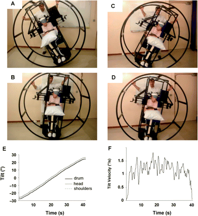
The VV, HV and PV were assessed in a darkened room. Subjects were seated, with head, trunk and limbs restrained by webbing and pads in a drum-like tilting apparatus (Fig. 1) facing along the rotational axis which was approximately at the level of the navel. The seated posture was adopted for testing because 46/81 patients could not stand alone; it is safer, more comfortable and allows comparison with previous studies in the literature invariably obtained in seated subjects. In the primary vertical orientation of the drum, the vertical positioning of body segments was obtained by the use of adjustable lateral wedges for the head, the trunk (with the arms) and the pelvis. Thighs, legs and feet were then strapped with pieces of foam between the limbs, and hands crossed over a pillow on the thighs. Drum rotational tilt was measured with an inertial inclinometer with a resolution of 0.5°. After several practice trials, verticality perception was always performed in the order VV, PV and HV. The subjects were kept at earth vertical between the tests, and also for VV and HV testing. To rule out any visual input during HV and PV measurements subjects were required to close their eyes supported by a sleeping eye mask. No feedback was given to subjects about their performance before the whole assessment was completed (about 45 min). For each modality, measurements started after 1 min in darkness. Between testing each modality, subjects’ eyes were open normally with the room lights on.
The wheel paradigm for measuring the postural vertical in disabled patients. The subject is randomly tilted to either side of true vertical between 15° to 45°, the wheel is then rolled immediately in the opposite direction until the subject reports having reached an upright position. Example of two trials in a patient with a right hemisphere stroke and left lateropulsion. (A) starting position on the right, the patient feels upright in (B), and (C) starting position on the left, the patient feels upright in (D). PV calculated as the average of 10 trials performed in a pseudo-random sequence was −11°. (E) manual rotation of the wheel with a subject strapped inside, measured using a precise movement analysis system (BTS Smart-e). Wheel rotation induces the same body rotation without unwanted movement of the head or trunk, which passively follow the wheel orientation. (F) the derivative of the wheel position signal (velocity) shows that the mean angular velocity was around 1–1.5°/s, except when the movement starts/stops for minor positional adjustments.
For VV testing, a 15-cm long luminous line was presented on a levelled computer screen at eye level, 1.2 m away from the nasion. Tube brightness was set to minimum and visual cues from the luminous line were excluded by placing a ‘vignetting’ window over the screen to mask its edges. The subjects were oriented upright in the drum which was set to vertical according to the inclinometer. The visual line was tilted in leftwards and rightwards rotation about its centre using keyboard arrows; tilt resolution was 0.2°. The tilts of the line were presented in a pseudo-random sequence balanced between leftwards and rightwards and after each tilt the subject/patient verbally controlled the operator's adjustment of the line back to his subjective VV until he was satisfied with its orientation. The subject then closed her/his eyes for a couple of seconds and a further test line was presented. Ten trials were performed and all stimulus tilt and vertical estimates were acquired.
Before starting PV assessment pilot measurements ensured that the subject could be passively tilted with the drum without significant differential displacements of body segments (head–trunk–limbs) (Fig. 1, bottom). For PV measurements the subject was randomly tilted to either side of true vertical between 15° to 45°, the wheel was then rolled in the opposite direction until the patient reported that he had reached an upright position. Small adjustments around this position were then performed if needed until the subject was satisfied. The wheel was rotated manually as steadily and smoothly as possible by two experimenters (DP+GM or DP+VC) at an approximate velocity of 1.5°/s to minimize semicircular canal stimulation (Seemungal et al., 2004; Sadeghi et al., 2007). The balanced design of the drum with a large radius ensured that the rotation was silent and smooth excluding bias from acoustic or vibration feedback (Fig. 1F). At the level of the head, tilting had both rotational and translational components. The latter component was weak (0.3 cm/s for a 1.8 m height subject) and below the stimulation threshold of the otoliths (Gianna et al., 1996). Hence all otolithic stimulation was due to gravitational tilt (4.9 m/s2 at 30° tilt from vertical). Ten trials were given in a pseudo-random sequence, five from left to right, and five from right to left.
For the HV a rod (radius 25 cm, terminating in an arrowhead, weight 150 g) pivoting about a horizontal central axis was manually rotated to vertical. Arm and trunk wedges on one side were released to allow the upper limb to manipulate the rod whose axis was centred at the level of the navel, at a 40–50 cm distance from the trunk. The inclination of the rod was measured against a levelled protractor to 0.5° accuracy. The rod was pseudo-randomly tilted (balanced between left and right) and subjects instructed to set the rod to vertical according to ‘how it felt’ in their hands. As a control for right and left stroke patients, 10 normal subjects performed the test with the left hand; hemisphere stroke patients used their contralesional hand and brainstem stroke patients the less affected hand. Since visual cues were excluded, tactile exploration of the rod was encouraged. Before computing each trial, subjects were asked whether they were satisfied with the chosen position of the rod and additional adjustments were performed accordingly. Ten trials were performed as for VV and PV estimates.
VV, HV and PV were calculated by averaging the 10 trials of each modality; data were normalized so that negative values indicated a contralesional tilt (leftward in normals).
Postural behaviour
Lateropulsion and pushing were diagnosed according to the scale for contraversive pushing (SCP) criteria (Karnath et al., 2000), with a total score ranging from 0 to 6. The SCP assesses: (i) Lateropulsion while sitting and standing; (ii) the use of the arm or the leg to extend the base of support in sitting and standing; (iii) resistance to passive correction of posture while sitting and standing. Lateropulsion is scored 0.25 for a mild contraversive body tilt without falling; 0.50 for a severe contraversive body tilt without falling; one for a severe contraversive body tilt with falling to the contralesional side. In the present study, which included hemispheric and brainstem lesions, patients were considered as having lateropulsion if they scored more than 0.5 on the SCP, indicating that they showed a severe body tilt in sitting or/and in standing, whatever the direction of the tilt, with or without falling. As proposed by Karnath et al. (2000), pushing was diagnosed if all three criteria of the SCP were present. That means that a patient with pushing would show a severe lateropulsion, use the arm of the leg to extend the area of physical contact to the ground and have resistance to passive correction of posture while sitting and standing. To improve the reliability of this scoring, the SCP was filled in collectively by the two or three physiotherapists who daily treated the patient, and were unaware of her/his verticality perception. Patients with a hemisphere stroke and clinically symmetrical sitting and standing posture (SCP ≤ 0.5) were considered as ‘upright’ (n = 45; age 52.7 ± 13.4 years, time since stroke 10.7 ± 7.4 weeks). The group of patients with a hemisphere stroke and a contralesional lateropulsion without pushing was called ‘contralist’, i.e. listing to the side opposite the stroke (n = 29; age 58 ± 12.3 years; time period since stroke 13.8 ± 8.7 weeks). The group of patients with an ipsilesional lateropulsion after a brainstem stroke was called ‘ipsilist’, i.e. listing towards the stroke side (n = 6; age 56.8 ± 12.1 years; time since stroke 15.2 ± 12 weeks). Patients with a hemisphere stroke and a contralesional lateropulsion plus pushing were called pushers (n = 6; age 62.7 ± 11.3; time since stroke 8.8 ± 7.4 weeks). These four groups of patients did not differ in age or time since stroke.
Statistical analyses
Statistical analyses were performed with the NCSS software. The normality of data distribution was tested using D’Agostino-Kurtosis test. VV, PV and HV distributions were normal in healthy subjects, upright patients and patients with lateropulsion. The SCP distribution was not normal. Equality of variances was tested using a Kolmogorov–Smirnov test. Comparisons between controls’ and patients’ groups were carried out with Aspin–Welch unequal-variance tests. Comparisons between the controls’ group and/or patients’ subgroups were carried out with Kruskal–Wallis tests corrected for ties when required (clinical data); with Aspin–Welch unequal-variance tests (verticality perception, one-factor); then with a two-factor ANOVA (lesion side and size, or group and modality of verticality perception). Post hoc analyses were performed using Kruskal–Wallis multiple-comparison tests or Bonferroni multiple-comparison tests. Frequency analyses were carried out with a Chi-square test (gender, lesion side and lesion location for subgroups of patients). Non-parametric correlations were performed to test possible relationship between the verticality estimates and the SCP. A principal component analysis (PCA) was also run to determine the optimal set of variables describing disorders of verticality. Unless otherwise indicated, the 0.05 level of significance was adopted throughout. Data are given in the text as: mean ± SD.
Results
Verticality perception in normals
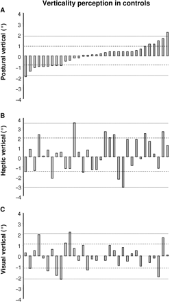
Data distribution of normals according to a decreasing order on PV values is displayed in Fig. 2. Normal subjects’ perceptual estimates of the vertical did not differ from the earth vertical: VV = −0.04 ± 1.1° (T = 0.23, P = 0.82) with −2.2° and +2.2° as minimal and maximal values; HV = 0.25 ± 1.7° (T = 0.87, P = 0.39) with −3.1° and +3.5° as minimal and maximal values; PV = 0.03 ± 0.9° (T = 0.21, P = 0.83) with −1.9° and +2.2° as minimal and maximal values. Since the distributions of these data were all Gaussian, and given the low ratio between the magnitude of verticality estimates in normals and the accuracy of measurements (ratio = 0.1, 0.2 and 0.5 for PV, VV and HV, respectively) the ranges of normality were defined as mean ± 2SD ± measurement accuracy. This precaution reduced the risk of including some patients with marginal estimates of verticality perception due to measurement uncertainty as abnormal. Defined thus, normal estimates of the perceptual vertical ranged from −2.5° to 2.5° for VV and PV and from −4.5° to 4.5° for HV. No difference between modalities was found by a one-factor ANOVA [F(2,96) = 0.48, P = 0.62].
Distribution of the orientation indices in healthy subjects. Individual data given in Figs 2 (A) PV, (B) HV and (C) VV. Each bar corresponds to an individual subject data. Dot lines indicate 1 and 2 SD.
Distribution of verticality data in stroke patients
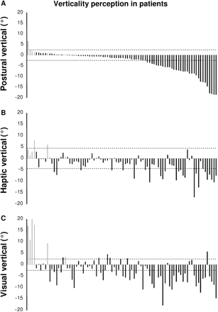
Individual data ranked according to a decreasing order on PV values are given in Fig. 3A (PV), 3B (HV) and 3C (VV).
Distribution of the orientation indices in patients, ranked in an increasing order according to PV. Each bar corresponds to an individual data, gray for patients with a brain stem stroke and black for patients with a hemisphere stroke. Dot lines represent the ranges of normality. (A) PV, (B) HV, (C) VV. Negative values indicate a contralesional tilt (side opposite the brain lesion).
PV
In patients PV (Fig. 3A) was on average more tilted than in controls (−3.5 ± 5.1°; T = 6.2, P < 10−6; power = 1) and with a more scattered distribution than that of controls (P < 10−3). Only one patient (brainstem stroke) showed an abnormal ipsilesional PV tilt (6°) whereas 34 patients (all with a hemisphere stroke) showed abnormal contralesional PV tilts (average −8.5 ± 2.8°).
HV
In patients HV (Fig. 3B) was on average more tilted than in controls (−2.7 ± 4.2°; T = −5.5, P < 10−6; power = 1), with a more scattered data distribution than controls (P < 10−3). Two patients (both with a large lateral lesion of the brainstem) showed an abnormal ipsilesional HV tilt over 4.5° (average 6°) whereas 26 patients (all with an hemisphere stroke) showed an abnormal HV tilt (−7.8 ± 2.8°).
VV
In patients, VV (Fig. 3C) was on average more tilted than in controls (−2.6 ± 6.3°; T = 3.7, P < 10−3; power = 0.95), with a more scattered distribution than controls (P < 10−6). Thirteen patients showed an abnormal ipsilesional VV tilt (>2.5°): all six patients with a brainstem stroke, and 7/80 with a hemisphere stroke (9%). The ipsilesional VV tilt was generally more pronounced in patients with a brainstem stroke (13 ± 6.4°); and less severe (3.6 ± 1.1°) in patients with a hemisphere stroke. Forty-four patients, all with a hemisphere stroke, showed abnormal contralesional VV tilts of various magnitude (average −7 ± 3.2°).
In overview, all brainstem patients showed an ipsilesional VV tilt and, with less consistency and magnitude, ipsilesional HV or PV tilts. No patient with a hemisphere stroke showed an ipsilesional HV or PV tilt whereas 9% showed a mild ipsilesional VV tilt. Among patients with hemisphere stroke abnormal contralesional tilt was found in 55% for VV, 42% for PV and 32.5% for HV.
A PCA with varimax orthogonal rotation was used to analyse the multidimensional components involved in verticality perception in the 86 stroke patients. About 87% of the data on verticality perception was accounted for by two factors: factor one mainly loaded by PV (VV −0.26, HV −0.70, PV −0.92) and factor two mainly loaded by VV (VV +0.94, HV +0.50, PV +0.19). Thus VV and PV were independent and together accounted for the majority of the variance of the data on verticality perception.
Among patients with a hemisphere stroke, 18 (22%) showed a transmodal contralesional tilt (i.e. PV + HV + VV), 21 (26%) showed no tilt of any modality (normal PV, HV and VV), and 41 (52%) showed one or two abnormal modality(ies) (Table 1). Their ages, sex ratio and delay from stroke onset were similar. Patients without any tilt in verticality perception had a smaller stroke than patients with one or more tilted verticality estimate(s). All patients with a transmodal tilt except one (who was the only patient with a left hemisphere stroke who showed pushing) had a stroke of the RH, with a much more extensive lesion than other patients. Strikingly, their verticality perception was much more tilted. All showed a severe contralesional lateropulsion and six showed pushing. However, this transmodal tilt in verticality perception was not sufficient to explain pushing since severe transmodal biases in verticality perception were also present in some patients with lateropulsion who were not pushers (Table 2).
Comparative analysis of patients without any tilt of verticality perception, patients with a transmodal tilt and those with a tilt of one or two modalities
| . | No tilt (n = 21) . | 1 or 2 tilt(s) (n = 41) . | Transmodal tilt (n = 18) . | . |
|---|---|---|---|---|
| Age (yrs) | 56.1 ± 2.9 | 53.7 ± 2.1 | 58.3 ± 3.1 | P = 0.42 |
| Gender | 7F 14M | 15F 26M | 3F 15M | P = 0.31 |
| Delay (wks) | 10.9 ± 1.7 | 11.5 ± 1.3 | 12.9 ± 1.9 | P = 0.96 |
| Lesion size (1–8 a.u.) | 3.9 ± 0.4 | 4.7 ± 0.3 | 6.3 ± 0.5 | P < 10−3† |
| Lesion side | 10L 11R | 15L 26R | 1L 17R | P = 0.01† |
| SCP (0–6 a.u.) | 0.2 ± 0.2 | 0.7 ± 0.2 | 2.5 ± 0.2 | P <10"−6† |
| VV (°) | −0.8 ± 0.8 | −3.8 ± 0.6 | −7.4 ± 0.9 | P <10"−6‡ |
| HV (°) | −0.8 ± 0.6 | −2.2 ± 0.4 | −8.3 ± 0.6 | P <10"−6‡ |
| PV (°) | −0.3 ± 0.8 | −3 ± 0.6 | −10 ± 0.8 | P <10"−6‡ |
Data are presented in the form mean ± SEM. Meaning of the abbreviations used: yrs = years; F = female, M = male; wks = weeks; L = left; R = right; a.u. = arbitrary units. † means that the group ‘transmodal tilt’ differs from the two others. ‡ means that the group ‘transmodal tilt’ differs from the two others and that the group ‘no tilt’ also differs from the two others. When a statistical difference was found between the three groups, patients without any tilt were compared to other patients with one or more tilted verticality estimate(s). Stroke size was found smaller (P = 0.02) in patients with a normal verticality perception.
Individual data of the 35 patients with a hemisphere stroke showing contralesional lateropulsion, without (n = 29) or with (n = 6) pushing behaviour
| Age (yrs) . | Gender . | Laterality . | Stroke side . | Delay (wks) . | Pushing . | SCP . | PV . | VV . | HV . | Transmodal tilt . |
|---|---|---|---|---|---|---|---|---|---|---|
| 60 | M | 0.6 | L | 25.7 | No | 1.5 | 1.2 | 0.2 | −3.9 | No |
| 70 | M | 1 | L | 15 | No | 1.5 | 0.7 | −7.4 | 0.3 | No |
| 71 | M | 0.9 | L | 12.9 | No | 0.75 | 0.4 | −3.2 | −6 | No |
| 48 | M | 1 | L | 11.4 | No | 1 | 0.3 | −9 | −7.2 | No |
| 50 | M | 0.9 | L | 30 | No | 0.75 | −1.6 | −5.6 | −2.5 | No |
| 64 | M | 0.3 | L | 20.3 | No | 0.75 | −2.1 | −3.8 | −2.7 | No |
| 49 | M | 1 | R | 16 | No | 0.75 | −3.6 | −4.9 | −10.4 | Yes |
| 65 | F | 1 | R | 11.4 | No | 0.75 | −4.2 | −8.3 | −2.6 | No |
| 20 | M | 1 | R | 9 | No | 3.25 | −4.3 | −10.3 | −2.7 | No |
| 74 | F | 1 | R | 8.9 | No | 1 | −4.7 | −0.1 | 0.1 | No |
| 71 | M | 1 | R | 4 | No | 3 | −5 | −5.8 | −3.9 | No |
| 35 | F | 0.6 | R | 2.9 | No | 0.75 | −5 | −4.9 | −7.8 | Yes |
| 67 | M | 1 | R | 4.3 | No | 0.75 | −5.5 | −18 | −9 | Yes |
| 63 | M | 1 | R | 4.3 | Yes | 3 | −5.6 | −8.2 | −5.6 | Yes |
| 75 | M | 1 | R | 17.1 | No | 1.5 | −5.7 | −9.5 | −2.5 | No |
| 45 | F | 1 | R | 7 | No | 2 | −6.4 | −10.9 | −2.9 | No |
| 69 | F | 1 | R | 2.5 | Yes | 3.5 | −6.9 | −7.5 | −9.5 | Yes |
| 67 | M | 1 | R | 20 | Yes | 5.5 | −7.2 | −3.3 | −5.7 | Yes |
| 58 | M | 1 | R | 29.5 | No | 2 | −7.4 | −4.6 | −5.1 | Yes |
| 49 | F | 1 | R | 16.4 | Yes | 3.25 | −7.7 | −7.5 | −7.6 | Yes |
| 68 | M | 1 | R | 8.6 | No | 1.5 | −7.8 | −3.8 | −8.3 | Yes |
| 51 | F | 1 | R | 4.3 | No | 2 | −7.8 | −7.7 | 3.8 | No |
| 54 | M | 1 | R | 5.1 | No | 0.75 | −8.6 | 1.5 | −0.6 | No |
| 53 | M | 1 | R | 7.1 | No | 1.25 | −8.7 | 2.1 | 1.1 | No |
| 55 | M | 0.8 | R | 22.5 | No | 1.75 | −8.7 | −4.8 | −17.1 | Yes |
| 52 | M | 1 | R | 25.7 | No | 2 | −9.6 | −9.6 | −6.5 | Yes |
| 56 | M | 0.9 | R | 6.3 | No | 2 | −10.8 | −7.9 | −12.7 | Yes |
| 62 | M | 1 | R | 13.9 | No | 2.25 | −12.5 | −12.2 | −1.1 | No |
| 66 | M | −0.2 | R | 12.9 | No | 4 | −12.7 | −6.2 | −4.5 | No |
| 60 | M | 1 | R | 12.9 | No | 2.25 | −13 | −5.7 | −5.8 | Yes |
| 75 | M | 1 | R | 17.1 | No | 4 | −14.7 | 3.2 | −9.4 | Yes |
| 50 | M | 1 | R | 5 | Yes | 3.25 | −18 | −5.3 | −7.1 | Yes |
| 78 | M | 1 | L | 4.5 | Yes | 6 | −18.2 | −7.4 | −9.4 | Yes |
| 52 | M | 1 | R | 33.4 | No | 2.75 | −18.7 | −8.8 | −10.5 | Yes |
| 56 | M | 1 | R | 4.9 | No | 0.75 | −18.7 | −14.8 | −7.5 | Yes |
Observations are ranked according to a decreasing order on PV. yrs = years; F = female; M = male; wks = weeks; L = left; R = right.
Bold values indicate patients with a transmodal tilt.
Influence of lesion location on verticality perception
Brainstem strokes
There were three extended lesions reaching the pons but not the midbrain, two lateral medullary lesions (Wallenberg syndrome) and one paramedian bulbar lesion. The smallest ipsilesional VV tilt was observed in the patient with the paramedian bulbar lesion. Two patients with an extended lateral lesion had an abnormal ipsilesional HV tilt and one an abnormal ipsilesional PV tilt of moderate magnitude (HV: average 6°; PV 3°).
Hemisphere strokes
First, we compared lesion location in patients with no tilt in verticality perception; patients with one or two tilted verticality estimate(s) and patients with a transmodal tilt (Table 3). The frequency of cortical areas involved increased with the number of vertical perception modalities that were tilted: lower in patients without any tilt in verticality perception than in patients with one or more tilted verticality estimate(s), and reaching 100% in patients with a transmodal tilt.
Comparative analysis of lesion location in patients without any tilt, patients with a transmodal tilt and those with one or two tilted verticality estimate(s)
| . | No tilt (n=21) . | 1 or 2 Tilt(s) (n=41) . | Transmodal tilt (n=18) . | . |
|---|---|---|---|---|
| Frontal cortex | 7 (33%) | 25 (61%) | 18 (100%) | P < 10−3 |
| Rolondo's cortex | 7 (33%) | 19 (46%) | 14 (78%) | P < 0.05 |
| Parietal cortex | 8 (38%) | 25 (61%) | 16 (89%) | P = 5*10−3 |
| Temporal cortex | 8 (38%) | 20 (49%) | 15 (83%) | P = 0.01 |
| Striatum | 14 (67%) | 29 (71%) | 14 (78%) | P = 0.74 |
| Internal capsule | 16 (76%) | 27 (66%) | 12 (67%) | P = 0.70 |
| Thalamus | 5 (24%) | 11 (27%) | 8 (44%) | P = 0.31 |
The percentages indicate, for every group, the frequency with which a given brain structure was damaged. When a statistical difference was found between the three groups, patients with no tilt were compared to other patients with one or more tilted verticality estimate(s). The cortex (Frontal P = 0.004; Rolando P = 0.07; Parietal P = 0.01 and Temporal P = 0.09) was less frequently damaged in patients with a normal verticality perception.
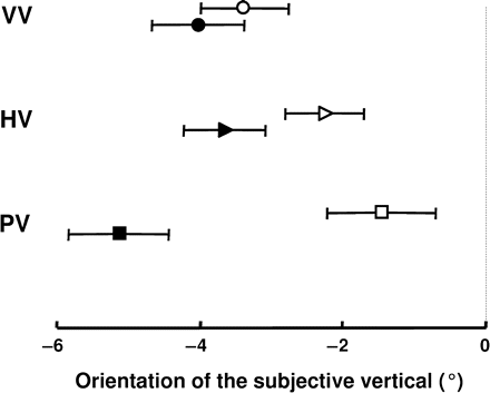
Then, verticality perception in patients with left and RH strokes, whose lesion sizes were similar (left 4.8 ± 1.8, right 4.8 ± 2.4; T = 0.07, P = 0.95), were compared. A difference was found between left and RH strokes, depending on the modality considered (Fig. 4). No significant difference between left and RH strokes was found for VV (T = 0.71, P = 0.48) or HV (T = 1.75, P = 0.08) whereas PV was more tilted in patients with a RH stroke than in those with a left hemisphere stroke (T = 3.55, P < 10−3). Since results showed that verticality perception was both influenced by lesion size (Table 2) and side (Fig. 4), the existence of a possible interaction between these two factors was explored using a two-factor ANOVA (side, size in three groups) in the 78 right-handed or ambidextrous patients with a hemisphere stroke. On VV the ANOVA did not show any side effect [F(1,72) = 0.6; P = 0.46], size effect [F(2,72) = 1.5; P = 0.23] or interaction [F(2,72) = 2.9; P = 0.06]. On HV the ANOVA also failed to show any side effect [F(1,72) = 2.7; P = 0.1], size effect [F(2,72) = 0.8; P = 0.44] or interaction [F(2,72) = 2.8; P = 0.07]. In contrast on PV the ANOVA confirmed the side effect [F(1,72) = 12.9; P < 10−3] and revealed a size effect [F(2,72) = 5.1; P = 0.008] as well as an interaction between the factors side and size [F(2,72) = 3.3; P = 0.04]. The tilt of the PV was not influenced by lesion size in the left hemisphere but increased with lesion size in the RH (−1.3°/−5.9°/−7.9°).
Comparative analysis of verticality estimates in patients with a left (open symbols) or a right (black symbols) hemisphere stroke. Data are given in the form mean values ± SE.
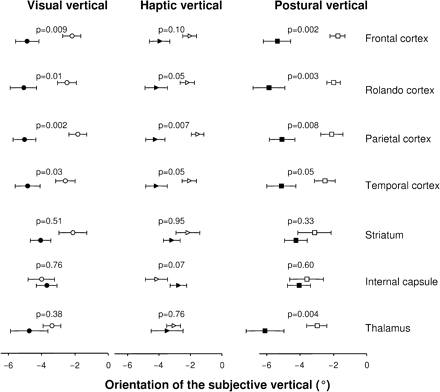
For each modality of perception comparisons were made of verticality estimates with involvement versus non-involvement of seven anatomical regions: frontal, Rolandic, parietal and temporal cortices, striatum, internal capsule and thalamus (Fig. 5). Due to the multiple comparisons we applied a Bonferroni correction to adjust the level of significance to 0.007 (0.05/7). In patients with parietal involvement, all modalities were more tilted than in patients in whom this area was spared. No other significant difference was found for VV or HV. The PV was also significantly more tilted when the frontal cortex, the Rolando's cortex and thalamus were involved. These findings indicate that the Rolando's cortex (presumably the superior parietal cortex) and the thalamus are key cerebral areas for the construction of the PV.
Comparisons of VV, HV and PV between patients in whom a given cerebral structure was involved by the stroke (black symbols) and patients in whom this cerebral structure was anatomically intact (spared by the stroke; open symbols). Data are given as means ± SE with a level of significance set at 0.007 (see main text).
Relationship between verticality perception and lateropulsion with or without pushing (Fig. 6)
Brainstem strokes
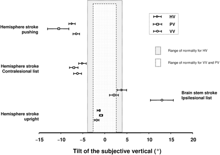
Patients with a brainstem stroke were compared to normal subjects with a two-factor ANOVA (group, modality). Verticality perception was more tilted in brainstem stroke than in normals [F(1,111) = 153.8, P < 10−6] with a difference between modalities [F(2,111) = 43.7, P < 10−6], as well as an interaction between group and modality factors [F(2,111) = 46.5, P < 10−6]. This interaction was due to VV, HV and PV in normals being of similar magnitude (see the section ‘Verticality perception in normals’) whereas a dissociation between modalities was found in patients with a brainstem stroke [F(2,15) = 11.4, P < 10−3]: post hoc analyses showed that VV was much more tilted than HV and PV which did not differ.
Orientation of the three modalities of verticality perception (VV in circles, HV in triangles and PV in squares) for each group of patients: upright (patients with a hemisphere stroke without lateropulsion), with an ipsilesional lateropulsion without pushing (brain stem stroke), with a contralesional lateropulsion without pushing (hemisphere stroke), with a contralesional lateropulsion plus pushing (hemisphere stroke). Limits of normality calculated in 33 normal age/sex matched subjects are represented: between dotted lines for VV and PV (from −2.5° to + 2.5°), in gray for HV (from −4.5° to +4.5°). Data are given in the form mean ± SE.
Hemisphere strokes
Patients with a hemisphere stroke were compared to normal subjects with a two-factor ANOVA (group = normal, upright, listed, pushing; modality = VV, HV, PV). There was a marked difference between groups [F(3,327) = 72.6, P < 10−6] with no significant difference between modalities [F(2,327) = 2.7, P = 0.06] and an interaction between group and modality factors [F(6,327) = 2.14, P = 0.05]. Post hoc analyses showed that verticality perception was more tilted in pushers than in all other groups; more tilted in listing patients than in upright patients and normal controls, and also more tilted in upright patients than in healthy subjects (although both were within the normal range).
The interaction between subject groups and verticality modality factors was analysed by comparing patient groups to the normals with a two-factor ANOVA (group, modality). In the 80 patients with a hemisphere stroke the correlation between verticality estimates and the degree of lateropulsion (SCP) was weak for VV (r = −0.33, P = 0.003), intermediate for HV (r = −0.49, P < 10−3) and much stronger for PV (r = −0.71, P < 10−6).
Patients considered as ‘upright’ (without listing) showed a greater inclination of verticality estimates than normals [F(2,128) = 29.6, P < 10−6], but with no difference between modalities [F(2,228) = 1.1, P = 0.23] nor interaction between group and modality factors [F(2,228) = 0.72, P = 0.49].
Patients with listing had a greater inclination of verticality estimates than normals [F(1,180) = 147.7, P < 10−6], but no difference between modalities [F(2,180) = 1.6, P = 0.21] nor interaction between group and modality factors [F(2,180) = 0.99, P = 0.37].
Pushers had a greater inclination of verticality estimates than normals [F(1,111) = 321.7, P < 10−6], as well as a difference between modalities [F(2,111) = 7.2, P = 0.001] and an interaction between group and modality factors [F(2,111) = 7.1, P = 0.001]. Post hoc analyses showed that all VV, HV and PV were much more tilted in pushers than in normals, and that their PV was more tilted than the VV or HV.
Discussion
VV in stroke
Verticality perception in stroke patients has generally been assessed using the VV (Dieterich and Brandt, 1992; Brandt et al., 1994; Kerkhoff and Zoelch, 1998; Yelnik et al., 2002). Reported abnormalities include a severe tilt due to ocular torsion in brainstem lesions (Friedmann, 1970; Dieterich and Brandt, 1992; Bronstein et al., 2003) or a moderate tilt away from the lesion side in hemispheric lesions (Brandt et al., 1994; Kerkhoff and Zoelch, 1998; Yelnik et al., 2002; Saj et al., 2005b; Johannsen et al., 2006; Bonan et al., 2007). Since extensive multi-sensory processing occurs in vestibulo-cortical and related areas, the moderate VV tilts observed in cortical lesions have been postulated to reflect a problem in verticality representation (Brandt et al., 1994). Together with Kerkhoff (1999), who found a correlation between the haptic and the visual verticals in some patients, our finding that the haptic, the visual and the PVs can tilt similarly in hemisphere stroke provides one of the first direct results in support of this notion.
Ipsilesional tilts of the VV in stroke are of two types. Large, usually ispilesional VV tilts occur in brainstem stroke, as in 6/6 of our brainstem patients and are due to ocular torsion caused by asymmetrical tonus in torsional vestibulo-ocular pathways (Dieterich and Brandt, 1992; Bronstein et al., 2003). Patients with upper brainstem lesions, absent in our current series, can show contralateral VV tilt due to mid brainstem decussation of torsional vestibulo-ocular pathways (Dieterich and Brandt, 1993b). Our study also confirms that <10% of patients with a hemisphere stroke can have small ipsilesional VV tilts (Dieterich and Brandt, 1993a; Brandt et al., 1994; Saj et al., 2005a). This latter finding does not result from an ipsilesional tilt in a high order representation of verticality since we found no hemispheric stroke patients with ipsilateral HV or PV tilts, but several patients with ipsilesional VV tilts and contralateral PV tilts (Fig. 3). On the basis of its low frequency, small magnitude and non-congruency with PV tilts, ipsilesional VV tilt remains a curiosity for future study.
Our study also confirmed that VV, PV and HV can be dissociated (Bisdorff et al., 1996b; Anastasopoulos et al., 1997; Karnath et al., 2000; Bronstein et al., 2003). Such dissociations were present in all our patients with brainstem lesions indicating that the existence of a unique representation of verticality is unlikely at this low anatomical level. According to previous studies (Bisdorff et al., 1996b; Bronstein et al., 2003) this is explained by the prominent ocular tilt/torsion created by interruption of vestibulo-ocular pathways, a main cause inducing tilts of the VV (Dieterich and Brandt, 1992), without concomitant HV or PV. Dissociations were also frequent in hemisphere stroke and these were mediated by different stroke locations (see below). However, none of the six patients with pushing showed dissociation between modalities of verticality perception (transmodal tilt). The corollary of these dissociations between VV, HV and PV is that they are differently related to the severity of the lateropulsion. Our study confirms that a VV tilt is a poor predictor of the postural impairment (Kerkhoff, 1999; Bonan et al., 2007), and reveals that the severity of the lateropulsion is closely associated to the magnitude of PV tilts.
PV, lateropulsion and pushing behaviour
Our data in brainstem stroke show severe VV tilt and lateropulsion and yet minor or no tilts of the PV. VV tilt and lateropulsion can therefore be explained by a low level effect, namely, involvement of vestibular nuclei and related structures directly affecting vestibulo-ocular and vestibulo-spinal projections respectively (Bisdorff et al., 1996b; Bronstein et al., 2003). In addition, the fact that PV is essentially intact contradicts previous interpretations (Dieterich and Brandt, 1992) invoking a high level processing origin for brainstem lateropulsion (e.g. as due to an abnormal internal representation of verticality). This is further supported by observations in normal subjects during vestibular galvanic stimulation in whom galvanic stimuli induced lateropulsion whilst perceptual PV estimates were not tilted (Bisdorff et al., 1996b). Thus vestibular nuclear lesions or stimulation (galvanic) interfere with postural control via direct vestibulo-spinal mechanisms rather than via high order representational mechanisms. Recent direct evidence of this has been given by imaging (Thomke et al., 2005).
Abnormal PV tilts in hemisphere stroke were always contralesional (Fig. 3). The degree of lateropulsion correlated with the magnitude of the PV bias, suggesting an implicit alignment of posture with a tilted PV. A similar mechanism has been shown in the pitch plane: frail elderly patients with postural retropulsion have a net backward tilt of their PV (around 8°) in addition to a transmodal disorder (PV + HV) of their verticality perception (Manckoundia et al., 2007). In the present study patients with pushing displayed the largest PV tilts together with HV and VV contralesional tilts. Therefore pushing may result if the patients struggle to orient to such a compelling set of cues to verticality, using their stronger limbs to push their body into a position in which they feel vertical. Lateropulsion or listing of hemispheric origin may lie on a continuum with pushing, that is, sharing a common mechanism of attempting to align body posture with an erroneous perception of verticality. This interpretation is consistent with the difficulty experienced in making a clinical diagnosis of pushing since the subjective manual assessment is a potential cause of error (lateropulsion with no resistance, no pushing of the examiner versus lateropulsion with resistance, patients push the examiner). This has been discussed in the study by Baccini et al. (2006) who reported a very low agreement on the diagnosis of pushing between the use of the SCP criteria and the clinical impression of an expert. An objective measure of the lateropulsion would be advantageous in resolving such issues (Gissot et al., 2007).
There is disagreement in findings between the few available studies on the PV in stroke. The bias we found was congruent in direction with that found by Perennou et al. (1998) in stroke patients during an active body orientation task with respect to vertical. In contrast, in five pushers Karnath et al. (2000) found biases of similar magnitude to those in our current study but opposite in direction, i.e. ipsilesional. On the other hand, Lafosse et al. (2004, 2007) described increased variance and small tilts of the PV of variable direction (2–6° ipsilesional) in neglect patients. The explanation of the difference found in PV polarity between Karnath study and ours is not due to the nature of the strokes studied, similar in size and location. An explanation could be sought in the interpretation of ‘upright’. A normal seated subject can indicate accurately if the long axis of his body is earth upright (Bisdorff et al., 1996b; Bronstein, 1999) because left-right tilts are mirror symmetrical in terms of forces acting on the body. Changes in body symmetry could be relevant to patients with hemiplegia who might feel more securely supported when lying on the side with good muscle control and sensation than if tilted to the hemiplegic side, just as they prefer to base their foot support on the side of lesion when standing. In the sense that ‘uprightness’ is the direction about which one is best balanced (Riccio et al., 1992) it is not surprising therefore that a patient might feel most stable, and therefore most ‘upright’, when tilted to the side of the brain lesion as found by Karnath et al. (2000). However we fail to see how such an ipsilesional bias of PV might explain a contralesional body lateropulsion in pushers, sitting or standing, with eyes open or closed. If patients feel upright in a given body orientation (corresponding to PV), they should not push themselves away from this spatial reference. In contrast, patients who extract their orientation from a multi-sensory combination of cues might have a different perception of subjective orientation, as we found. The ‘switch’ between devoting attention to contact force support and more integrative mechanisms of spatial orientation for estimates of PV could be related to the stage of recovery. In support of this possibility is that Karnath et al. (2000) tested patients within 3 weeks of onset whereas ours were tested about 8 weeks after the stroke. A related possibility is that the effects of perceptual functions temporally change as the stroke evolves, as do the effects of stroke on sensory-motor mechanisms, but it would be unlikely that such reversal in verticality processing would take place without a parallel reversal of the associated pushing behaviour. The fact that time of testing did not influence the direction of the PV tilt in our study in which some patients including pushers were tested early after the stroke (Table 2) argues against this interpretation. On the other hand, differences between our data and those of Lafosse et al. (2004, 2007) could relate to differences in time-to-testing. Their data were obtained on average 4–18 months after stroke and their results may be interpreted as reflecting an improved status. Since tactile/contact and gravitational/visceral mechanisms are key factors for the perception of the PV (Mittelstaedt, 1992, 1998; Pérennou et al., 1998; Anastasopoulos and Bronstein, 1999) as well as proprioception (Bisdorff et al., 1996a; Barbieri et al., 2008), a potential flaw of the studies previously published on PV in stroke patients (Pérennou et al., 1998, 2002; Karnath et al., 2000; Lafosse et al., 2004, 2007) should also be noted in that the legs, and apparently the head, were not restrained during subject tilt. Passive or active movements of the head and the limbs during tilt could provide biased cues to verticality perception. In summary, our finding of severe contralesional PV tilts suggests that a major component in the pusher syndrome is an implicit active body postural alignment with the perceived vertical. Technical and timing issues may have marred some results in previous studies.
Two recent studies have explored the possibility that lateropulsion and/or pushing after stroke could be caused by a biased representation of the long body axis, possibly in interaction with a biased representation of the vertical. They concluded that lateropulsion or pushing cannot be caused by a bias in the representation of the long body axis (Barra et al., 2007) nor a mismatch between the representation of the long body axis and that of the vertical (Barra et al., 2008). This strengthens the idea that, in hemisphere stroke, lateropulsion and pushing may be caused only by a severe contralesional tilt in the vertical reference system.
Lesion location
Our study brings three novel findings. Firstly, transmodal biases in verticality perception were almost exclusively found (94%) in RH strokes suggesting that a representation of verticality integrated across sensory modalities is elaborated in the RH. Secondly, RH strokes were associated with larger tilts of the PV than left hemisphere strokes, and these PV tilts were closely linked both to the size and the location (superior parietal cortex and thalamus) of the stroke. Finally, lesions in all central nervous system (CNS) areas could produce dissociations between modalities of verticality perception with the exception of parietal cortex where lesions biased all modalities of verticality perception. These findings support the view that the RH has a predominant role in the control of the vertical orientation of the body, an essential component for successful postural control (Pérennou, 2006). These findings also support the view that the parietal cortex plays a critical role in the multimodal perception of the vertical, which is a condition for elaborating an internal model of verticality. Our study, consistently with previous findings (Masdeu and Gorelick, 1988; Brandt et al., 1994; Karnath et al., 2005; Johannsen et al., 2006), also reveals that a normal postural perception of the vertical, a pre-condition for normal body orientation with respect to gravity, requires the integrity of neural circuits centred around the superior parietal cortex i.e. the primary somatosensory cortex and the thalamus. Such connectivity between several distant brain structures, such as the thalamus and the parietal cortex, partly explains why the PV is more sensitive than other modalities to the size of the lesion, especially in the RH. Dieterich et al. (2005) have shown the functional importance of the posterolateral thalamus as a relay station for vestibular input to the cortex, with a predominance of the RH. In agreement with Anastasopoulos and Bronstein (1999) our study shows that the functional role of thalamo-parietal projections may be extended to the processing of the somaesthetic graviceptive information.
Although the majority of our patients had a degree of abnormality in verticality perception, one patient in four showed normal VV, HV and PV. Our study revealed that this (infrequent) normality was mainly due to lesion size and location, smaller and more frequently sparing the cortex.
Conclusion
This study shows that brainstem strokes induce ipsilesional tilts in verticality estimates but this tilt occurs mostly, and is disproportionately large, for VV measurements. In contrast, hemisphere strokes provoke for all sensory modalities mostly contralesional tilts, concordant or dissociated across sensory modality. A significant finding is that large contralesional tilts of PV are associated with postural disability and severe lateropulsion. Pushers have a transmodal (i.e. across sensory modalities) contralesional tilt and a large contralesional PV tilt (always larger than −6°). The findings suggest that a major component in the pusher behaviour is an implicit active body postural alignment with the perceived vertical. The RH plays a predominant role in the perception of the PV, and a critical role for the elaboration of an internal model of verticality.
Acknowledgements
For this study DP was supported by La Fondation de l'Avenir; programs ETO-300 and ETA-382.
References
Abbreviations:
- RH
Right hemisphere
- PV
postural vertical
- HV
haptic vertical
- VV
visual vertical
- ipsilist
the patients with an ipsilesional lateropulsion
- contralist
the patients with a contralesional lateropulsion
- SCP
Scale for Contraversive Pushing
- ANOVA
analysis of variance
- PCA
principal component analysis

