
Main menu

  • HOME
  • CONTENT
    • Early Release
    • Featured
    • Current Issue
    • Issue Archive
    • Blog
    • Collections
    • Podcast
  • TOPICS
    • Cognition and Behavior
    • Development
    • Disorders of the Nervous System
    • History, Teaching and Public Awareness
    • Integrative Systems
    • Neuronal Excitability
    • Novel Tools and Methods
    • Sensory and Motor Systems
  • ALERTS
  • FOR AUTHORS
  • ABOUT
    • Overview
    • Editorial Board
    • For the Media
    • Privacy Policy
    • Contact Us
    • Feedback
  • SUBMIT

User menu

Search

  • Advanced search
eNeuro

eNeuro

Advanced Search


  • HOME
  • CONTENT
    • Early Release
    • Featured
    • Current Issue
    • Issue Archive
    • Blog
    • Collections
    • Podcast
  • TOPICS
    • Cognition and Behavior
    • Development
    • Disorders of the Nervous System
    • History, Teaching and Public Awareness
    • Integrative Systems
    • Neuronal Excitability
    • Novel Tools and Methods
    • Sensory and Motor Systems
  • ALERTS
  • FOR AUTHORS
  • ABOUT
    • Overview
    • Editorial Board
    • For the Media
    • Privacy Policy
    • Contact Us
    • Feedback
  • SUBMIT
PreviousNext
Review, Integrative Systems

P2X7 Receptor and Purinergic Signaling: Orchestrating Mitochondrial Dysfunction in Neurodegenerative Diseases

Alexsandra S. Zelentsova, Alexei V. Deykin, Vladislav O. Soldatov, Anastasia A. Ulezko, Alina Y. Borisova, Veronika S. Belyaeva, Marina Y. Skorkina and Plamena R. Angelova
eNeuro 14 November 2022, 9 (6) ENEURO.0092-22.2022; DOI: https://doi.org/10.1523/ENEURO.0092-22.2022
Alexsandra S. Zelentsova
1Belgorod State University, Belgorod 308015, Russia
  • Find this author on Google Scholar
  • Find this author on PubMed
  • Search for this author on this site
  • ORCID record for Alexsandra S. Zelentsova
Alexei V. Deykin
1Belgorod State University, Belgorod 308015, Russia
  • Find this author on Google Scholar
  • Find this author on PubMed
  • Search for this author on this site
Vladislav O. Soldatov
1Belgorod State University, Belgorod 308015, Russia
  • Find this author on Google Scholar
  • Find this author on PubMed
  • Search for this author on this site
Anastasia A. Ulezko
1Belgorod State University, Belgorod 308015, Russia
  • Find this author on Google Scholar
  • Find this author on PubMed
  • Search for this author on this site
Alina Y. Borisova
1Belgorod State University, Belgorod 308015, Russia
  • Find this author on Google Scholar
  • Find this author on PubMed
  • Search for this author on this site
Veronika S. Belyaeva
1Belgorod State University, Belgorod 308015, Russia
  • Find this author on Google Scholar
  • Find this author on PubMed
  • Search for this author on this site
Marina Y. Skorkina
1Belgorod State University, Belgorod 308015, Russia
  • Find this author on Google Scholar
  • Find this author on PubMed
  • Search for this author on this site
Plamena R. Angelova
2Quine Square Institute of Neurology, London WC1N 3BG, United Kingdom
  • Find this author on Google Scholar
  • Find this author on PubMed
  • Search for this author on this site
  • Article
  • Figures & Data
  • Info & Metrics
  • eLetters
  • PDF
Loading

Visual Abstract

Figure
  • Download figure
  • Open in new tab
  • Download powerpoint

Abstract

Mitochondrial dysfunction is one of the basic hallmarks of cellular pathology in neurodegenerative diseases. Since the metabolic activity of neurons is highly dependent on energy supply, nerve cells are especially vulnerable to impaired mitochondrial function. Besides providing oxidative phosphorylation, mitochondria are also involved in controlling levels of second messengers such as Ca2+ ions and reactive oxygen species (ROS). Interestingly, the critical role of mitochondria as producers of ROS is closely related to P2XR purinergic receptors, the activity of which is modulated by free radicals. Here, we review the relationships between the purinergic signaling system and affected mitochondrial function. Purinergic signaling regulates numerous vital biological processes in the CNS. The two main purines, ATP and adenosine, act as excitatory and inhibitory neurotransmitters, respectively. Current evidence suggests that purinergic signaling best explains how neuronal activity is related to neuronal electrical activity and energy homeostasis, especially in the development of Alzheimer’s and Parkinson’s diseases. In this review, we focus on the mechanisms underlying the involvement of the P2RX7 purinoreceptor in triggering mitochondrial dysfunction during the development of neurodegenerative disorders. We also summarize various avenues by which the purine signaling pathway may trigger metabolic dysfunction contributing to neuronal death and the inflammatory activation of glial cells. Finally, we discuss the potential role of the purinergic system in the search for new therapeutic approaches to treat neurodegenerative diseases.

  • α-synuclein
  • exosomes
  • mitochondrion
  • oxidative stress
  • purinergic metabolome
  • tau protein

Significance Statement

We wanted to understand the role of purinergic signaling system in the development of mitochondrial dysfunction in neurodegenerative diseases. This is important because the development of neurodegenerative diseases is closely connected with the mitochondrial dysfunction of nervous cells and purinoreceptors can by triggers in the neuroinflammations. Our results provide a guide on what the potential role of purinergic signaling in the development of neuroinflammation in Alzheimer’s and Parkinson’s diseases is. We found the dual-functional role of purinoreceptor (P2X7), which is involved in both cell proliferation and apoptotic cell death. Investigation of these mechanisms allows researchers to focus their attention on the search for pharmacological targets.

Introduction

Although there is a number of known causative factors, mitochondrial dysfunction serves as a principle driver of neurodegenerative disorders (Mattson et al., 2008; Negroni et al., 2015; Pickrell and Youle, 2015; Abramov et al., 2017; Sukhorukov et al., 2020.; Kaur et al., 2021; Onyango et al., 2021). Mitochondria provide the cell’s energy sources and also participate in the control of secondary messenger levels such as Ca2+ ions and reactive oxygen species (ROS; Roger et al., 2017; Giorgi et al., 2018). Consequently, mitochondrial dysfunction facilitates a decrease in ATP production, Ca2+ dyshomeostasis, and ROS generation. It is known that mitochondria are the primary source of ROS and generate superoxide at the initial site in the respiratory chain under conditions of ischemia and hypoxia. ROS attack macromolecules within the neuronal plasma membrane which leads to their oxidative modification and destruction (Subramaniam and Chesselet, 2013). In the earlier stage of Alzheimer’s disease, alterations of mitochondrial dynamics and mitophagy occur, but the principle mechanisms underlying these changes have yet to be studied in detail (Tapias, 2019).

The metabolic regulation system mediated by purinergic signaling plays a separate role in the neuronal response to oxidative stress. A large amount of experimental data on the modulating effect of ROS on purinergic signaling has been accumulated (Fujita et al., 2009; Espada et al., 2010; Cieślak and Wojtczak, 2018; Maiolino et al., 2019). In particular, a modulating effect of ROS on P2XR purinergic receptor channels has been demonstrated (Mason et al., 2004), which indicates the participation of ROS not only acts as agents triggering oxidative stress but also as neurotransmitters. P2RX7 is unique among purinergic receptors since it is involved in mechanisms of neuroinflammation and is responsible for ATP-dependent cell lysis (Falzone et al., 2018).

This review aims to summarize our current understanding of the role of the P2RX7 receptor and purinergic signaling in the development of mitochondrial dysfunction in neurodegenerative diseases. An understanding of these mechanisms will make it possible to develop and supplement new and existing therapeutic approaches in the treatment of neurodegenerative disorders. Indeed, the potential of purinergic receptor mechanisms as therapeutic targets for the treatment of neurologic and degenerative diseases has already been demonstrated in several studies (Burnstock, 2006; Engel et al., 2016; Woods et al., 2016).

The Concept of Purinergic Signaling and Mitochondrial Physiology

ATP release is regulated by the mechanism of autocrine feedback under normal physiological conditions (Corriden and Insel, 2010; Burnstock, 2015). Extracellular ATP (eATP), associated with purine and pyrimidine nucleotides, perform their functions by transmitting signals through P2 receptors. These receptors are widely represented on various cells of the body (Atkinson et al., 2002).

In the physiology of purine signaling, the following key elements are prominent: ATP release from cells in response to the activation of the cell surface receptor, autocrine activation of P2 receptors, ATP hydrolysis and the formation of adenosine by ectonucleotidases (Bagatini et al., 2018), activation of P1 receptors, and the removal and conversion of adenosine (Junger, 2011).

Families of purinergic receptors and the biology of the P2X7 receptor

A role for the ATP molecule acting as a powerful signal activated at the cell surface via families of purinergic receptors has been previously identified (Campwala and Fountain, 2013). Based on pharmacology and function, purinergic receptors were classified into P1 and P2 families. Adenosine is an essential ligand for the P1 family, and ATP, ADP, and other ligands are for the P2 family.

P1 receptors include four subtypes: A1, A2A, A2B, and A3 (North and Jarvis, 2013; von Kügelgen and Hoffmann, 2016). P2 receptors are divided into two subtypes: ionotropic (P2X) and metabotropic (P2Y). Seven subtypes of ionotropic P2X receptors and eight subtypes of metabotropic P2Y receptors are known (White and Burnstock, 2006). The structural features of purinergic receptors, the architecture of the subunits of homomeric and heteromeric channels, and signal transduction mechanisms are presented in several reviews (Piirainen et al., 2011; Compan, 2012; Hattori and Gouaux, 2012; Baroja-Mazo et al., 2013; North, 2016; Kroemer and Verkhratsky, 2021; Sarti et al., 2021). The structural and schematic models of P2X7 receptor, the most studied member of P2X receptors family, are presented in Figure 1.

Figure 1.
  • Download figure
  • Open in new tab
  • Download powerpoint
Figure 1.

Schematic models of P2X7 receptor. Extracellular ATP is a P2RX7 receptor agonist, as well as a substrate for 5′-ectonucleotidase, which hydrolyzes ATP to adenosine and temporarily generates ADP, which is a P2Y receptor agonist. Adenosine, the product of hydrolysis of adenine nucleotides, activates adenosine receptors or P1 receptors. P2X receptors are ionotropic, and their activation opens the cation channel, which leads to cell hyperpolarization because of the outflow of K+ and the influx of extracellular Na+ and Ca2+. Figure Contributions: Alexsandra S. Zelentsova prepared the structure of P2X7 receptors and several sites for ligands. Plamena R. Angelova developed the ATP-binding site and activation of P2X7 receptor by ATP molecule.

Purinergic P2X receptors are activated by extracellular ATP (Burnstock and Williams, 2000). All members of this family share a common structure, including the transmembrane domain, a large extracellular loop, and intracellular N and C termini (North, 2002). The sequences from the N terminus to the second transmembrane domain are relatively identical. The C terminus is specific for individual P2X receptors and includes consensus binding motifs for protein kinases and other regions that may be involved in intracellular signaling. The P2RX7 is unique in the P2RX family. First, it is activated by a relatively unusually high concentration of eATP (1 mm below the physiological concentration). Second, it allows the penetration of large molecules up to 900 Da, and additionally, it is responsible for eATP-dependent cell lysis, a response that depends on the presence of the C-terminal sequences (Falzone et al., 2018). P2RX7 is associated with lipid rafts, which depend on palmitoylation of Cys residues located in the C-terminal region (Feng et al., 2006). The binding of ATP or its agonists to P2X7 causes a rapid uptake of Ca2+ and exposure of phosphatidylserine (PtdSer) to the cell followed by the release of microvesicles, pore formation, and pyroptosis (Savio et al., 2018). P2RX7 mRNA is expressed by almost all cells. P2RX7 protein can be expressed but persists in the cytoplasm in an inactivated form (Benzaquen et al., 2019). The P2RX7 gene is located on chromosome 12q24 and consists of 13 exons. P2RX7-A mRNA is formed by constitutive splicing, but 12 additional transcripts can be generated by alternative splicing (Cheewatrakoolpong et al., 2005).

The purinergic metabolome in mitochondria

Mitochondria are dynamically linked and can exchange material with one another and with other cellular organelles such as lysosomes and endoplasmic reticulum (ER; Marchi et al., 2014; Soto-Heredero et al., 2017). Mitochondrial fusion is occurred through GTF hydrolysis and is coordinated by the mitofusins MFN1 and MFN2, and the optic atrophy 1 (OPA1) protein located on the outer mitochondrial membrane (OMM; Wai and Langer, 2016). MFN2 is also present on the ER and controls binding of the mitochondria to ER (de Brito and Scorrano, 2008; Naon et al., 2016; Basso et al., 2018). OPA1 is anchored to the inner mitochondrial membrane (IMM) and is responsible for mitochondrial fusion (Delettre et al., 2000). The mitochondrial-specific lipid, cardiolipin, is attached to OPA1 and plays a critical role in inner mitochondrial membrane fusion (Ban et al., 2017; Tilokani et al., 2018). In the regulation of mitochondrial dynamics, proteolytic processing plays a key role, as reviewed by Dietz et al. (2019). Deficiency or loss of fused proteins leads to fragmentation of mitochondria (Ichishita et al., 2008; Kanazawa et al., 2008).

In mitochondrial physiology, an important role is attributed to the purinergic metabolome, which includes the linkage of purines with purinergic receptors and nucleotidases, which is regulated by purinosomes (An et al., 2008). Purinosomes are mesoscale assemblies formed to protect unstable intermediate products and increase metabolic flux during de novo synthesis of purines (Zhao et al., 2013). These structures are dynamic and reversible in response to the depletion of purines (Zhao et al., 2015). Their formation is cell cycle dependent, and is regulated by G-coupled-proteins agonists (GPCR) and casein kinase 2 (Chan et al., 2015). An increase in the overall number of cells containing purinosomes, positively correlates with the degree of purine deficiency in Lesch-Nyhan disease (R. Fu et al., 2015).

Purinosomes are colocalized with mitochondria, supplying the immediate demand for purines via microtubule-driven interactions (Savio et al., 2021). This spatial interaction depends on regular mitochondrial activity and is linked with the mTOR signaling pathway (French et al., 2016; Pedleyet al., 2018). A variety of kinase cascades are involved in purinosome regulation, including the PI3/AKT cascade in the mTOR signal pathway (French et al., 2013).

Mitochondrial physiology is closely associated with intracellular communication and development of different neurodegenerative diseases. It has been shown that whole mitochondrion transfer between cells confers neuroprotective activity (Hayakawa et al., 2016). Mitochondrial stress has also been demonstrated to stimulate the release of molecules such as damage-associated molecular pattern (DAMP) proteins, which generate strong proinflammatory activity (Sliter et al., 2018). PINK and PARKIN are implicated in general control over the quality of mitochondria (Y. Yang et al., 2006), and both have been observed to be mutated in familial cases of PD (Trinh and Farrer, 2013). The mitochondrion is a target for PINK1, but it breaks down very quickly in the cytosol through proteolytic pathways by cleavage of the N terminus (Kondapalli et al., 2012; Lazarou et al., 2012; Yamano and Youle, 2013). PARKIN ubiquitinates some proteins on the surface of mitochondria that are then recognized as autophagy adaptor proteins and delivered to the autophagosome (Chen and Dorn, 2013; Sarraf et al., 2013).

The functional activity of the mitochondrial network depends on protein folding. Incorrectly folded proteins in the mitochondrial matrix induce mitophagy (Jin and Youle, 2013). Nevertheless, mitochondria can form mitovesicles that protect them from mitophagy by removing misfolded proteins. Sugiura et al. (2014) described a model of mitovesicle formation using electron microscopy. Phosphorylated ubiquitin on the mitochondrial membrane can act as a signaling mechanism for common cellular responses to mitochondrial stress. Soubannier et al. (2012) described the formation of mitovesicles enriched with the oxidized protein under conditions of mitochondrial stress, while the nature of the protein load depended on the type of induced stress.

Studies on the proteome of mitovesicles have shown that it is enriched with components of catabolic pathways, electron transport chain proteins, subunits of the pyruvate dehydrogenase complex and Krebs cycle proteins, catabolism of ketone bodies, β-oxidation of fatty acids, and metabolism of neurotransmitters. Concurrently, mitovesicles lack cytosolic mitochondrial peptides, iron-sulfur clusters, proteins of ubiquinone biosynthesis, mitoribosome components, mitochondrial tRNA (mtRNA), enzymes of metabolism and replication of mtDNA, and enzymes of transcription and translation (D’Acunzo et al., 2021). Moreover, recent experimental data revealed different mitovesicles profile derivates within neurons and astrocytes (Fecher et al., 2019). Specifically, sideroflexin-5 (Sfxn5) and monoamine oxidase type B (MAO-B) are particular to astrocytes, while the homolog of the NipSnap1 protein, the OCIA domain-containing protein 2 (OCIAD2), mitochondrial calcium uniporter (MCU), and monoamine oxidase type A (MAO-A) are predominant in neurons (D’Acunzo et al., 2021).

Mitophagy, mitochondrial homeostasis, and neurodegeneration diseases

Mitophagy plays an important physiological role in maintaining mitochondrial homeostasis (Palikaras et al., 2018), regulates such aspects of mitochondrial physiology as dynamics, biogenesis, transport, and recruitment of autophagosomes to eliminate defective mitochondria (Harper et al., 2018). Several molecular pathways of mitophagy regulated through ubiquitin dependent or independent signaling cascades are described in modern works (Khaminets et al., 2016). Ubiquitin-dependent mitophagy is regulated by PINK1 kinase according to modern experimental data (Pickles et al., 2018). At physiological conditions, PINK1 is transported to the inner mitochondrial membrane (IMM) of healthy mitochondria and is cleaved by presenilino-associated rhomboid protein (PARL) to a 52-kDa fragment that is released into the cytosol. The PINK1 fragment is then rapidly ubiquitinated and cleaved by the ubiquitin-proteasome system (UPS). However, various stimuli that cause mitochondrial damage lead to the accumulation of 63-kDa PINK1 on the outer mitochondrial membrane (OMM). PINK1 is then activated by autophosphorylation during mitophagy via PARKIN anchored to OMM and facilitating its E3 ligase activity (Palikaras et al., 2018; Ryan et al., 2021). Then, PARKIN and ubiquitin are phosphorylated, resulting in the assembly of ubiquitin-linked chains K6-, K11-, K48-, and K63- on the OMM (Heo et al., 2015). Parkin builds ubiquitin chains on damaged mitochondria for lysosomal degradation. Mitophagy mediated by the PINK1/Parkin pathway plays an important role in the mitochondrial quality control system and in the elimination of misfolded and unwanted proteins deposited in mitochondria (Cen et al., 2021).

Other ubiquitin E3 ligases such as Gp78 (M. Fu et al., 2013), SMURF1 (Orvedahl et al., 2011), SIAH1 (Szargel et al., 2016), MUL1 (Lokireddy et al., 2012), and ARIH1 (Villa et al., 2017) are also involved in the regulation of mitophagy. After localization on the surface of the OMM, these ligases generate ubiquitin chains, triggering the recursion of autophagy adapters such as optinervin (OPTN), nuclear dot protein 52 (NDP52), and p62 (Lazarou et al., 2015).

Impaired mitophagy mechanisms are closely associated with the development of neurodegenerative diseases including Alzheimer’s disease and Parkinson’s disease (PD; Park et al., 2021). The development of PD is associated with the occurrence of mutations in the PINK1-PARKIN pathway, which inhibits mitophagy (Youle and Narendra, 2011). PARKIN mutations associated with PD prevent the recruitment of PARKIN to mitochondria and the accumulation of damaged mitochondria (Fig. 2). This increases ROS production thereby contributing to the development of PD pathologies (Pickrell and Youle, 2015).

Figure 2.
  • Download figure
  • Open in new tab
  • Download powerpoint
Figure 2.

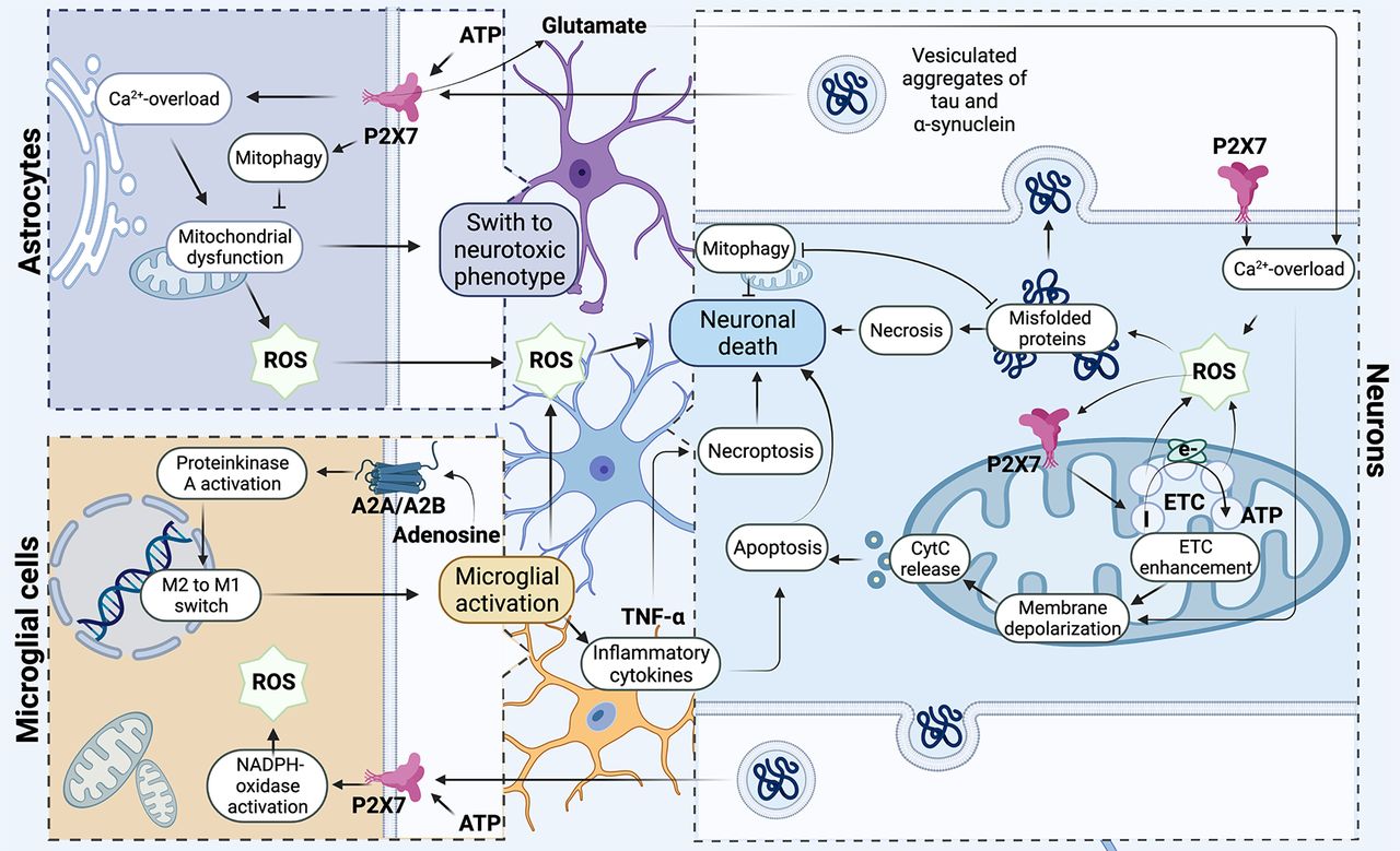
Participation of the P2X7 receptor in the development of mitochondrial degeneration in neurons. The P2X7 purinergic receptor is involved in modulating the redox potential, triggering the production of hydrogen peroxide through the mechanism of Ca2+ release from intracellular stores from the primary microglia. At the same time, in neurons, mitochondria are the primary source of ROS and generate superoxide at the initial site in the respiratory chain under conditions of ischemia and hypoxia. ROS initiates increase of P2X7 expression on the mitochondrial membrane and activity of Complex I, hence increasing mitochondrial polarization, which leads to an elevation of a cascade of unfavorable processes: depolarization of the mitochondrial membrane, calcium overload, and release of cytochrome c, which ultimately leads to cell death by apoptosis way. Under conditions of excessive accumulation of ROS, the process of incorrect aggregation of α-synuclein and tau protein triggered. Oligomeric proteins are involved in coordinating neuroinflammation via the direct interaction of extracellular oligomers with the P2X7 receptor of microglia, which triggers NADPH oxidase activation. Oligomeric proteins trigger the development of mitochondrial dysfunction, localizing in mitochondria and reducing their oxygen consumption and significantly increasing extracellular production of hydrogen peroxide by astrocytes. Purinergic receptors play a key role in the process of neuroinflammation and can trigger activation of microglia. In activated microglial cells, P2X7 is directly involved in triggering the production of TNFα cytokine that can be involved in the process of neuronal death both through apoptosis and through necroptosis; in addition, this receptor stimulates ROS production and triggers mitochondrial dysfunction. Adenosine receptors are involved in the regulation of microglial polarization. Activation of microglia closely connected with purine receptors P1 associated with G-proteins. Activation of adenosine receptor (AR) regulates the activity of protein kinase A (PKA) and participates in the switching of phenotype M1/M2 in the microglial cells. Figure Contributions: Alexsandra S. Zelentsova prepared the mechanism developing of neuronal death. Alexei V. Deykin developed the involvement of oligomeric proteins in the organization of neuroinflammation. Vladislav O. Soldatov performed microglial activation. Anastasia A. Ulezko developed forming ROS by mitochondrion. Alina Y. Borisova developed the pathogenetic mechanisms between microglia, astrocytes, and neuron. Veronika S. Belyaeva performed the formation of unregular folding oligomeric proteins. Marina Y. Skorkina performed the mechanisms of organization of neuroinflammation. Plamena R. Angelova developed the activation of the microglial neuron and the role of P2X7 receptor in neuroinflammation and death of neuron ny necroptosis and apoptosis.

Moreover, α-synuclein disrupts mitophagy through different signaling pathways. In neurons of PD patients, α-synuclein interacts with Miro through its N terminus and increases the level of Miro protein on OMM, which leads to delayed mitophagy. These data indicate that Miro is a target of α-Syn-associated mitochondrial damage (Liu et al., 2019). In addition, overexpression of the A53T α-synuclein mutant leads to p38 MAPK activation and induces PARKIN phosphorylation at Serine131, disrupting PARKIN function and mitophagy (Hirota et al., 2015). In a mouse model with A53T α-synuclein overexpression, mitophagy and neuronal death have been shown to increase on accumulation of α-synuclein in mitochondria (Lonskaya et al., 2013; Rojas-Charr et al., 2014).

Apparently, one of the crucial mechanisms of α-synuclein-induced mitochondrial dysfunction and increased free radical production is activation of the purinergic receptor P2X7. Obviously, α-synuclein is able to directly bind to the transmemrane domain of P2X7 (Fig. 1). The stimulatory effect of α-synuclein on P2X7 and a significant mobilization of Ca2+ were proven on the SHSY5Y neuronal cell model (Wilkaniec et al., 2017).

Direct involvement of the P2X7 purinergic receptor in increased oxidative stress through stimulation of this receptor by extracellular α-synuclein was shown on microglial cells (Jiang et al., 2015). Reduced mitochondrial respiration through mitochondrial depolarization and disruption of mitochondrial Complex I activity followed by increased free radical production are hypothesized to be key molecular events activated by α-synuclein (Devi et al., 2008; Chinta et al., 2010; Reeve et al., 2015). Extracellular α-synuclein induces mitochondrial depolarization with a subsequent increase in the level of mitochondrial superoxide as well as dysregulation of mitochondrial redox homeostasis. Moreover, this effect of α-synuclein largely depends on the activation of P2X7 that induces the Panx-1 recruitment and the formation of pores permeable to large molecules up to 900 Da in size (Wilkaniec et al., 2017).

AMPK is a key P2X7R signaling modulator that triggers mitophagy and mitochondrial division in microglia. On microglial cells, the mechanism of AMPK activation through ROS and calcium signaling pathways during P2X7R stimulation, leading to mitochondrial division, induction of mitophagy, and activation of the nuclear transcription factor TFEB has been proven (Sekar et al., 2018). AMPK regulates the activity of PINK and PARKIN and thus participates in the initiation of mitophagy (Herzig and Shaw, 2018). AMPK regulates PINK and PARKIN activity and thus participates in the initiation of mitophagy (Herzig and Shaw, 2018). Notwithstanding, сalcium signaling activated by P2X7R stimulation may be the molecular mechanism for the α-synuclein-dependent decrease in AMPK activity. The negative effect of P2X7R predominantly involves lysosomal disturbances in microglial cells (Takenouchi et al., 2009; Sekar et al., 2018). However, a study by Wilkaniec et al. (2020) demonstrated inhibition of AMPK activity followed by inhibition of Ulk-1 under conditions of α-synuclein-dependent P2X7 stimulation in nerve cells. It is possible that various cell types can activate different cellular pathways on P2X7 stimulation depending on the receptor isoform (Kaczmarek-Hajek et al., 2018). Thus, mitophagy is the central mechanism in maintaining cellular homeostasis and has a neuroprotective function that protects neurons from the accumulation of defective proteins.

Purinergic Signaling and Redox Homeostasis of the Cell

Purinergic signaling affects the activity of antioxidant enzyme systems, contributing to a change in the redox potential of cells (Piacenza et al., 2019; Coutinho-Silva and Savio, 2021). Reactive oxidants regulate physiological and pathophysiological processes by acting as signaling molecules aimed at cell division and their programmed death (Torres et al., 2013), but uncontrolled production has been shown to contribute to the development of oxidative stress (Rana, 2021).

P2X7 receptor-like inductor of oxidative stress

The P2X7 receptor is involved in modulating the redox potential and can act as an inducer of production of hydrogen peroxide (H2O2) through the mechanism of Ca+2 release from intracellular depots in the primary microglia of transgenic mice with Alzheimer’s disease. These results were confirmed using P2X7 inhibitors (polyphosphonamides and oxidized ATP), which blocked the production of H2O2 and P2X7 agonists (BzATP) that stimulated ROS through the activation of NADPH oxidase (Parvathenani et al., 2003). Also, the P2X7 agonist BzATP was shown to stimulate the phosphorylation of MAPK (Pfeiffer et al., 2007). The work of Zhang and colleagues demonstrated the accumulation of intracellular ROS and the subsequent activation of the NLRP3 inflammasome in macrophages from mice of the J774.1 line when the P2X7 receptor was activated (Zhang et al., 2021).

ROS and the sensitivity of Krebs cycle enzymes

The inactivation of enzymes of the citric acid cycle is one of the negative effects of ROS. Аconitase and α-ketoglutarate dehydrogenase complex (α-KGDHC) are particularly sensitive to oxidative stress (Sadek et al., 2002). Aconitase is most susceptible to ROS which is related to the sulfur-iron complex (Tretter and Adam-Vizi, 2000). α-KGDHC is closely associated with the inner mitochondrial membrane through Complex I of the respiratory chain, and may be a target for ROS because of the close spatial proximity to sites generating ROS (Berndt et al., 2012). Concomitantly, α-KGDHC can also generate H2O2 itself and is a source of reactive oxygen species.

A previous study also demonstrated the production of hydrogen peroxide by isolated α-KGDHC with coenzyme A (HS-CoA) and thiamine pyrophosphate in the absence of nicotinamide adenine oxidized (NAD; Tretter and Adam-Vizi, 2004). In contrast, NADH stimulated the formation of H2O2 by α-KGDHC in the absence of α-ketoglutarate or HS-CoA in the medium (Tretter and Adam-Vizi, 2004).

α-KGDHC is considered one of the key enzymes which limits the rate of the Krebs cycle; accordingly, a decrease in the activity of this enzyme complex initiates a cascade of unfavorable processes: depolarization of the mitochondrial membrane, calcium overload, and release of cytochrome c, which ultimately leads to cell death (Huang et al., 2003). Pathophysiological reactions are triggered within the mitochondria of neurons with reduced activity of the α-KGDHC which faces a high demand for ATP in the cell. Calcium is not sufficiently exported out of the cell into the extracellular space when the cytosolic level of ATP is low, as a result, its concentration increases, which leads to the overloading of mitochondria with calcium, which against the background of reduced activity of the α-KGDHC can initiate apoptosis. This cascade has also been described in the abundant loss of dopaminergic neurons in the substantia nigra (SN) of patients with Parkinson’s disease (Fiskum et al., 2003).

Oxidative stress as a major factor in neurodegenerative disease

ROS-induced oxidative stress is considered by researchers as a major factor in the pathogenesis of Alzheimer’s disease (Cenini and Voos, 2019). In Alzheimer’s disease, the reduction in α-KGDH activity ranges from 25% to 75%; at the same time, there is a correlation with cognitive decline (Boveris and Navarro, 2008). In experimental animal models, as well as in patients with Alzheimer’s disease, a close relationship between oxidative stress and mitochondrial dysfunction has been observed. Transgenic mice with overexpression of human APP (Tg mAPP mice) and progressive accumulation of amyloid-β (Aβ) peptide in synaptic mitochondria exhibited the development of mitochondrial synaptic dysfunction as a result of impaired respiratory activity and mitochondrial axonal transport (Du et al., 2010). A 3xTg-AD mouse model shows that disruption of mitochondrial bioenergetics, together with increased levels of oxidative stress, represents early phenomena that occur before the observed development of Aβ plaques (Hauptmann et al., 2009; Yao et al., 2011).

Some Aspects of Neuroinflammation

Exosomes and neuroinflammation

In recent work, extracellular vesicles have been recognized as playing an essential role in the development of neurodegenerative diseases. Vesicles are a heterogeneous family of endocytosed microvesicles originating from cell membranes circulating in the biological fluids of the organism as a result of the separation of membrane material from any cell type in the body (Paolicelli et al., 2019). Extracellular vesicles perform a regulatory role by an endocrine-like mechanism consisting of the modulation of gene expression in cells located remotely from each other (Budnik et al., 2016; Krämer-Albers and Hill, 2016). Specifically, the role of mobile vesicles produced by microglial cells has been established in the development of cytokine-mediated inflammatory responses in different parts of the brain (Frühbeis et al., 2013). Exosomes are involved in the development of neurodegenerative pathophysiological processes in nervous tissue and transport. Such proteins include α-synuclein, τ and β-amyloid, and pathogenic prion proteins (Ngolab et al., 2017; Y Wang et al., 2017; Zheng et al., 2017).

Exosomes together with purinergic receptors play a key role in the process of neuroinflammation and can trigger apoptosis (Chen et al., 2014). Extracellular ATP is a stimulator of vesiculation in microglial cells bearing P2X7 purinergic receptors. In a mouse model with subclinical inflammation, it was shown that extracellular vesicles released by ATP-stimulated microglia cause a strong inflammatory reaction in glial cells in vitro and can propagate an inflammatory response among microglia (Verderio et al., 2012).

In a study by Drago et al. (2017), ATP-stimulated vesiculation of microglial cells (1 h in 1 mm ATP) was shown. Several enzymes necessary for glycolysis (glucose-6-phosphate isomerase), lactate formation (lactate dehydrogenase, malate dehydrogenase), plus an enzyme of the oxidative branch of the pentose phosphate pathway (transketolase), an enzyme of glutamine metabolism (glutamate dehydrogenase 1) and an enzyme of fatty acid synthesis (acetyl-CoA carboxylase-β) were found in the composition of ATP-stimulated exosomes. ATP-stimulated exosomes also contain proteins of the cytoskeleton and chaperones that interact with the P2X7 C terminus (Gu et al., 2009). In addition, microglial exosomes can function as independent metabolic units and have the potential to increase glucose-based energy outside of mitochondria in recipient cells (Iraci et al., 2017).

The P2RX7 receptor of exosomes as a signal for tau protein spread

Tau protein is known to be associated with microtubules: it interacts with α and β-tubulin subunits helping microtubule assembly. Six isoforms of tau protein have been described which are formed by alternative mRNA splicing and phosphorylation of multiple sites (Kumar et al., 2015). Phosphorylation can lead to oligomerization and the resulting aggregates are implicated in cell-mediated transport which underlies the basis of disease spreading in tauopathies (Kundel et al., 2018).

In a recent study, it was shown that insoluble aggregates of tau proteins can induce ROS production by activation of NADPH oxidase in a calcium-dependent way (Esteras et al., 2021). Furthermore, the activation of NADPH oxidase in combination with membrane-active properties of tau protein aggregates causes neuronal death (Olguín-Albuerne and Morán, 2015).

The main role in the distribution of pathologic tau protein is attributed to exosomes released by microglia during the development of Alzheimer’s disease (AD) (Asai et al., 2015). Among the many constituent exosome molecules, the researchers focused on P2X7 (P2RX7) purinoreceptors which are predominantly expressed in microglia and represent an ATP-induced Na+/Ca2+ channel (Kaczmarek-Hajek et al., 2018). The P2RX7 receptor is thought to have a central role in the progression of AD disease based on the increased expression of P2RX7 in the proximity of amyloid-β (Aβ) in the amyloid plaques of AD patients and in animal models of AD (Parvathenani et al., 2003; McLarnon et al., 2006).

Treatment of the BV2 cell line with α-synuclein increases the secretion of exovesicles expressing the MHC Class II and TNF-α molecules on the surface as well as inducing neuronal apoptosis (Chang et al., 2013). In the case of AD, it has been shown that the Aβ40 and Aβ42 species involved in amyloid formation are present in exosomes (Rajendran et al., 2006). Microglial cells modulate the clearance of Aβ because of its internalization and degradation. Microvesicles from microglia in the presence of excess amyloid are neurotoxic and also have myelotoxic effects (Agosta et al., 2014). Moreover, it has been described that inflammatory microglia release miRNA-enriched microvesicles capable of regulating the level of synaptic proteins in recipient neurons leading to the loss of excitatory synapses, indicating a new mechanism by which microglial cells can mediate synaptic changes in neurodegeneration (Prada et al., 2018).

Exosomes associated with microglia seem to play a role not only in the regulation of synaptic proteins and Aβ levels but also in the spread of tau pathology. In two different mouse models of AD, tau, a neuronal protein associated with microtubules that abnormally accumulates in degenerating neurons, was selectively absorbed by microglia (Guo et al., 2021). Nevertheless, microglia also released tau protein along with exosomes which were captured by cortical neurons thereby contributing to the spread of tau pathology (Vogels et al., 2019). Exosomes can participate in tau transmission demonstrating a certain amount of tau protein is released from neurons through exosomes and partly through synaptic activity (Y. Wang et al., 2017). Depletion of microglia or of the production of exosomes led to a decrease in the spread of mutant tau (Asai et al., 2015). Recent publications have presented data indicating that the spread of tau protein occurs together with low-density lipoproteins, receptor-like proteins, or proteoglycan heparan sulfate (Puangmalai et al., 2020; Rauch et al., 2020).

Pharmacological blockade of P2RX7 by GSK1482160 at an early stage in P301S mice significantly reduced tau accumulation in the hippocampus and exosomes as well as improved hippocampal memory (Ruan et al., 2020). Moreover, the ATP-induced secretion of exosomes from mouse microglia was significantly suppressed by the P2RX7 GSK1482160 inhibitor, whereas it did not affect the secretion of tau or exosomes of primary mouse neurons or astrocytes in vitro. These results indicate the functional role of P2RX7 in the secretion of microglial exosomes and the progression of tau pathology (Ruan et al., 2020).

The stimulation of the P2X7R receptor has a double effect in AD: activation of a nonamyloidogenic neuroprotective pathway and/or overactivation of glial cells causing an excessive proinflammatory response (Martin and Delarasse, 2018; Martin et al., 2019b). The work of Sanz and colleagues demonstrated the release of IL-1β by Aβ plaques during the activation of P2X7R (Sanz et al., 2009). It was found that short-term stimulation of the receptor activates nonamyloidogenic proteolytic cleavage of the precursor protein of the amyloid peptide: α-secretase cuts the APP in the Aβ peptide sequence, which prevents the formation of neurotoxic Aβ peptides and produces a soluble fragment of sAPPα, which is neurotrophic and has neuroprotective effects (Delarasse et al., 2011; Darmellah et al., 2012; Ortega et al., 2013; Heneka et al., 2015; Miras-Portugal et al., 2015; Sáez-Orellana et al., 2016; Hunter et al., 2018; Illes et al., 2019; Martin et al., 2019a). Consequently, activation of P2X7R may exhibit neuroprotective action.

The effect of genetic knock-out of P2X7R in TgAPP/PS1 mice was evaluated to study the role of P2X7R in the development of AD. Using this model, it was revealed that P2X7R deficiency reduces the pathologic effects of the amyloid protein, improves synaptic plasticity in the hippocampus, and normalizes memory. This effect is realized through the recruitment of immune cells in the CNS (Martin et al., 2017; Martin et al., 2019b). The absence of P2X7R reduces the set of cytotoxic CD8 T-lymphocytes which correlates with a decrease in the level of CCL3. The expression of CCL3 and its CCR5 receptor is elevated in the brains of patients with AD and AD mouse models. In these mice, overexpression of CCR5 leads to cognitive impairment. Conversely, inhibition in the hippocampus improves memory (Zhou et al., 2016; Martin and Delarasse, 2018).

α-Synuclein and neuroinflammation

α-Synuclein is mainly located at presynaptic terminals in the brain and participates in vesicular transport, neurotransmitter release, and synaptic plasticity (Stefanis, 2012). Furthermore, the localization of α-synuclein in the nucleus and the membranes of the endoplasmic reticulum-associated with mitochondria has been previously described (Surguchov, 2015). α-Synuclein in monomeric form takes part in synaptic signal transduction: it binds to F0/F1-ATP synthase and increases the ATP synthesis rate in mitochondria under physiological conditions (Ludtmann et al., 2016). Tau protein joins microtubules and is involved in their stabilization and growth processes (Y. Wang and Mandelkow, 2016).

Oligomeric α-synuclein is involved in coordinating neuroinflammation via the direct interaction of extracellular oligomers with the P2X7 receptor of microglia (Hou et al., 2019), which triggers NADPH oxidase activation (Q. Wang et al., 2015). On this basis, it was shown that inhibition of NADPH oxidase by apocynin prevents learning and memory disorders in a model of Parkinson’s disease (PD) in mice (Hou et al., 2019).

Oligomeric α-synuclein binds to F0/F1-ATP synthase causing oxidation of its β-subunit and also lipid peroxidation of the mitochondrial membrane, which in combination with calcium overload, leads to the formation and opening of additional mitochondrial permeability channels and, as a consequence, cell death (Ludtmann et al., 2018). α-Synuclein fibrils also cause neurotoxicity and cell death through activation of nitric oxide synthase (NOS), resulting in DNA damage and activation of polymerase-1 (PARP-1). PARP-1 accelerates α-synuclein fibrillation (Kam et al., 2018). According to the experimental data presented in several works, oligomeric α-synuclein is involved in the initiation of apoptosis (Angelova et al., 2015), necrosis (Reeve et al., 2015), and ferroptosis (Angelova et al., 2020).

Experimental molecular docking data confirm the interaction between α-synuclein and the BH3 domain of BAX, and it has been suggested, that the α-synuclein-BAX complex migrates into mitochondria and initiates apoptotic cell death (Dewson and Kluck, 2009; Chakrabarti et al., 2020).

Purinergic Signaling Pathway and Mitochondrial Disruption

Mitochondrial dysfunction of neurons, astrocytes and microglia is a key factor in the development of neurodegenerative diseases. One of the central signaling mechanisms for the development of mitochondrial dysfunction in neurons and neuroglia is the mobilization of intracellular calcium which can be triggered by the P2X7 purine receptor, as well as through adenosine receptors (ARs). The P2X7 receptor is predominantly localized on the plasma membrane of astrocytes and microglia (Tsuda, 2017). In neurons, its localization on the membrane has not been proven; however, there is evidence that neuronal P2X7R exists as false immunologic signals, but also as potentially operational subunits that regulate neuronal function (Anderson and Nedergaard, 2006; Sperlágh and Illés, 2006). Data have been presented that functional P2X7R is also expressed in immortalized dopaminergic neurons (SN4741 cells) obtained from the substantia nigra of transgenic mouse embryos and performs a function in neuronal differentiation (Jun et al., 2007). In several studies, P2X7R intracellular localization was indicated on the nuclear membrane (Atkinson et al., 2002), mitochondria (Sarti et al., 2021), and intracellular phagosome (Kuehnel et al., 2009). P2X7Rs are thought to induce apoptosis in glial cells through caspase activation and necrosis (Zhao et al., 2018). P2X7Rs control microglial activation and proliferation, potentially leading to a destructive cycle of neuroinflammation and neurodegeneration (Monif et al., 2010).

The P2X7 purinergic receptor is involved in modulating the redox potential, triggering the production of hydrogen peroxide through the mechanism of Ca2+ release from intracellular stores (Piacenza et al., 2019; Coutinho-Silva and Savio, 2021). Excessive accumulation of ROS against the background of dysregulation of the antioxidant system leads to damage to enzymes of the Krebs cycle (aconitase, α-CTGD), oxidation of proteins, DNA, and lipids, thereby disrupting mitochondrial energy which ultimately acts as a pathogenetic mechanism in the development of neurodegenerative diseases (see Fig. 2) and predetermines the aging of the brain and its performance (Rana, 2021).

Mitochondrial dysfunction of astrocytic and microglial cells is associated with their activation. Disruption of calcium homeostasis, overproduction of ROS, and triggering of astrocyte death cascades have all been described in activated astrocytes (Gollihue and Norris, 2020). Oligomeric α-synuclein acts as a factor in the activation of astrocytes and triggers the development of mitochondrial dysfunction, localizing in mitochondria and reducing their oxygen consumption (Braidy et al., 2013). It was found that the accumulation of α-synuclein aggregates in the trans-region of the Golgi complex also disrupts mitochondrial dynamics and leads to fragmentation of mitochondria (Verkhratsky et al., 2016). Fibrillar α-synuclein induces mitochondrial dysfunction of astrocytes and significantly increases the extracellular production of hydrogen peroxide by cells (Chavarría et al., 2018). Moreover, astrocytes can be activated by fragments released from microglia, which are viewed as neurotoxic signaling mediators that inhibit neuronal mitochondrial respiration, significantly increasing neuronal death (Joshi et al., 2019). The process of astrocytic mitophagy is important for the survival of neurons, and astrocytes are directly involved in the spheroid-mediated transmytophagy of dopaminergic neurons. It has been experimentally confirmed that damaged mitochondria during degeneration of dopaminergic neurons are preserved in spheroids where the mitophagy process is initiated but not completed, and subsequently, these spheroids penetrate astrocytic cells and undergo further degradation (Morales et al., 2020, 2021). Consequently, neuron-astrocyte transmitophagy is crucial for preventing the release of mitochondria into the extracellular environment during neuronal death. In addition, astroglia activated by microglia have been found in the brains of patients with different neurodegenerative diseases (Hickman et al., 2018), including PD (Banati et al., 1998), AD (Maat-Schieman et al., 1994), and Huntington’s disease (Tai et al., 2007). Researchers point to the existence of two phenotypes of microglial cells: proinflammatory M1 and neuroprotective M2 types (Kwon and Koh, 2020). Microglial activation is also known to be associated with P1 purine receptors connected with G-proteins (see Fig. 2).

The impaired P2X2 receptor and purinergic signaling across different neurodegenerative diseases are summarized in Table 1.

View this table:
  • View inline
  • View popup
Table 1

Receptors P2X2 family and neurodegenerative diseases

Extracellular ATP is cleaved by ectonucleotidases with the formation of AMP which under the influence of the substrate ecto-5′-nucleotidase (CD73) decomposes to extracellular adenosine. Adenosine can be released from cells into the extracellular environment and act on P1 receptors located in neurons and is considered like a neuromodulator. So far, four subtypes of P1 families have been found: A1, A2A, A2B, and A3. Subtypes A1 and A3 bind to Gi-coupled GPCRs thereby inactivating adenylate cyclase and decreasing intracellular cAMP levels. A2A and A2B bind to Gs-coupled GPCRs, activating adenylate cyclase and increasing intracellular cAMP levels (Yakovlev et al., 2022). Therefore, activation of the adenosine receptor (AR) regulates the activity of protein kinase A (PKA). In addition, protein kinase C (PKC) can be activated through the mobilization of intracellular calcium-mediated by the A2B receptor (Alnouri et al., 2015). Mitochondrial homeostasis in neurons is a key factor in preventing neurodegeneration (Li et al., 2020). Despite adenosine receptors being activated in response to extracellular stimuli, they are also found in mitochondria (Valenzuela et al., 2016; Melser et al., 2017), where they are involved in controlling oxidative stress and mitochondrial performance. Adenosine receptors are involved in the regulation of microglial polarization. A2AR activation leads to increased release of nitric oxide by activated microglia. This study demonstrated that the effect was dependent on the presence of astroglia although both A2AR expression and NO synthase II immunoreactivity were observed in microglia only (Saura et al., 2005).

An important aspect to consider in neurodegenerative disease development is that vesicular transport is used as a means of intercellular communication. Researchers have assigned exosomes, together with receptors of the purinergic signaling system, a key role in the formation of neuroinflammation and the initiation of apoptosis. Extracellular ATP stimulates the vesiculation of microglial cells carrying on their surface P2X7 receptors of the purinergic signaling system (Verderio et al., 2012); these experimental studies have thus provided evidence of the P2RX7 receptor in the progression of AD disease.

P2X7 is directly involved in the regulation of various cell death pathways, such as apoptosis, pyroptosis, necrosis, and autophagy, by the fact that this receptor can regulate cell death via caspase-8 and caspase-9, stimulating AFC production, mitochondrial dysfunction (see Fig. 2), cytochrome c release, and caspase-3/7 activation (Bidula et al., 2019). The involvement of the P2X7 receptor in triggering necroptosis is mediated by its long-term stimulation on glial cells and their release of TNFα (Salas et al., 2013).

Necroptosis, as a type of programmed cell death of necrosis independent on caspases, is activated in response to activation of death receptors and depends on activation of receptor-interacting kinase 3 (RIPK3; activated by RIPK1) and pseudokinase, which acts as the main effector of necroptosis (MLKL). MLKL oligomerizes under the influence of RIPK3 and moves to the lipid rafts of the plasma membrane with pore formation, causing influx of Na+ ions and cell death (Su, 2015; Grootjans et al., 2017; Liu, 2019). It has been proven that RIP3 leads to a switch in cellular metabolism which is accompanied by an increase in the production of mitochondrial ROS and ultimately of cell death. Necroptosis is characterized by rapid membrane degradation accompanied by the release of intracellular contents, that is, damage-associated molecular patterns (DAMPs) such as highly mobile group 1 protein (HMGB1), heat shock proteins, DNA, and RNA, which activate the pattern recognitions receptors (PRRs) to further stimulate the inflammatory response (Negroni, 2015).

Various triggers, including TNF, Fas, TNF-associated apoptosis-inducing ligand (TRAIL), interferon (IFN), lipopolysaccharide, DNA damage, viral infection, and cancer cell suppressant drugs, are believed to trigger necroptosis in cells (Holler et al., 2000; Jouan-Lanhouet et al., 2012; Grootjans et al., 2017; Xu et al., 2021). Fas, TNF-induced, and TRAIL-induced necroptosis requires the activity of RIPK1 kinase, which has several domains and can activate various cell death pathways (Cai et al., 2014; H Wang et al., 2014; Hribljan et al., 2019). Inhibitor of necroptosis is caspase-8. Necroptosis represents, a tightly regulated process, the malfunction in the regulation of which is associated with the development of human diseases, which are accompanied by cell loss and inflammatory reactions (Chen et al., 2019).

Thus, necroptosis is involved in the development of neurodegenerative diseases such as AD, ALS, PD. Increased levels of markers of necroptosis, including RIPK1, MLKL, necrosome complex, and MLKL oligomer, in these diseases have been noted (Zheng-gang and Jiao, 2020). Because of these findings, the possibility of inhibiting necroptosis by interfering with and reducing the activity of necrosome components is being considered as new therapeutic targets for treatment (Caccamo et al., 2017).

Purinergic Receptors as Therapeutic Targets for the Treatment of Alzheimer’s and Parkinson’s Diseases

Purinergic signaling pathways are now considered as therapeutic targets in the treatment of various pathologies. In the difficult treatment of neurodegenerative diseases, successes can be realized, provided that new therapeutic targets are identified.

P2X7 receptors as therapeutic targets

The P2X7 receptor is promising as a therapeutic target in the development of Parkinson’s disease. A study by Oliveira-Giacomelli et al. (2019) found that ATP and its metabolites were extensively released in an animal model of Parkinson’s disease induced by 6-hydroxydopamine (6-OHDA). Previous work has examined the effects of pharmacological antagonists on the P2X7 receptors in preventing as well as altering hemi-Parkinson’s behavior and dopaminergic deficiency. The P2X7 receptor antagonist brilliant blue G (BBG) at a dose of 75 mg/kg restored the dopaminergic nigrostriatal pathway in rats (Carmo et al., 2014; Y. Wang et al., 2017; Oliveira-Giacomelli et al., 2019). Therapeutic strategies in the treatment of Alzheimer’s disease are also aimed at suppressing neuroinflammation, organized with the participation of microglial cells.

Microglial cells and astrocytes overexpress the P2X7R receptor which has been shown to be responsible for NLRP3 (cryopyrin, part of the NOD-like receptor family) inflammasome activation and promotes the release of the proinflammatory cytokine interleukin-1β (IL-1β). IL-1β was in turn found in the brains of AD patients and in a mouse model with developing amyloid lesions (TgAPP/PS13 transgenic mice; Marcoli et al., 2008; Bhaskar et al., 2010; Cervetto et al., 2013; Ficker et al., 2014; Di Virgilio et al., 2017; Martin et al., 2017; Martin and Delarasse, 2018; Martin et al., 2019a).

At present, the idea of developing a drug that can act on the NMDA and P2X7 receptors simultaneously would present a useful and promising treatment strategy for Alzheimer’s disease. Both receptors exhibit neuroprotective and cytotoxic effects that make them ideal candidates not only for stopping the progression of the disease but also for alleviating symptoms. Hence, the most appropriate antagonist would be a compound that exhibits inhibitory activity in the low micromolar range, thus preserving the physiological function of the receptors while inhibiting them when overactivated. Karoutzou and colleagues used amino adamantyl carbohydrazide as a putative dual inhibitor. These investigators also synthesized, characterized, and evaluated several new adamantyl carbohydrazides as potential dual NMDAR and P2X7R antagonists. Three derivatives showed low micromolar activity as P2X7R antagonists, and only one compound, designated “9g,” showed some activity as an NMDA receptor antagonist. New compound 9g for dual P2X7 and NMDA receptors with low inhibitory potenciec has been designed (Karoutzou et al., 2018).

The P2Y6 receptor

PD induced by 1-methyl-4-phenylpyridine (MPP) is known to increase UDP/P2Y6R levels on SH-SY5Y-derived neuronal cells. Pharmacological inhibition of P2Y6R or knock-down of P2Y6R using small interfering RNA (siRNA) prevented MPP-induced increases in levels of ROS, superoxide-anions, inducible nitric oxide synthase (iNOS), malondialdehyde (MDA), and superoxide dismutase 1 (SOD1). Apyrase and MRS2578 have also been investigated as potential pharmacological inhibitors (Qian et al., 2018).

MRS2578 being a selective P2Y6 receptor antagonist prevents the death of dopaminergic neurons in SH-SY5Y-derived neurons both in vitro and in vivo in rat substantia nigra. In addition, authors found that both treatments were accompanied by a decrease in microglial activation in the substantia nigra. Thus, antagonism of P2Y6 receptors has clear neuroprotective effects (Oliveira-Giacomelli et al., 2019).

A2A receptors

There is a wide range of pharmacotherapeutic agents available for the treatment of Parkinson’s disease. These include dopamine precursors with peripheral dopa decarboxylase inhibitors, dopamine agonists, cholinolytics, monoamine oxidase B (MAO-B) inhibitors, and catechol-o-methyltransferase (COMT) inhibitors. Nevertheless, the gold standard of treatment is still levodopa/carbidopa combination therapy, because levodopa has been seen to provide lasting improvement in mobility and carbidopa helps maintain appropriate levels of levodopa in the substantia nigra (SN; Hayes, 2019; Armstrong and Okun, 2020; Sahoo et al., 2020). However, the therapeutic effects begin to wane and are gradually outweighed by side effects such as on-off fluctuations with or without dyskinesia, clinically characterized by the absence and recurrence of motor symptoms with continued use of this combination (Armstrong and Okun, 2020; Sahoo et al., 2020). In addition to existing therapies, researchers have suggested the inclusion of drugs aimed at inhibiting A2A purinergic receptors, which inhibit the amplification of signaling through A2A receptors and reduce signaling through D2 dopaminergic receptors (Sahoo et al., 2020; Singh et al., 2021). These changes explain the positive effect of A2A receptor antagonists on enhancing motor function without progression of levodopa-induced dyskinesias (Fuxe et al., 2015; Navarro et al., 2016). Accordingly, it is logical to use selective A2A receptor antagonists such as istradefylline as additional agents to alleviate these.

Istradefylline (KW-6002) is a xanthine-based compound with increased selectivity for A2A receptors against A1 receptors (Poewe et al., 2012), and in a rat model of lipopolysaccharide (LPS)-induced neuroinflammation, it showed an anti-inflammatory effect of this compound (Gołembiowska et al., 2013). In addition to istradefylline, several powerful and highly selective inhibitors of adenosine A2A receptors have been developed (Shook and Jackson, 2011). The drugs istradefylline, preladenate, and tozadenate have been tested in phase 2b and phase 3 clinical trials and were potent and highly selective A2A receptor antagonists with good CNS penetration (Brooks et al., 2010; Pawsey et al., 2013; Pinna, 2014; LeWitt et al., 2020). Preladenate, however, did not make it to the end of phase 3 clinical trials in the treatment of PD because the compound did not outperform placebo in reducing the “off” state, and tozadenate was excluded from the phase 3 clinical trial in the  year 2018 because of serious side effects, hematologic toxicity such as agranulocytosis (Hauser et al., 2014, 2015; Pinna, 2014; Pinna et al., 2018; LeWitt et al., 2020).

A2A receptor antagonists can also directly or indirectly affect microglia and inflammatory processes. Gyoneva et al. (2014) processed brain slices of mice injected with 1-methyl-4-phenyl-1,2,3,6-tetrahydropyridine (MPTP) and concluded that preladenate adequately promotes microglial responses to tissue damage as well as enhancing the therapeutic effect of low dose l-DOPA (Gyoneva et al., 2014).

Information on preclinical and clinical trials of substances showing antagonism to purine receptors is presented in Table 2.

View this table:
  • View inline
  • View popup
Table 2

Purine receptor antagonists in Parkinson’s disease

Discussion: Perspectives and Challenges

The undoubted interest in P2X7R as a potential target for therapeutic agents is associated with certain successes in the treatment of inflammatory processes. However, the dual-functional role of P2X7, which is involved in both cell proliferation and apoptotic cell death shown in some studies, requires further study to understand the mechanisms involved in intracellular signal transmission with the participation of this receptor and imposes some restrictions on the use of these blockers in the development of neurodegenerative diseases. In addition, the presence of multiple isoforms of P2X7R in humans and experimental animals requires a meticulous approach when using certain receptor antagonists, since they are not always effective in some polymorphic types of receptors. An important aspect to consider is a detailed study of the mechanisms of the inflammatory response, since the nonselectivity of the antagonists chosen could lead to an imbalance in immune homeostasis.

A promising future direction is the search for therapeutic targets to provide microglial-cell-mediated neuroprotection. In this context, the therapeutic targets are adenosine receptors which provide a mix of phenotypes from proinflammatory (M1) to neuroprotective (M2). This approach to maintaining the balance of M1/M2 can ensure the survival of neurons in conditions of chronic inflammation.

Astroglia may also provide another course for further research. In astroglia, misfolded oligomeric proteins can result in a dysfunctional phenotype. In particular, α-synuclein is transferred from neurons to astrocytes during the development of neurodegenerative diseases. As a result, as mitochondrial dysfunction develops, the process of mitophagy is disrupted, and ER stress proceeds, which exacerbates neurodegenerative processes. Therefore, one of the tactics in maintaining the viability of neurons in conditions of neurodegeneration is the preservation of astrocytic function, which will be key for the survival of neurons. Further studies of the mechanisms of involvement of astrocytes in the development of neurodegenerative diseases will be important in the development of new therapeutic approaches.

Conclusion

The purinergic signaling system is directly involved in the regulation of intracellular redox homeostasis and triggers a cascade of reactions that produce various forms of reactive oxygen and nitrogen. Excessive accumulation of ROS against the background of dysregulation of the antioxidant system leads to the oxidation of intracellular proteins, DNA, and lipids, which ultimately leads to disruption of mitochondrial energy. In modern research, extensive work is currently being devoted to describing the potential role of purinergic signaling in the development of various neurodegenerative diseases (Alzheimer’s, Parkinson’s, and Huntington’s diseases, multiple and amyotrophic lateral sclerosis). A common characteristic of these diseases is neuronal degeneration, which occurs against the background of neuroinflammation with sequential neurodegeneration because of the activation of P2X7Rs in microglia. In addition, the P2X7 receptor has specific binding sites for extracellular misfolded proteins that accompany the development of Alzheimer’s and Parkinson’s diseases and can spread between neurons and astrocytes by a prion-like mechanism.

The purinergic signaling system is the central trigger element in the development of mitochondrial dysfunction that accompanies neurodegenerative diseases. Among the whole family of purinergic receptors, P2RX7 is of decisive importance in future studies in terms of studying the involvement of this process in trophic, cytoprotective, and neuroinflammatory aspects (Fig. 3). Now, under what conditions one or another signaling path turned on is not clear, or what mutual signaling paths can switch one or another signal transmission path. It is necessary to find out what common substrates can be involved in the implementation of the opposite functions of this receptor.

Figure 3.
  • Download figure
  • Open in new tab
  • Download powerpoint
Figure 3.

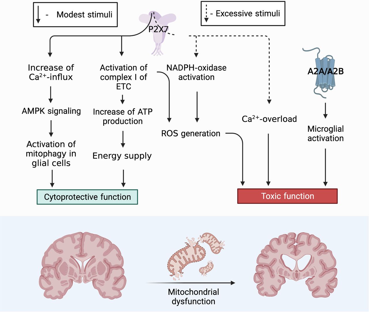
Role of P2X7 receptor in the functional activity of neurons. Toxic function: P2X7 triggers an intracellular signaling pathway for the activation of free radical oxidation processes and the accumulation of ROS, as a result, mitochondria are actively involved in the process of ROS production. ROS via AMPK signaling pathway can trigger mitophagy, thereby controlling the presence of defective mitochondria in the neuron removing them and ensuring cell viability. However, with excessive accumulation of ROS and a decrease in the activity of the antioxidant systems of the cell, the correct functioning of the PARKIN protein is blocked, which leads to inhibition of the mitophagy process. Misfolding of proteins, defective mitochondria accumulate in the cell. One of the defective proteins is α-synuclein that in turn has binding sites for the P2X7 receptor stimulating it and thereby increasing the Ca2+ concentration in the cell. An elevated level of Ca2+ blocks AMPK activity, after which the death of neurons can follow the path of necrosis. Cytoprotective function: P2X7 triggers mitophagy throught the Ca2+ increases which activation AMPK signaling pathway. It eliminates defective mitochondria thereby contributing to the survival of neuroglia and maintaining its viability in conditions of short-term stimulation. P2X7, localized on the outer mitochondrial membrane, captures the concentration of ATP in the cell and, with a decrease in its production, enhances the work of Complex I (NADH-DH) of the electron transport chain, as a result of which oxidative phosphorylation is stimulated and ATP production in the cell increases. Figure Contributions: Alexei V. Deykin described the cytoprotective function of P2X7 through activating AMPK-signaling pathway. Vladislav O. Soldatov performed the cytoprotective function by activation Complex I of ETC. Marina Y. Skorkina described the toxic function through ROS. Plamena R. Angelova developed the toxic function through Ca2+ overload.

Based on our generalizations, the primary role of P2X7 localized on neurons and glia in the formation of an inflammatory focus during prolonged uncontrolled stimulation is obvious. Under these conditions, P2X7 triggers an intracellular signaling pathway for the activation of free radical oxidation processes and the accumulation of ROS; as a result, mitochondria are actively involved in the process of ROS production. ROS via AMPK signaling pathway can trigger mitophagy, thereby controlling the presence of defective mitochondria in the neuron removing them and ensuring cell viability. However, with excessive accumulation of ROS and a decrease in the activity of the antioxidant systems of the cell, the correct functioning of the PARKIN protein is blocked, which leads to inhibition of the mitophagy process. In case of misfolding of proteins, defective mitochondria accumulate in the cell. One of the defective proteins is α-synuclein that in turn has binding sites for the P2X7 receptor stimulating it and thereby increasing the Ca2+ concentration in the cell. An elevated level of Ca2+ blocks AMPK activity, after which the death of neurons can follow the path of necrosis.

The cytoprotective function of P2X7 for glial cells has been described. With short-term stimulation of P2X7, the concentration of Ca2+ increases and activating AMPK signaling pathway. It triggers mitophagy and eliminates defective mitochondria thereby contributing to the survival of neuroglia and maintaining its viability.

The trophic function of the P2X7 receptor localized on intracellular membranes is an interesting and little studied aspect. In particular, P2X7, localized on the outer mitochondrial membrane, captures the concentration of ATP in the cell and, with a decrease in its production, enhances the work of Complex I (NADH-DH) of the electron transport chain, as a result of which oxidative phosphorylation is stimulated and ATP production in the cell increases. Thus, this receptor performs a trophic and sensory function by controlling the level of ATP inside the cell. In this regard, especially promising in terms of studying the mechanisms of development of neurodegenerative diseases is the search for signaling mediators involved in interactions between the P2X7 purinoreceptor localized on the neuron plasma membrane and on the membranes of intracellular organelles, such as mitochondria, phagosomes, and the nuclear membrane. Now is not clear the interaction between these receptors and through which signaling mediators they provide coordination in signal transmission.

Future research should focus on finding a set of signaling mediators that cause the P2X7 receptor to use different signaling pathways depending on the microenvironment and the concentration of signaling ligands. It is necessary to understand under what conditions the inflammatory role, trophic or cytoprotective function of the receptor, is realized, as well as how this receptor can be involved in the formation of the mitochondrial network and the fusion of intracellular membranes.

Purinergic receptors have multifaceted properties in the CNS: they not only promote neurotransmission and neuromodulation but also promote chemotaxis. Investigation of the role of the purinergic signaling system and understanding of the intracellular signaling cascades triggered by this system in neuropathological conditions allows researchers to focus their attention on the search for pharmacological targets that would minimize the harmful effects of pathologies and significantly improve the quality of life of patients. Therapeutic strategies in the treatment of neurodegenerative disease are aimed at suppressing neuroinflammation, organized with the participation of microglial cells and the P2RX7 receptor.

Footnotes

  • The authors declare no competing financial interests.

  • This study was supported by a grant from the Belgorod region for the government support implementation of innovative technologies into promotion, within the frameworks of full-cycle technological projects. Molecular studies were supported by the Ministry of Science and Higher Education of the Russian Federation Grant 075-15-2021-1346. Animal procedures were supported by the State Task of the Laboratory of Genome Editing for Biomedicine and Animal Health Grant FZWG-2021-0016.

This is an open-access article distributed under the terms of the Creative Commons Attribution 4.0 International license, which permits unrestricted use, distribution and reproduction in any medium provided that the original work is properly attributed.

References

  1. ↵
    Abramov AY, Berezhnov V, Fedotova RI, Zinchenko VP, Dolgacheva LP (2017) Interaction of misfolded proteins and mitochondria in neurodegenerative disorders. Biochem Soc Trans 45:1025–1033. doi:10.1042/BST20170024 pmid:28733489
    OpenUrlAbstract/FREE Full Text
  2. ↵
    Agosta F, Dalla Libera D, Spinelli EG, Finardi A, Canu E, Bergami A, Bocchio Chiavetto L, Baronio M, Comi G, Martino G, Matteoli M, Magnani G, Verderio C, Furlan R (2014) Myeloid microvesicles in cerebrospinal fluid are associated with myelin damage and neuronal loss in mild cognitive impairment and Alzheimer disease. Ann Neurol 76:813–825. doi:10.1002/ana.24235 pmid:25087695
    OpenUrlCrossRefPubMed
  3. ↵
    Alnouri MW, Jepards S, Casari A, Schiedel AC, Hinz S, Müller CE (2015) Selectivity is species-dependent: characterization of standard agonists and antagonists at human, rat, and mouse adenosine receptors. Purinergic Signal 11:389–407. doi:10.1007/s11302-015-9460-9 pmid:26126429
    OpenUrlCrossRefPubMed
  4. ↵
    An S, Kumar R, Sheets ED, Benkovic SJ (2008) Reversible compartmentalization of de novo purine biosynthetic complexes in living cells. Science 320:103–106. doi:10.1126/science.1152241 pmid:18388293
    OpenUrlAbstract/FREE Full Text
  5. ↵
    Anderson CM, Nedergaard M (2006) Emerging challenges of assigning P2X7 receptor function and immunoreactivity in neurons. Trends Neurosci 29:257–262. doi:10.1016/j.tins.2006.03.003 pmid:16564580
    OpenUrlCrossRefPubMed
  6. ↵
    Angelova PR, Horrocks MH, Klenerman D, Gandhi S, Abramov AY, Shchepinov MS (2015) Lipid peroxidation is essential for α-synuclein-induced cell death. J Neurochem 133:582–589. doi:10.1111/jnc.13024 pmid:25580849
    OpenUrlCrossRefPubMed
  7. ↵
    Angelova PR, Choi ML, Berezhnov AV, Horrocks MH, Hughes CD, De S, Rodrigues M, Yapom R, Little D, Dolt KS, Kunath T, Devine MJ, Gissen P, Shchepinov MS, Sylantyev S, Pavlov EV, Klenerman D, Abramov AY, Gandhi S (2020) Alpha synuclein aggregation drives ferroptosis: an interplay of iron, calcium and lipid peroxidation. Cell Death Differ 27:2781–2796. doi:10.1038/s41418-020-0542-z pmid:32341450
    OpenUrlCrossRefPubMed
  8. Anwar S, Pons V, Rivest S (2020) Microglia purinoceptor P2Y6: an emerging therapeutic target in CNS diseases. Cells 9:1595. doi:10.3390/cells9071595
    OpenUrlCrossRef
  9. ↵
    Armstrong MJ, Okun MS (2020) Diagnosis and treatment of Parkinson disease: a review. JAMA 323:548–560. doi:10.1001/jama.2019.22360 pmid:32044947
    OpenUrlCrossRefPubMed
  10. ↵
    Asai H, Ikezu S, Tsunoda S, Medalla M, Luebke J, Haydar T, Wolozin B, Butovsky O, Kügler S, Ikezu T (2015) Depletion of microglia and inhibition of exosome synthesis halt tau propagation. Nat Neurosci 18:1584–1593. doi:10.1038/nn.4132 pmid:26436904
    OpenUrlCrossRefPubMed
  11. ↵
    Atkinson L, Milligan LC, Buckley NJ, Deuchars J (2002) Purinergic receptors: an ATP-gated ion channel at the cell nucleus. Nature 420:42. doi:10.1038/420042a pmid:12422208
    OpenUrlCrossRefPubMed
  12. ↵
    Bagatini MD, Dos Santos AA, Cardoso AM, Mânica A, Reschke CR, Carvalho FB (2018) The impact of purinergic system enzymes on noncommunicable, neurological, and degenerative diseases. J Immunol Res 2018:4892473. doi:10.1155/2018/4892473 pmid:30159340
    OpenUrlCrossRefPubMed
  13. ↵
    Ban T, Ishihara T, Kohno H, Saita S, Ichimura A, Maenaka K, Oka T, Mihara K, Ishihara N (2017) Molecular basis of selective mitochondrial fusion by heterotypic action between OPA1 and cardiolipin. Nat Cell Biol 19:856–863. doi:10.1038/ncb3560 pmid:28628083
    OpenUrlCrossRefPubMed
  14. ↵
    Banati RB, Daniel SE, Blunt SB (1998) Glial pathology but absence of apoptotic nigral neurons in long-standing Parkinson’s disease. Mov Disord 13:221–227. doi:10.1002/mds.870130205 pmid:9539333
    OpenUrlCrossRefPubMed
  15. ↵
    Baroja-Mazo A, Barberà-Cremade M, Pelegrín P (2013) The participation of plasma membrane hemichannels to purinergic signaling. Biochim Biophys Acta 1828:79–93. doi:10.1016/j.bbamem.2012.01.002 pmid:22266266
    OpenUrlCrossRefPubMed
  16. ↵
    Basso E, Rigotto G, Zucchetti A, Pozzan T (2018) Slow activation of fast mitochondrial Ca2+ uptake by cytosolic Ca2. J Biol. Chem 293:14081–17094.
    OpenUrl
  17. ↵
    Benzaquen J, Heeke S, Hreich J, Douguet L, Marquette CH, Hofman P, Vouret-Craviari V (2019) Alternative splicing of P2RX7 pre-messenger RNA in health and diseases: myth or reality? Biomed J 42:141–154. doi:10.1016/j.bj.2019.05.007 pmid:31466708
    OpenUrlCrossRefPubMed
  18. ↵
    Berndt N, Bulik S, Holzhütter H-G (2012) Kinetic modeling of the mitochondrial energy metabolism of neuronal cells: the impact of reduced α-ketoglutarate dehydrogenase activities on ATP production and generation of reactive oxygen species. Int J Cell Biol 2012:757594. doi:10.1155/2012/757594
    OpenUrlCrossRefPubMed
  19. ↵
    Bhaskar K, Konerth M, Kokiko O, Cochran N, Cardona A, Ransohoff RM, Lamb BT (2010) Regulation of tau pathology by the microglial fractalkine receptor. Neuron 68:19–31. doi:10.1016/j.neuron.2010.08.023 pmid:20920788
    OpenUrlCrossRefPubMed
  20. ↵
    Bidula S, Dhuna K, Helliwell R, Stokes L (2019) Positive allosteric modulation of P2X7 promotes apoptotic cell death over lytic cell death responses in macrophages. Cell Death Dis 10:882. doi:10.1038/s41419-019-2110-3 pmid:31767863
    OpenUrlCrossRefPubMed
  21. ↵
    Boveris A, Navarro A (2008) Brain mitochondrial dysfunction in aging. IUBMB Life 60:308–314. doi:10.1002/iub.46 pmid:18421773
    OpenUrlCrossRefPubMed
  22. ↵
    Braidy N, Gai WP, Xu YH, Sachdev P, Guillemin GJ, Jiang XM, Ballard JWO, Horan MP, Fang ZM, Chong BH, Chan DKY (2013) Uptake and mitochondrial dysfunction of alpha-synuclein in human astrocytes, cortical neurons and fibroblasts. Transl Neurodegener 2:20. doi:10.1186/2047-9158-2-20 pmid:24093918
    OpenUrlCrossRefPubMed
  23. ↵
    Brooks DJ, Papapetropoulos S, Vandenhende F, Tomic D, He P, Coppell A, O’Neill G (2010) An open-label, positron emission tomography study to assess adenosine A2A brain receptor occupancy of vipadenant (BIIB014) at steady-state levels in healthy male volunteers. Clin Neuropharmacol 33:55–60. doi:10.1097/WNF.0b013e3181d137d2 pmid:20375654
    OpenUrlCrossRefPubMed
  24. ↵
    Budnik V, Ruiz-Cañada C, Wendler F (2016) Extracellular vesicles round off communication in the nervous system. Nat Rev Neurosci 17:160–172. doi:10.1038/nrn.2015.29 pmid:26891626
    OpenUrlCrossRefPubMed
  25. ↵
    Burnstock G (2006) Pathophysiology and therapeutic potential of purinergic signaling. Pharmacol Rev 58:58–86. doi:10.1124/pr.58.1.5 pmid:16507883
    OpenUrlAbstract/FREE Full Text
  26. ↵
    Burnstock G (2015) Physiopathological roles of P2X receptors in the central nervous system. Curr Med Chem 22:819–844. doi:10.2174/0929867321666140706130415 pmid:25005189
    OpenUrlCrossRefPubMed
  27. ↵
    Burnstock G, Williams M (2000) P2 purinergic receptors: modulation of cell function and therapeutic potential. J Pharmacol Exp Ther 295:862–869. pmid:11082418
    OpenUrlFREE Full Text
  28. ↵
    Caccamo A, Branca C, Piras IS, Ferreira E, Huentelman MJ, Liang WS, Readhead B, Dudley JT, Spangenberg EE, Green KN, Belfiore R, Winslow W, Oddo S (2017) Necroptosis activation in Alzheimer’s disease. Nat Neurosci 20:1236–1246. doi:10.1038/nn.4608 pmid:28758999
    OpenUrlCrossRefPubMed
  29. ↵
    Cai Z, Jitkaew S, Zhao J, Chiang H-C, Choksi S, Liu J, Ward Y, Wu L-G, Liu Z-G (2014) Plasma membrane translocation of trimerized MLKL protein is required for TNF-induced necroptosis. Nat Cell Biol 16:55–65. doi:10.1038/ncb2883 pmid:24316671
    OpenUrlCrossRefPubMed
  30. ↵
    Campwala H, Fountain SJ (2013) Constitutive and agonist stimulated ATP secretion in leukocytes. Commun Integr Biol 6:e23631. doi:10.4161/cib.23631 pmid:23713132
    OpenUrlCrossRefPubMed
  31. ↵
    Carmo MR, Menezes AP, Nunes AC, Pliássova A, Rolo AP, Palmeira CM, Cunha RA, Canas PM, Andrade GM (2014) The P2X7 receptor antagonist Brilliant Blue G attenuates contralateral rotations in a rat model of Parkinsonism through a combined control of synaptotoxicity, neurotoxicity and gliosis. Neuropharmacology 81:142–152. doi:10.1016/j.neuropharm.2014.01.045 pmid:24508709
    OpenUrlCrossRefPubMed
  32. ↵
    Cen X, Zhang M, Zhou M, Ye L, Xia H (2021) Mitophagy regulates neurodegenerative diseases. Cells 10:1876. doi:10.3390/cells10081876
    OpenUrlCrossRef
  33. ↵
    Cenini G, Voos W (2019) Mitochondria as potential targets in Alzheimer disease therapy: an update. Front Pharmacol 10:902. doi:10.3389/fphar.2019.00902 pmid:31507410
    OpenUrlCrossRefPubMed
  34. ↵
    Cervetto C, Alloisio S, Frattaroli D, Mazzotta MC, Milanese M, Gavazzo P, Passalacqua M, Nobile M, Maura G, Marcoli M (2013) The P2X7 receptor as a route for non-exocytotic glutamate release: dependence on the carboxyl tail. J Neurochem 124:821–831. doi:10.1111/jnc.12143 pmid:23293841
    OpenUrlCrossRefPubMed
  35. ↵
    Chakrabarti SS, Sunder VS, Kaur U, Bala S, Sharma P, Kiran M, Rawal RK, Chakarabarti S (2020) Identifying the mechanisms of α-synuclein-mediated cytotoxicity in Parkinson’s disease: new insights from a bioinformatics-based approach. Future Neurol 15:FNL49.
    OpenUrl
  36. ↵
    Chan CY, Zhao H, Pugh RJ, Pedley AM, French J, Jones SA, Zhuang X, Jinnah H, Huang TJ, Benkovic SJ (2015) Purinosome formation as a function of the cell cycle. Proc Natl Acad Sci U S A 112:1368–1373. doi:10.1073/pnas.1423009112 pmid:25605889
    OpenUrlAbstract/FREE Full Text
  37. ↵
    Chang C, Lang H, Geng N, Wang J, Li N, Wang X (2013) Exosomes of BV-2 cells induced by alpha-synuclein: important mediator of neurodegeneration in PD. Neurosci Lett 548:190–195. doi:10.1016/j.neulet.2013.06.009 pmid:23792198
    OpenUrlCrossRefPubMed
  38. ↵
    Chavarría C, Rodríguez-Bottero S, Quijano C, Cassina P, Souza JM (2018) Impact of monomeric, oligomeric and fibrillar alpha-synuclein on astrocyte reactivity and toxicity to neurons. Biochem J 475:3153–3169. doi:10.1042/BCJ20180297 pmid:30185433
    OpenUrlAbstract/FREE Full Text
  39. ↵
    Cheewatrakoolpong B, Gilchrest H, Anthes JC, Greenfeder S (2005) Identification and characterization of splice variants of the human P2X7 ATP channel. Biochem Biophys Res Commun 332:17e27–27. doi:10.1016/j.bbrc.2005.04.087 pmid:15896293
    OpenUrlCrossRefPubMed
  40. ↵
    Chen J, Zhao Y, Liu Y (2014) The role of nucleotides and purinergic signaling in apoptotic cell clearance – implications for chronic inflammatory diseases. Front Immunol 5:656. doi:10.3389/fimmu.2014.00656 pmid:25566266
    OpenUrlCrossRefPubMed
  41. ↵
    Chen J, Kos R, Garssen J, Redegeld F (2019) Molecular insights into the mechanism of necroptosis: the necrosome as a potential therapeutic target. Cells 8:1486. doi:10.3390/cells8121486
    OpenUrlCrossRef
  42. ↵
    Chen Y, Dorn GW (2013) II PINK1-phosphorylated mitofusin 2 is a Parkin receptor for culling damaged mitochondria. Science 340:471–475. doi:10.1126/science.1231031 pmid:23620051
    OpenUrlAbstract/FREE Full Text
  43. ↵
    Chinta SJ, Mallajosyula JK, Rane A, Andersen JK (2010) Mitochondrial α-synuclein accumulation impairs complex I function in dopaminergic neurons and results in increased mitophagy in vivo. Neurosci Lett 486:235–239. doi:10.1016/j.neulet.2010.09.061 pmid:20887775
    OpenUrlCrossRefPubMed
  44. ↵
    Cieślak M, Wojtczak A (2018) Role of purinergic receptors in the Alzheimer’s disease. Purinergic Signal 14:331–344. doi:10.1007/s11302-018-9629-0 pmid:30362042
    OpenUrlCrossRefPubMed
  45. ↵
    Compan V, Ulmann L, Stelmashenko O, Chemin J, Chaumont S, Rassendren F (2012) P2X2 and P2X5 subunits define a new heteromeric receptor with P2X7-like properties. J Neurosci 32:4284–4296. doi:10.1523/JNEUROSCI.6332-11.2012 pmid:22442090
    OpenUrlAbstract/FREE Full Text
  46. ↵
    Corriden R, Insel PA (2010) Basal release ATP: an autocrine-paracrine mechanism for cell regulation. Sci Signal 3:re1. doi:10.1126/scisignal.3104re1 pmid:20068232
    OpenUrlAbstract/FREE Full Text
  47. ↵
    Coutinho-Silva R, Savio LEB (2021) Purinergic signalling in host innate immune defence against intracellular pathogens. Biochem Pharmacol 187:114405. doi:10.1016/j.bcp.2021.114405 pmid:33406411
    OpenUrlCrossRefPubMed
  48. ↵
    D’Acunzo P, Pérez-González R, Kim Y, Hargash T, Miller C, Alldred MJ, Erdjument-Bromage H, Penikalapati SC, Pawlik M, Saito M, Saito M, Ginsberg SD, Neubert TA, Goulbourne CN, Levy E (2021) Mitovesicles are a novel population of extracellular vesicles of mitochondrial origin altered in Down syndrome. Sci Adv 7:eabe5085. doi:10.1126/sciadv.abe5085
    OpenUrlFREE Full Text
  49. ↵
    Darmellah A, Rayah A, Auger R, Cuif M-H, Prigent M, Arpin M, Alcover A, Delarasse C, Kanellopoulos JM (2012) Ezrin/radixin/moesin are required for the purinergic P2X7 receptor (P2X7R)-dependent processing of the amyloid precursor protein. J Biol Chem 287:34583–34595. doi:10.1074/jbc.M112.400010 pmid:22891241
    OpenUrlAbstract/FREE Full Text
  50. ↵
    de Brito O, Scorrano L (2008) Mitofusin 2 tethers endoplasmic reticulum to mitochondria. Nature 456:605–610. doi:10.1038/nature07534 pmid:19052620
    OpenUrlCrossRefPubMed
  51. ↵
    Delarasse C, Auger R, Gonnord P, Fontaine B, Kanellopoulos JM (2011) The purinergic receptor P2X7 triggers alpha-secretase-dependent processing of the amyloid precursor protein. J Biol Chem 286:2596–2606. doi:10.1074/jbc.M110.200618 pmid:21081501
    OpenUrlAbstract/FREE Full Text
  52. ↵
    Delettre C, Lenaers G, Griffoin JM, Gigarel N, Lorenzo C, Belenguer P, Pelloquin L, Grosgeorge J, Turc-Carel C, Perret E, Astarie-Dequeker C, Lasquellec L, Arnaud B, Ducommun B, Kaplan J, Hamel CP (2000) Nuclear gene OPA1, encoding a mitochondrial dynamin-related protein, is mutated in dominant optic atrophy. Nat Genet 26:207–210. doi:10.1038/79936 pmid:11017079
    OpenUrlCrossRefPubMed
  53. ↵
    Devi L, Raghavendran V, Prabhu BM, Avadhani NG, Anandatheerthavarada HK (2008) Mitochondrial import and accumulation of alpha-synuclein impair complex I in human dopaminergic neuronal cultures and Parkinson disease brain. J Biol Chem 283:9089–9100. doi:10.1074/jbc.M710012200 pmid:18245082
    OpenUrlAbstract/FREE Full Text
  54. ↵
    Dewson G, Kluck RM (2009) Mechanisms by which Bak and Bax permeabilise mitochondria during apoptosis. J Cell Sci 122:2801–2808.
    OpenUrlAbstract/FREE Full Text
  55. ↵
    Dietz J, Bohovych I, Viana MP, Khalimonchuk O (2019) Proteolytic regulation of mitochondrial dynamics. Mitochondrion 49:289–304. doi:10.1016/j.mito.2019.04.008 pmid:31029640
    OpenUrlCrossRefPubMed
  56. ↵
    Di Virgilio F, Dal Ben D, Sarti AC, Giuliani AL, Falzoni S (2017) The P2X7 receptor in infection and inflammation. Immunity 47:15–31. doi:10.1016/j.immuni.2017.06.020 pmid:28723547
    OpenUrlCrossRefPubMed
  57. ↵
    Drago F, Lombardi M, Prada I, Gabrielli M, Joshi P, Cojoc D, Franck J, Fournier I, Vizioli J, Verderio C (2017) ATP modifies the proteome of extracellular vesicles released by microglia and influences their action on astrocytes. Front Pharmacol 8:910. doi:10.3389/fphar.2017.00910 pmid:29321741
    OpenUrlCrossRefPubMed
  58. ↵
    Du H, Guo L, Yan S, Sosunov AA, McKhann GM, Yan SS (2010) Early deficits in synaptic mitochondria in an Alzheimer’s disease mouse model. Proc Natl Acad Sci U S A 107:18670–18675. doi:10.1073/pnas.1006586107
    OpenUrlAbstract/FREE Full Text
  59. ↵
    Engel T, Alves M, Sheedy C, Henshall DC (2016) ATPergic signalling during seizures and epilepsy. Neuropharmacology 104:140–153.
    OpenUrlCrossRefPubMed
  60. ↵
    Espada S, Ortega F, Molina-Jijón E, Rojo AI, Pérez-Sen R, Pedraza-Chaverri J, Miras-Portugal MT, Cuadrado A (2010) The purinergic P2Y(13) receptor activates the Nrf2/HO-1 axis and protects against oxidative stress-induced neuronal death. Free Radic Biol Med 49:416–426. doi:10.1016/j.freeradbiomed.2010.04.031 pmid:20447456
    OpenUrlCrossRefPubMed
  61. ↵
    Esteras N, Kundel F, Amodeo GF, Pavlov EV, Klenerman D, Abramov AY (2021) Insoluble tau aggregates induce neuronal death through modification of membrane ion conductance, activation of voltage-gated calcium channels and NADPH oxidase. FEBS J 288:127–141. doi:10.1111/febs.15340 pmid:32338825
    OpenUrlCrossRefPubMed
  62. ↵
    Falzone ME, Malvezzi M, Lee BC, Accardi A (2018) Known structures and unknown mechanisms of TMEM16 scramblases and channels. J Gen Physiol 150:933–947. doi:10.1085/jgp.201711957 pmid:29915161
    OpenUrlAbstract/FREE Full Text
  63. ↵
    Fecher C, Trovò L, Müller SA, Snaidero N, Wettmarshausen J, Heink S, Ortiz O, Wagner I, Kühn R, Hartmann J, Karl RS, Konnerth A, Korn T, Wurst W, Merkler D, Lichtenthaler S, Perocchi F, Misgeld T (2019) Cell-type-specific profiling of brain mitochondria reveals functional and molecular diversity. Nat Neurosci 22:1731–1742. doi:10.1038/s41593-019-0479-z pmid:31501572
    OpenUrlCrossRefPubMed
  64. ↵
    Feng YH, Li X, Wang L, Zhou L, Gorodeski GI (2006) A truncated P2X7 receptor variant (P2X7-j) endogenously expressed in cervical cancer cells antagonizes the full-length P2X7 receptor through hetero-oligomerization. J Biol Chem 281:17228–17237. doi:10.1074/jbc.M602999200 pmid:16624800
    OpenUrlAbstract/FREE Full Text
  65. Ferreira DG, Batalha VL, Vicente Miranda H, Coelho JE, Gomes R, Gonçalves FQ, Real JI, Rino J, Albino-Teixeira A, Cunha RA (2017) Adenosine A2A receptors modulate α-synuclein aggregation and toxicity. Cerebral Cortex 27:718–730.
    OpenUrlCrossRef
  66. ↵
    Ficker C, Rozmer K, Kató E, Andó RD, Schumann L, Krügel U, Franke H, Sperlágh B, Riedel T, Illes P (2014) Astrocyte-neuron interaction in the substantia gelatinosa of the spinal cord dorsal horn via P2X7 receptor-mediated release of glutamate and reactive oxygen species. Glia 62:1671–1686. doi:10.1002/glia.22707 pmid:24895290
    OpenUrlCrossRefPubMed
  67. ↵
    Fiskum G, Starkov A, Polster BM, Chinopoulos C (2003) Mitochondrial mechanisms of neural cell death and neuroprotective interventions in Parkinson’s disease. Ann N Y Acad Sci 991:111–119. doi:10.1111/j.1749-6632.2003.tb07469.x pmid:12846980
    OpenUrlCrossRefPubMed
  68. ↵
    French JB, Zhao H, An S, Niessen S, Deng Y, Cravatt BF, Benkovic SJ (2013) Hsp70/Hsp90 chaperone machinery is involved in the assembly of the purinosome. Proc Natl Acad Sci U S A 110:2528–2533. doi:10.1073/pnas.1300173110 pmid:23359685
    OpenUrlAbstract/FREE Full Text
  69. ↵
    French JB, Jones SA, Deng H, Pedley AM, Kim D, Chan CY, Hu H, Pugh RJ, Zhao H, Zhang Y, Huang TJ, Fang Y, Zhuang X, Benkovic SJ (2016) Spatial colocalization and functional link of purinosomes with mitochondria. Science 351:733–737. doi:10.1126/science.aac6054 pmid:26912862
    OpenUrlAbstract/FREE Full Text
  70. ↵
    Frühbeis C, Froёhlich D, Kuo WP, Kraёmer-Albers EM (2013) Extracellular vesicles as mediators of neuron-glia communication. Front Cell Neurosci 7:182. pmid:24194697
    OpenUrlCrossRefPubMed
  71. ↵
    Fu M, St-Pierre P, Shankar J, Wang PTC, Joshi B, Nabi IR (2013) Regulation of mitophagy by the Gp78 E3 ubiquitin ligase. Mol Biol Cell 24:1153–1162. doi:10.1091/mbc.E12-08-0607 pmid:23427266
    OpenUrlAbstract/FREE Full Text
  72. ↵
    Fu R, Sutcliffe D, Zhao H, Huang X, Schretlen DJ, Benkovic S, Jinnah HA (2015) Clinical severity in Lesch-Nyhan disease: the role of residual enzyme and compensatory pathways. Mol Genet Metab 114:55–61. doi:10.1016/j.ymgme.2014.11.001 pmid:25481104
    OpenUrlCrossRefPubMed
  73. ↵
    Fujita T, Piuz I, Schlegel W (2009) Negative elongation factor NELF controls transcription of immediate early genes in astimulus-specific manner. Exp Cell Res 315:274–284.
    OpenUrlCrossRefPubMed
  74. ↵
    Fuxe K, Guidolin D, Agnati LF, Borroto-Escuela DO (2015) Dopamine heteroreceptor complexes as therapeutic targets in Parkinson’s disease. Expert Opin Ther Targets 19:377–398. doi:10.1517/14728222.2014.981529 pmid:25486101
    OpenUrlCrossRefPubMed
  75. Fuzzati-Armentero MT, Cerri S, Levandis G, Ambrosi G, Montepeloso E, Antoninetti G, Blandini F, Baqi Y, Müller CE, Volpini R (2015) Dual target strategy: Combining distinct non-dopaminergic treatments reduces neuronal cell loss and synergistically modulates l- DOPA-induced rotational behavior in a rodent model of Parkinson’s disease. J Neurochem 134:740–747.
    OpenUrl
  76. Gan M, Moussaud S, Jiang P, McLean PJ (2015) Extracellular ATP induces intracellular alpha-synuclein accumulation via P2X1 receptor-mediated lysosomal dysfunction. Neurobiology of Aging 36:1209–1220.
    OpenUrlCrossRefPubMed
  77. Gillespie RJ, et al. (2009) Antagonists of the human A2A adenosine receptor. 4. Design, synthesis, and preclinical evaluation of 7-Aryltriazolo[4,5-d]pyrimidines. J Med Chem 52:33–47.
    OpenUrlCrossRefPubMed
  78. ↵
    Giorgi C, Marchi S, Pinton P (2018) The machinerie, regulation and cellular functions of mitochondrial calcium. Nat Rev Mol Cell Biol 19:713–730. doi:10.1038/s41580-018-0052-8 pmid:30143745
    OpenUrlCrossRefPubMed
  79. ↵
    Gołembiowska K, Wardas J, Noworyta-Sokołowska K, Kamińska K, Górska A (2013) Effects of adenosine receptor antagonists on the in vivo LPS-induced inflammation model of Parkinson’s disease. Neurotox Res 24:29–40. doi:10.1007/s12640-012-9372-1 pmid:23296550
    OpenUrlCrossRefPubMed
  80. ↵
    Gollihue JL, Norris CM (2020) Astrocyte mitochondria: central players and potential therapeutic targets for neurodegenerative diseases and injury. Ageing Res Rev 59:101039. doi:10.1016/j.arr.2020.101039 pmid:32105849
    OpenUrlCrossRefPubMed
  81. ↵
    Grootjans S, Berghe TV, Vandenabeele P (2017) Initiation and execution mechanisms of necroptosis: an overview. Cell Death Differ 24:1184–1195. doi:10.1038/cdd.2017.65 pmid:28498367
    OpenUrlCrossRefPubMed
  82. ↵
    Gu BJ, Rathsam C, Stokes L, Mcgeachie AB, Wiley JS (2009) Extracellular ATP dissociates nonmuscle myosin from P2X(7) complex: this dissociation regulates P2X(7) pore formation. Am J Physiol Cell Physiol 297:430–439.
    OpenUrl
  83. ↵
    Guo M, Hao Y, Feng Y, Li H, Mao Y, Dong Q, Cui M (2021) Microglial exosomes in neurodegenerative disease. Front Mol Neurosci 14:630808. doi:10.3389/fnmol.2021.630808 pmid:34045943
    OpenUrlCrossRefPubMed
  84. ↵
    Gyoneva S, Shapiro L, Lazo C, Garnier-Amblard E, Smith Y, Miller GW, Traynelis SF (2014) Adenosine A2A receptor antagonism reverses inflammation-induced impairment of microglial process extension in a model of Parkinson’s disease. Neurobiol Dis 67:191–202. doi:10.1016/j.nbd.2014.03.004 pmid:24632419
    OpenUrlCrossRefPubMed
  85. ↵
    Harper JW, Ordureau A, Heo JM (2018) Building and decoding ubiquitin chains for mitophagy. Nat Rev Mol Cell Biol 19:93–108. doi:10.1038/nrm.2017.129 pmid:29358684
    OpenUrlCrossRefPubMed
  86. ↵
    Hattori M, Gouaux E (2012) Molecular mechanism of ATP binding and ion channel activation in P2X receptors. Nature 485:207–212. doi:10.1038/nature11010 pmid:22535247
    OpenUrlCrossRefPubMed
  87. ↵
    Hauptmann S, Scherping I, Dröse S, Brandt U, Schulz KL, Jendrach M, Leuner K, Ecker A, Müller WE (2009) Mitochondrial dysfunction: an early event in Alzheimer pathology accumulates with age in AD transgenic mice. Neurobiol Aging 30:1574–1586. doi:10.1016/j.neurobiolaging.2007.12.005 pmid:18295378
    OpenUrlCrossRefPubMed
  88. ↵
    Hauser RA, Olanow CW, Kieburtz KD, Pourcher E, Docu-Axelerad A, Lew M, Kozyolkin O, Neale A, Resburg C, Meya U, Kenney C, Bandak S (2014) Tozadenant (SYN115) in patients with Parkinson’s disease who have motor fluctuations on levodopa: a phase 2b, double-blind, randomised trial. Lancet Neurol 13:767–776. doi:10.1016/S1474-4422(14)70148-6 pmid:25008546
    OpenUrlCrossRefPubMed
  89. ↵
    Hauser RA, Stocchi F, Rascol O, Huyck SB, Capece R, Ho TW, Sklar P, Lines C, Michelson D, Hewitt D (2015) Preladenant as an adjunctive therapy with levodopa in Parkinson disease: two randomized clinical trials and lessons learned. JAMA Neurol 72:1491–1500. doi:10.1001/jamaneurol.2015.2268 pmid:26523919
    OpenUrlCrossRefPubMed
  90. ↵
    Hayakawa K, Esposito E, Wang X, Terasaki Y, Liu Y, Xing C, Ji X, Lo EH (2016) Transfer of mitochondria from astrocytes to neurons after stroke. Nature 535:551–555. doi:10.1038/nature18928 pmid:27466127
    OpenUrlCrossRefPubMed
  91. ↵
    Hayes MT (2019) Parkinson’s disease and parkinsonism. Am J Med 132:802–807. doi:10.1016/j.amjmed.2019.03.001 pmid:30890425
    OpenUrlCrossRefPubMed
  92. ↵
    Heneka MT, et al. (2015) Neuroinflammation in Alzheimer’s disease. Lancet Neurol 14:388–405. doi:10.1016/S1474-4422(15)70016-5
    OpenUrlCrossRefPubMed
  93. ↵
    Heo JM, Ordureau A, Paulo JA, Rinehart J, Harper JW (2015) The PINK1-PARKIN mitochondrial ubiquitylation pathway drives a program of OPTN/NDP52 recruitment and TBK1 activation to promote mitophagy. Mol Cell 60:7–20. doi:10.1016/j.molcel.2015.08.016 pmid:26365381
    OpenUrlCrossRefPubMed
  94. ↵
    Herzig S, Shaw RJ (2018) AMPK: guardian of metabolism and mitochondrial homeostasis. Nat Rev Mol Cell Biol 19:121–135. doi:10.1038/nrm.2017.95 pmid:28974774
    OpenUrlCrossRefPubMed
  95. ↵
    Hickman S, Izzy S, Sen P, Morsett L, El Khoury J (2018) Microglia in neurodegeneration. Nat Neurosci 21:1359–1369. doi:10.1038/s41593-018-0242-x pmid:30258234
    OpenUrlCrossRefPubMed
  96. ↵
    Hirota Y, Yamashita S, Kurihara Y, Jin X, Aihara M, saigusa T, Kang D, Kanki T (2015) Mitophagy is primarily due to alternative autophagy and requires the MAPK1 and MAPK14 signaling pathways. Autophagy 11:332–343. doi:10.1080/15548627.2015.1023047 pmid:25831013
    OpenUrlCrossRefPubMed
  97. ↵
    Holler N, Zaru R, Micheau O, Thome M, Attinger A, Valitutti S, Bodmer JL, Schneider P, Seed B, Tschopp J (2000) Fas triggers an alternative, caspase-8-independent cell death pathway using the kinase RIP as effector molecule. Nat Immunol 1:489–495. doi:10.1038/82732 pmid:11101870
    OpenUrlCrossRefPubMed
  98. ↵
    Hou L, Sun F, Huang R, Sun W, Zhang D, Wang Q (2019) Inhibition of NADPH oxidase by apocynin prevents learning and memory deficits in a mouse Parkinson’s disease model. Redox Biol 22:101134. doi:10.1016/j.redox.2019.101134 pmid:30798073
    OpenUrlCrossRefPubMed
  99. ↵
    Hribljan V, Lisjak D, Petrović DJ, Mitrečić D (2019) Necroptosis is one of the modalities of cell death accompanying ischemic brain stroke: from pathogenesis to therapeutic possibilities. Croat Med J 60:121–126. pmid:31044583
    OpenUrlPubMed
  100. ↵
    Huang HM, Ou HC, Xu H, Chen HL, Fowler C, Gibson GE (2003) Inhibition of α-ketoglutarate dehydrogenase complex promotes cytochrome C release from mitochondria, caspase-3 activation, and necrotic cell death. J Neurosci Res 74:309–317. doi:10.1002/jnr.10756 pmid:14515360
    OpenUrlCrossRefPubMed
  101. ↵
    Hunter S, Smailagic N, Brayne C (2018) Aβ and the dementia syndrome: simple versus complex perspectives. Eur J Clin Investig 48:13025.
    OpenUrl
  102. ↵
    Ichishita R, Tanaka K, Sugiura Y, Sayano T, Mihara K, Oka T (2008) An RNAi screen for mitochondrial proteins required to maintain the morphology of the organelle in Caenorhabditis elegans. J Biochem 143:449–454. doi:10.1093/jb/mvm245 pmid:18174190
    OpenUrlCrossRefPubMed
  103. ↵
    Illes P, Rubini P, Huang L, Tang Y (2019) The P2X7 receptor: a new therapeutic target in Alzheimer’s disease. Expert Opin Ther Targets 23:165–176. doi:10.1080/14728222.2019.1575811 pmid:30691318
    OpenUrlCrossRefPubMed
  104. ↵
    Iraci N, Gaude E, Leonardi T, Costa ASH, Cossetti C, Peruzzotti-Jametti L, Bernstock JD, Saini HK, Gelati M, Vescovi AL, Bastos C, Faria N, Occhipinti LG, Enright A, Frezza C, Pluchino S (2017) Extracellular vesicles are independent metabolic units with asparaginase activity. Nat Chem Biol 13:951–955. doi:10.1038/nchembio.2422 pmid:28671681
    OpenUrlCrossRefPubMed
  105. ↵
    Jiang T, Hoekstra J, Heng X, Kang W, Ding J, Liu J, Chen S, Zhang J (2015) P2X7 receptor is critical in α-synuclein–mediated microglial NADPH oxidase activation. Neurobiol Aging 36:2304–2318. doi:10.1016/j.neurobiolaging.2015.03.015 pmid:25983062
    OpenUrlCrossRefPubMed
  106. ↵
    Jin SM, Youle R (2013) The accumulation of misfolded proteins in the mitochondrial matrix is sensed by PINK1 to induce PARK2/Parkin-mediated mitophagy of polarized mitochondria. Autophagy 9:1750–1757. doi:10.4161/auto.26122 pmid:24149988
    OpenUrlCrossRefPubMed
  107. ↵
    Joshi AU, Minhas PS, Liddelow SA, Haileselassie B, Andreasson KI, Dorn GW 2nd., Rosen DM (2019) Fragmented mitochondria released from microglia trigger A1 astrocytic response and propagate inflammatory neurodegeneration. Nat Neurosci 22:1635–1648. doi:10.1038/s41593-019-0486-0 pmid:31551592
    OpenUrlCrossRefPubMed
  108. ↵
    Jouan-Lanhouet S, Arshad MI, Piquet-Pellorce C, Martin-Chouly C, Le Moigne-Muller G, Van Herreweghe F, Takahashi N, Sergent O, Lagadic-Gossmann D, Vandenabeele P, Samson M, Dimanche-Boitrel MT (2012) TRAIL induces necroptosis involving RIPK1/RIPK3-dependent PARP-1 activation. Cell Death Differ 19:2003–2014. doi:10.1038/cdd.2012.90 pmid:22814620
    OpenUrlCrossRefPubMed
  109. ↵
    Jun DJ, Kim J, Jung SY, Song R, Noh JH, Park YS, Ryu SH, Kim JH, Kong YY, Chung JM, Kim KT (2007) Extracellular ATP mediates necrotic cell swelling in SN4741 dopaminergic neurons through P2X7 receptors. J Biol Chem 282:37350–37358. doi:10.1074/jbc.M707915200 pmid:17962183
    OpenUrlAbstract/FREE Full Text
  110. ↵
    Junger WG (2011) Immune cell regulation by autocrine purinergic signalling. Nat Rev Immunol 11:201–212. doi:10.1038/nri2938 pmid:21331080
    OpenUrlCrossRefPubMed
  111. Jurga AM, Piotrowska A, Makuch W, Przewlocka B, Mika J (2017) Blockade of P2X4 receptors inhibits neuropathic pain-related behavior by preventing MMP-9 activation and, consequently, pronociceptive interleukin release in a rat model. Front Pharmacol 8:48.
    OpenUrl
  112. ↵
    Kaczmarek-Hajek K, Zhang J, Kopp R, Grosche A, Rissiek B, Saul A, Bruzzone S, Engel T, Jooss T, Krautloher A, Schuster S, Magnus T, Stadelmann C, Sirko S, Koch-Nolte F, Eulenburg V, Nicke A (2018) Re-evaluation of neuronal P2X7 expression using novel mouse models and a P2X7-specific nanobody. Elife 7:e36217. doi:10.7554/eLife.36217
    OpenUrlCrossRef
  113. ↵
    Kam TI, et al. (2018) Poly(ADP-ribose) drives pathologic a-synuclein neurodegeneration in Parkinson’s disease. Science 362:eaat8407. doi:10.1126/science.aat8407
    OpenUrlAbstract/FREE Full Text
  114. ↵
    Kanazawa T, Zappaterra MD, Hasegawa A, Wright AP, Newman–Smith ED, Buttle KF, McDonald K, Mannella CA, Bliek AM (2008) The C. elegans Opa1 homologue EAT-3 is essential for resistance to free radical. PLoS Genet 4:e1000022. doi:10.1371/journal.pgen.1000022
    OpenUrlCrossRefPubMed
  115. ↵
    Karoutzou O, Kwak SH, Lee SD, Martínez-Falguera D, Sureda FX, Vázquez S, Kim YC, Barniol-Xicota M (2018) Towards a novel class of multitarget-directed ligands: dual P2X7-NMDA receptor antagonists. Molecules 23:230. doi:10.3390/molecules23010230
    OpenUrlCrossRef
  116. ↵
    Kaur I, Behl T, Sehgal A, Sing S, Sharma N, Aleya L, Bungau S (2021) Connecting the dots between mitochondrial dysfunction and Parkinson’s disorder: focus mitochondria-targeting therapeutic paradigm in mitigating the disease severity. Environ Sci Pollut Res 28:37060–37081. doi:10.1007/s11356-021-14619-6
    OpenUrlCrossRef
  117. ↵
    Khaminets A, Behl C, Dikic I (2016) Ubiquitin-dependent and independent signals in selective autophagy. Trends Cell Biol 26:6–16. doi:10.1016/j.tcb.2015.08.010 pmid:26437584
    OpenUrlCrossRefPubMed
  118. ↵
    Kondapalli C, Kazlauskaite A, Zhang N, Woodroof HI, Campbell DG, Gourlay R, Burchell L, Walden H, Macartney TJ, Deak M, Knebel A, Alessi DR, Muqit MMK (2012) PINK1 is activated by mitochondrial membrane potential depolarization and stimulates Parkin E3 ligase activity by phosphorylating Serine 65. Open Biol 2:120080. doi:10.1098/rsob.120080 pmid:22724072
    OpenUrlCrossRefPubMed
  119. ↵
    Krämer-Albers EM, Hill AF (2016) Extracellular vesicles: interneural shuttles of complex messages. Curr Opin Neurobiol 39:101–107. doi:10.1016/j.conb.2016.04.016 pmid:27183381
    OpenUrlCrossRefPubMed
  120. ↵
    Kroemer G, Verkhratsky A (2021) Mitochondrial localization and function of the purinergic receptor P2X7. Function (Oxf) 2:zqab006. doi:10.1093/function/zqab006
    OpenUrlCrossRef
  121. ↵
    Kuehnel MP, Rybin V, Anand PK, Anes E, Griffiths G (2009) Lipids regulate P2X7-receptor-dependent actin assembly by phagosomes via ADP translocation and ATP synthesis in the phagosome lumen. J Cell Sci 122:499–504. doi:10.1242/jcs.034199 pmid:19174471
    OpenUrlAbstract/FREE Full Text
  122. ↵
    Kumar P, Jha NK, Jha SK, Ramani K, Ambasta RK (2015) Tau phosphorylation, molecular chaperones, and ubiquitin E3 ligase: clinical relevance in Alzheimer’s disease. J Alzheimers Dis 43:341–361. doi:10.3233/JAD-140933 pmid:25096626
    OpenUrlCrossRefPubMed
  123. ↵
    Kundel F, Hong L, Falcon B, McEwan WA, Michaels TCT, Meisl G, Esteras N, Abramov AY, Knowles TJP, Goedert M, Klenerman D (2018) Measurement of tau filament fragmentation provides insights into prion-like spreading. ACS Chem Neurosci 9:1276–1282. doi:10.1021/acschemneuro.8b00094 pmid:29590529
    OpenUrlCrossRefPubMed
  124. ↵
    Kwon HS, Koh S-H (2020) Neuroinflammation in neurodegenerative disorders: the roles of microglia and astrocytes. Transl Neurodegener 9:42. doi:10.1186/s40035-020-00221-2 pmid:33239064
    OpenUrlCrossRefPubMed
  125. ↵
    Lazarou M, Jin SM, Kane LA, Youle RJ (2012) Role of PINK1 binding to the TOM complex and alternate intracellular membranes in recruitment and activation of the E3 ligase Parkin. Dev Cell 22:320–333. doi:10.1016/j.devcel.2011.12.014 pmid:22280891
    OpenUrlCrossRefPubMed
  126. ↵
    Lazarou M, Sliter DA, Kane LA, Sarraf SA, Wang C, Burman JL, Sideris DP, Fogel AI, Youle RJ (2015) The ubiquitin kinase PINK1 recruits autophagy receptors to induce mitophagy. Nature 524:309–314. doi:10.1038/nature14893 pmid:26266977
    OpenUrlCrossRefPubMed
  127. ↵
    LeWitt PA, Aradi SD, Hauser RA, Rascol O (2020) The challenge of developing adenosine A2A antagonists for Parkinson disease: istradefylline, preladenant, and tozadenant. Parkinsonism Relat Disord 80:S54–S63. doi:10.1016/j.parkreldis.2020.10.027
    OpenUrlCrossRef
  128. ↵
    Li W, Kui L, Demetrios T, Gong X, Tang M (2020) A glimmer of hope: maintain mitochondrial homeostasis to mitigate Alzheimer’s disease. Aging Dis 11:1260–1275. doi:10.14336/AD.2020.0105 pmid:33014536
    OpenUrlCrossRefPubMed
  129. ↵
    Liu J, Liu W, Li R, Yang H (2019) Mitophagy in Parkinson’s disease: from pathogenesis to treatment. Cells 8:712. doi:10.3390/cells8070712
    OpenUrlCrossRef
  130. ↵
    Lokireddy S, Wijesoma LW, Teng S, Bonala S, Gluckman PD, McFarlane C, Sharma M, Kambadur R (2012) The ubiquitin ligase Mul1 induces mitophagy in skeletal muscle in response to muscle-wasting stimuli. Cell Metab 16:613–624. doi:10.1016/j.cmet.2012.10.005 pmid:23140641
    OpenUrlCrossRefPubMed
  131. ↵
    Lonskaya I, Desforges NM, Hebron ML, Moussa CE (2013) Ubiquitination increases parkin activity to promote autophagic α-synuclein clearance. PLoS One 8:e83914. doi:10.1371/journal.pone.0083914 pmid:24386307
    OpenUrlCrossRefPubMed
  132. ↵
    Ludtmann MHR, Angelova PR, Ninkina NN, Gandhi S, Buchman VL, Abramov AY (2016) Monomeric alpha-synuclein exerts a physiological role on brain ATP synthase. J Neurosci 36:10510–10521. doi:10.1523/JNEUROSCI.1659-16.2016
    OpenUrlAbstract/FREE Full Text
  133. ↵
    Ludtmann MHR, Angelova PR, Horrocks MH, Choi ML, Rodrigues M, Baev AY, Berezhnov AV, Yao Z, Little D, Banushi B (2018) α-Synuclein oligomers interact with ATP Synthase and open the permeability transition pore in Parkinson’s disease. Nat. Commun 9:2293.
    OpenUrlCrossRefPubMed
  134. ↵
    Maat-Schieman M, Rozemuller A, Van Duinen S, Haan J, Eikelenboom P, Roos R (1994) Microglia in diffuse plaques in hereditary cerebral hemorrhage with amyloidosis (Dutch). An immunohistochemical study. J Neuropathol Exp Neurol 53:483–491. doi:10.1097/00005072-199409000-00007 pmid:7521904
    OpenUrlCrossRefPubMed
  135. ↵
    Maiolino M, O’Nelli N, Lariccia V, Amoroso S, Sylantyev S, Angelova PR, Abramov AY (2019) Inorganic polyphosphate regulates AMPA and NMDA receptors and protects against glutamate excitotoxicity via activation of P2Y receptors. J Neurosci 39:6038–6048. doi:10.1523/JNEUROSCI.0314-19.2019 pmid:31147524
    OpenUrlAbstract/FREE Full Text
  136. Marcellino D, Suárez-Boomgaard D, Sánchez-Reina MD, Aguirre JA, Yoshitake T, Yoshitake S, Hagman B, Kehr J, Agnati LF, Fuxe K, Rivera A (2010) On the role of P2X7 receptors in dopamine nerve cell degeneration in a rat model of Parkinson’s disease: studies with the P2X7 receptor antagonist A-438079. J Neural Transm 117:681–687.
    OpenUrlCrossRefPubMed
  137. ↵
    Marchi S, Patergnani S, Pinton P (2014) The endoplasmic reticulum-mitochondria connection: one touch, multiple functions. Biochim Biophys Acta 1837:461–469. doi:10.1016/j.bbabio.2013.10.015 pmid:24211533
    OpenUrlCrossRefPubMed
  138. ↵
    Marcoli M, Cervetto C, Paluzzi P, Guarnieri S, Alloisio S, Thellung S, Nobile M, Maura G (2008) P2X7 pre-synaptic receptors in adult rat cerebrocortical nerve terminals: a role in ATP-induced glutamate release. J Neurochem 105:2330–2342. doi:10.1111/j.1471-4159.2008.05322.x pmid:18315565
    OpenUrlCrossRefPubMed
  139. ↵
    Mason HS, Bourke S, Kemp PJ (2004) Selective modulation of ligand-gated P2X purinoceptor channels by acute hypoxia is mediated by reactive oxygen species. Mol Pharmacol 66:1525–1535.
    OpenUrlAbstract/FREE Full Text
  140. ↵
    Martin E, Delarasse C (2018) Complex role of chemokine mediators in animal models of Alzheimer’s disease. Biomed J 41:34–40. doi:10.1016/j.bj.2018.01.002 pmid:29673550
    OpenUrlCrossRefPubMed
  141. ↵
    Martin E, Boucher C, Fontaine B, Delarasse C (2017) Distinct inflammatory phenotypes of microglia and monocyte-derived macrophages in Alzheimer’s disease models: effects of aging and amyloid pathology. Aging Cell 16:27–38. doi:10.1111/acel.12522 pmid:27723233
    OpenUrlCrossRefPubMed
  142. ↵
    Martin E, Amar M, Dalle C, Youssef I, Boucher C, Duigou C, Brückner M, Prigent A, Sazdovitch V, Halle A, Kanellopoulos IM, Fontaine B, Delatour B, Delarasse C (2019a) New role of P2X7 receptor in an Alzheimer’s disease mouse model. Mol Psychiatry 24:108–125. doi:10.1038/s41380-018-0108-3 pmid:29934546
    OpenUrlCrossRefPubMed
  143. ↵
    Martin E, Kanellopoulos J, Fontaine B, Delatour B, Delarasse C (2019b) Le récepteur P2X7, une nouvelle cible thérapeutique dans la maladie d’Alzheimer. Med Sci (Paris) 35:97–99. doi:10.1051/medsci/2019017 pmid:30774071
    OpenUrlCrossRefPubMed
  144. ↵
    Mattson MP, Gleichmann M, Cheng A (2008) Mitochondria in neuroplasticity and neurological disorders. Neuron 60:748–766. doi:10.1016/j.neuron.2008.10.010 pmid:19081372
    OpenUrlCrossRefPubMed
  145. ↵
    McLarnon JG, Ryu JK, Walker DG, Choi HB (2006) Upregulated expression of purinergic P2X(7) receptor in Alzheimer disease and amyloid-beta peptide-treated microglia and in peptide-injected rat hippocampus. J Neuropathol Exp Neurol 65:1090–1097. doi:10.1097/01.jnen.0000240470.97295.d3 pmid:17086106
    OpenUrlCrossRefPubMed
  146. ↵
    Melser S, Zottola ACP, Serrat R, Puente N, Grandes P, Marsicano G, Hebert-Chatelain E (2017) Functional analysis of mitochondrial CB1 cannabinoid receptors (mtCB1) in the brain. Biofilms 593:143–174.
    OpenUrl
  147. ↵
    Miras-Portugal MT, Diaz-Hernandez JI, Gomez-Villafuertes R, Diaz-Hernandez M, Artalejo AR, Gualix J (2015) Role of P2X7 and P2Y2 receptors on α-secretase-dependent APP processing: control of amyloid plaques formation “in vivo” by P2X7 receptor. Comput Struct Biotechnol J 13:176–181. doi:10.1016/j.csbj.2015.02.005 pmid:25848496
    OpenUrlCrossRefPubMed
  148. ↵
    Monif M, Burnstock G, Williams DA (2010) Microglia: proliferation and activation driven by the P2X7 receptor. Int J Biochem Cell Biol 42:1753–1756. doi:10.1016/j.biocel.2010.06.021 pmid:20599520
    OpenUrlCrossRefPubMed
  149. ↵
    Morales I, Sanchez A, Puertas-Avendaño R, Rodriguez-Sabate C, Perez-Barreto A, Rodriguez M (2020) Neuroglial transmitophagy and Parkinson’s disease. Glia 68:2277–2299. doi:10.1002/glia.23839 pmid:32415886
    OpenUrlCrossRefPubMed
  150. ↵
    Morales I, Puertas-Avendaño R, Sanchez A, Perez-Barreto A, Rodriguez-Sabate C, Rodriguez M (2021) Astrocytes and retrograde degeneration of nigrostriatal dopaminergic neurons in Parkinson’s disease: removing axonal debris. Transl Neurodegener 10:43. doi:10.1186/s40035-021-00262-1 pmid:34727977
    OpenUrlCrossRefPubMed
  151. ↵
    Naon D, Zaninello M, Giacomello M, Varanita T, Grespi F, Lakshminaranayan S, Serafini A, Semenzato M, Herkenne S, Hernández-Alvarez MI, Zorzano A, De Stefani D, Dorn 2bd GW, Scorrano L (2016) Critical reappraisal confirms that Mitofusin 2 is an endoplasmic reticulum-mitochondria tether. Proc Natl Acad Sci U S A 113:11249–11254. doi:10.1073/pnas.1606786113
    OpenUrlAbstract/FREE Full Text
  152. ↵
    Navarro G, Borroto-Escuela DO, Fuxe K, Franco R (2016) Purinergic signaling in Parkinson’s disease. Relevance for treatment. Neuropharmacology 104:161–168. doi:10.1016/j.neuropharm.2015.07.024 pmid:26211977
    OpenUrlCrossRefPubMed
  153. ↵
    Negroni A, Cucchiara S, Stronati L (2015) Apoptosis, necrosis, and necroptosis in the gut and intestinal homeostasis. Mediators Inflamm 2015:250762. doi:10.1155/2015/250762
    OpenUrlCrossRefPubMed
  154. ↵
    Ngolab J, Trinh I, Rockenstein E, Mante M, Florio J, Trejo M, Masliah D, Adame A, Masliah E, Rissman RA (2017) Brain-derived exosomes from dementia with Lewy bodies propagate α-synuclein pathology. Acta Neuropathol Commun 5:46. doi:10.1186/s40478-017-0445-5 pmid:28599681
    OpenUrlCrossRefPubMed
  155. ↵
    North RA (2002) Molecular physiology of P2X receptors. Physiol Rev 82:1013–1067. doi:10.1152/physrev.00015.2002 pmid:12270951
    OpenUrlCrossRefPubMed
  156. ↵
    North RA (2016) P2X receptors. Philosoph Trans R Soc B 371:2015427.
    OpenUrl
  157. ↵
    North RA, Jarvis MF (2013) P2X receptors as drug targets. Mol Pharmacol 83:759–769. doi:10.1124/mol.112.083758 pmid:23253448
    OpenUrlAbstract/FREE Full Text
  158. ↵
    Olguín-Albuerne M, Morán J (2015) ROS produced by NOX2 control in vitro development of cerebellar granule neurons development. ASN Neuro 7:1759091415578712. doi:10.1177/1759091415578712
    OpenUrlCrossRefPubMed
  159. Oliveira-Giacomelli Á, Naaldijk Y, Sardá-Arroyo L, Gonçalves MCB, Corrêa-Velloso J, Pillat MM, de Souza HDN, Ulrich H (2018) Purinergic receptors in neurological diseases with motor symptoms: targets for therapy. Front Pharmacol 9:325. pmid:29692728
    OpenUrlPubMed
  160. ↵
    Oliveira-Giacomelli Á, Albino CM, de Souza HDN, Corrêa-Velloso J, de Jesus Santos AP, Baranova J, Ulrich H (2019) P2Y6 and P2X7 receptor antagonism exerts neuroprotective/neuroregenerative effects in an animal model of Parkinson’s disease. Front Cell Neurosci 13:476.
    OpenUrl
  161. Ollà I, Santos-Galindo M, Elorza A, Lucas JJ (2020) P2X7 receptor upregulation in Huntington’s disease brains. Front Mol Neurosci 13:567430. doi:10.3389/fnmol.2020.567430 pmid:33122998
    OpenUrlCrossRefPubMed
  162. ↵
    Onyango IG, Bennett JP, Stokin GB (2021) Regulation of neuronal bioenergetics as a therapeutic strategy in neurodegenerative disease. Neural Regen Res 16:1467–1482. doi:10.4103/1673-5374.303007 pmid:33433460
    OpenUrlCrossRefPubMed
  163. ↵
    Ortega F, Stott J, Visser SAG, Bendtsen C (2013) Interplay between α-, β-, and γ-secretases determines biphasic amyloid-β protein level in the presence of a γ-secretase inhibitor. J Biol Chem 288:785–792. doi:10.1074/jbc.M112.419135 pmid:23152503
    OpenUrlAbstract/FREE Full Text
  164. ↵
    Orvedahl A, Sumpter RJr, Xiao G, Ng A, Zou Z, Tang Y, Narimatsu M, Gilpin C, Sun Q, Roth M, Forst CV, Wrana JL, Zhang YE, Luby-Phelps K, Xavier RJ, Xie Y, Levine B (2011) Image-based genome-wide siRNA screen identifies selective autophagy factors. Nature 480:113–117.
    OpenUrlCrossRefPubMed
  165. ↵
    Palikaras K, Lionaki E, Tavernarakis N (2018) Mechanisms of mitophagy in cellular homeostasis, physiology and pathology. Nat Cell Biol 20:1013–1022. doi:10.1038/s41556-018-0176-2 pmid:30154567
    OpenUrlCrossRefPubMed
  166. ↵
    Paolicelli RC, Bergamini G, Rajendran L (2019) Cell-to-cell communication by extracellular vesicles: focus on microglia. Neuroscience 405:148–157. doi:10.1016/j.neuroscience.2018.04.003 pmid:29660443
    OpenUrlCrossRefPubMed
  167. ↵
    Park GH, Park JH, Chung KC (2021) Precise control of mitophagy through ubiquitin proteasome system and deubiquitin proteases and their dysfunction in Parkinson’s disease. BMB Rep 54:592–600. pmid:34674795
    OpenUrlPubMed
  168. ↵
    Parvathenani LK, Tertyshnikova S, Greco CR, Roberts SB, Robertson B, Posmantur R (2003) P2X7 mediates superoxide production in primary microglia and is up-regulated in a transgenic mouse model of Alzheimer’s disease. J Biol Chem 278:13309–13317. doi:10.1074/jbc.M209478200 pmid:12551918
    OpenUrlAbstract/FREE Full Text
  169. ↵
    Pawsey S, Donaldson K, Warrington S, Tranter E (2013) A phase I single and repeated dose pharmacokinetic study of oral V81444, a selective non-xanthine adenosine A2A receptor antagonist. J Neurol. Sci 333:e135. doi:10.1016/j.jns.2013.07.450
    OpenUrlCrossRef
  170. ↵
    Pedley AM, Karras GI, Zhang X, Lindquist S, Benkovic SJ (2018) Role of HSP90 in the regulation of de novo purine biosynthesis. Biochemistry 57:3217–3221. doi:10.1021/acs.biochem.8b00140 pmid:29553718
    OpenUrlCrossRefPubMed
  171. ↵
    Piacenza L, Trujillo M, Radi R (2019) Reactive species and pathogen antioxidant networks during phagocytosis. J Exp Med 216:501–516. doi:10.1084/jem.20181886 pmid:30792185
    OpenUrlAbstract/FREE Full Text
  172. ↵
    Pickles S, Vigié P, Youle RJ (2018) Mitophagy and quality control mechanisms in mitochondrial maintenance. Curr Biol 28:R170–R185. doi:10.1016/j.cub.2018.01.004 pmid:29462587
    OpenUrlCrossRefPubMed
  173. ↵
    Pickrell AM, Youle RJ (2015) The roles of PINK1, Parkin, and mitochondrial fidelity in Parkinson’s disease. Neuron 85:257–273. doi:10.1016/j.neuron.2014.12.007 pmid:25611507
    OpenUrlCrossRefPubMed
  174. ↵
    Piirainen H, Ashok Y, Nanekar RT, Jaakola VP (2011) Structural features of adenosine receptors: from crystal to function. Biochim Biophys Acta 1808:1233–1244. doi:10.1016/j.bbamem.2010.05.021 pmid:20595055
    OpenUrlCrossRefPubMed
  175. ↵
    Pinna A (2014) Adenosine A2A receptor antagonists in Parkinson’s disease: progress in clinical trials from the newly approved istradefylline to drugs in early development and those already discontinued. CNS Drugs 28:455–474. doi:10.1007/s40263-014-0161-7 pmid:24687255
    OpenUrlCrossRefPubMed
  176. ↵
    Pinna A, Serra M, Morelli M, Simola N (2018) Role of adenosine A2A receptors in motor control: relevance to Parkinson’s disease and dyskinesia. J Neural Transm (Vienna) 125:1273–1286. doi:10.1007/s00702-018-1848-6 pmid:29396609
    OpenUrlCrossRefPubMed
  177. ↵
    Pfeiffer ZA, Guerra AN, Hill LM, Gavala ML, Prabhu U, Aga M, Hall DJ, Bertics PJ (2007) Nucleotide receptor signaling in murine macrophages is linked to reactive oxygen species generation. Free Radic Biol Med 42:1506–1516. doi:10.1016/j.freeradbiomed.2007.02.010 pmid:17448897
    OpenUrlCrossRefPubMed
  178. ↵
    Poewe W, Mahlknecht P, Jankovic J (2012) Emerging therapies for Parkinson’s disease. Curr Opin Neurol 25:448–459. doi:10.1097/WCO.0b013e3283542fde pmid:22772874
    OpenUrlCrossRefPubMed
  179. ↵
    Prada I, Gabrielli M, Turola E, Iorio A, D’Arrigo G, Parolisi R, De Luca M, Pacifici M, Bastoni M, Lombardi M, Legname G, Cojoc D, Buffo A, Furlan R, Peruzzi F, Verderio C (2018) Glia-to-neuron transfer of miRNAs via extracellular vesicles: a new mechanism underlying inflammation-induced synaptic alterations. Acta Neuropathol 135:529–550. doi:10.1007/s00401-017-1803-x pmid:29302779
    OpenUrlCrossRefPubMed
  180. ↵
    Puangmalai N, Bhatt N, Montalbano M, Sengupta U, Gaikwad S, Ventura F, McAllen S, Ellsworth A, Garcia S, Kayed R (2020) Internalization mechanisms of brain-derived tau oligomers from patients with Alzheimer’s disease, progressive supranuclear palsy and dementia with Lewy bodies. Cell Death Dis 11:314. doi:10.1038/s41419-020-2503-3 pmid:32366836
    OpenUrlCrossRefPubMed
  181. ↵
    Qian Y, Xu S, Yang X, Xiao Q (2018) Purinergic receptor P2Y6 contributes to 1-methyl-4-phenylpyridinium-induced oxidative stress and cell death in neuronal SH-SY5Y cells. J Neurosci Res 96:253–264. doi:10.1002/jnr.24119 pmid:28752899
    OpenUrlCrossRefPubMed
  182. ↵
    Rajendran L, Honsho M, Zahn TR, Keller P, Geiger KD, Verkade P, Simons K (2006) Alzheimer’s disease beta-amyloid peptides are released in association with exosomes. Proc Natl Acad Sci U S A 103:11172–11177. doi:10.1073/pnas.0603838103 pmid:16837572
    OpenUrlAbstract/FREE Full Text
  183. ↵
    Rana SVS (2021) Mitochondrial stress by toxic elements. An overview. Austin J Pharmacol Ther 9:1155.
    OpenUrl
  184. ↵
    Rauch JN, Luna G, Guzman E, Audouard M, Challis C, Sibih YE, Leshuk C, Hernandez I, Wegmann S, Hyman BT, Gradinaru V, Kampmann M, Kosik K (2020) LRP1 is a master regulator of tau uptake and spread. Nature 580:381–385. doi:10.1038/s41586-020-2156-5 pmid:32296178
    OpenUrlCrossRefPubMed
  185. ↵
    Reeve AK, Ludtmann MH, Angelova PR, Simcox EM, Horrocks MH, Klenerman D, Gandhi S, Turnbull DM, Abramov AY (2015) Aggregated alpha-Synuclein and complex I deficiency: exploration of their relationship in differentiated neurons. Cell Death Dis 6:e1820. pmid:26181201
    OpenUrlCrossRefPubMed
  186. ↵
    Roger AJ, Muñoz-Gómez SA, Kamikawa R (2017) The origin and diversification of mitochondria. Curr Biol 27:R1177–R1192. doi:10.1016/j.cub.2017.09.015 pmid:29112874
    OpenUrlCrossRefPubMed
  187. ↵
    Rojas-Charr L, Cookson MR, Niño A, Arboleda H, Arboleda G (2014) Downregulation of PINK1 influences mitochondrial fusion-fission machinery and sensitizes to neurotoxins in dopaminergic cells. Neurotoxicology 44:140–148. doi:10.1016/j.neuro.2014.04.007 pmid:24792327
    OpenUrlCrossRefPubMed
  188. ↵
    Ruan Z, Delpech JC, Kalavai SV, Enoo AA, Hu Van J, Ikezu S, Ikezu T (2020) P2RX7 inhibitor suppresses exosome secretion and disease phenotype in P301S tau transgenic mice. Mol Neurodegener 15:47. doi:10.1186/s13024-020-00396-2 pmid:32811520
    OpenUrlCrossRefPubMed
  189. ↵
    Ryan BJ, Hoek S, Fon EA, Wade-Martins R (2021) Mitochondrial dysfunction and mitophagy in Parkinson’s disease: from mechanism to therapy. Trends Biochem Sci 40:200–210. doi:10.1016/j.tibs.2015.02.003 pmid:25757399
    OpenUrlCrossRefPubMed
  190. ↵
    Sadek HA, Humphries KM, Szweda PA, Szweda LI (2002) Selective inactivation of redox-sensitive mitochondrial enzymes during cardiac reperfusion. Arch Biochem Biophys 406:222–228. doi:10.1016/s0003-9861(02)00446-0 pmid:12361710
    OpenUrlCrossRefPubMed
  191. ↵
    Sáez-Orellana F, Godoy PA, Bastidas CY, Silva-Grecchi T, Guzmán L, Aguayo LG, Fuentealba J (2016) ATP leakage induces P2XR activation and contributes to acute synaptic excitotoxicity induced by soluble oligomers of β-amyloid peptide in hippocampal neurons. Neuropharmacology 100:116–123. doi:10.1016/j.neuropharm.2015.04.005 pmid:25896766
    OpenUrlCrossRefPubMed
  192. ↵
    Sahoo AK, Gupta D, Singh A (2020) Istradefylline: a novel drug for ‘off’ episodes in Parkinson’s disease. Drugs Ther Perspect 36:208–212. doi:10.1007/s40267-020-00718-w
    OpenUrlCrossRef
  193. Sako W, Murakami N, Motohama K, Izumi Y, Kaji R (2017) The effect of istradefylline for Parkinson’s disease: A meta-analysis. Sci Rep 7:18018.
    OpenUrl
  194. ↵
    Salas E, Carrasquero L, Olivos-Ore L, Bustillo D, Artalejo A, Miras-Portugal M T, Delicado E (2013) Purinergic P2X7 receptors mediate cell death in mouse cerebellar astrocytes in culture. Pharmacol Exp Ther 347:802–815.
    OpenUrlAbstract/FREE Full Text
  195. ↵
    Sanz JM, Chiozzi P, Ferrari D, Colaianna M, Idzko M, Falzoni S, Fellin R, Trabace L, Di Virgilio F (2009) Activation of microglia by amyloid β requires P2X7 receptor expression. J Immunol 182:4378–4385. doi:10.4049/jimmunol.0803612 pmid:19299738
    OpenUrlAbstract/FREE Full Text
  196. ↵
    Sarraf SA, Raman M, Guarani-Pereira V, Sowa ME, Huttlin EL, Gygi SP, Harper JW (2013) Landscape of the PARKIN-dependent ubiquitylome in response to mitochondrial depolarization. Nature 496:372–376. doi:10.1038/nature12043 pmid:23503661
    OpenUrlCrossRefPubMed
  197. ↵
    Sarti AC, Vultaggio-Poma V, Falzoni S, Missiroli S, Giuliani AL, Boldrini P, Bonora M, Faita F, Di Lascio N, Kusmic C, Solini A, Novello S, Morari M, Rossato M, Wieckowski MR, Giorgi C, Pinton P, Di Virgilio F (2021) Mitochondrial P2X7 receptor localization modulates energy metabolism enhancing physical performance. Function 2:zqab005.
    OpenUrl
  198. ↵
    Saura J, Angulo E, Ejarque A, Casadó V, Tusell JM, Moratalla R, Chen JF, Schwarzschild MA, Lluis C, Franco R (2005) Adenosine A2A receptor stimulation potentiates nitric oxide release by activated microglia. J Neurochem 95:919–929. doi:10.1111/j.1471-4159.2005.03395.x pmid:16092928
    OpenUrlCrossRefPubMed
  199. ↵
    Savio LEB, Mello de AP, Silva CG, Coutinho-Silva R (2018) The P2X7 receptor in inflammatory diseases: angel or demon? Front Pharmacol 9:52. doi:10.3389/fphar.2018.00052 pmid:29467654
    OpenUrlCrossRefPubMed
  200. ↵
    Savio LEB, Leite-Aguiar RL, Alex VS, Silva RC, Wyse ATS (2021) Purinergic signaling in the modulation of redox biology. Redox Biol 47:102137. doi:10.1016/j.redox.2021.102137 pmid:34563872
    OpenUrlCrossRefPubMed
  201. ↵
    Sekar P, Huang DY, Hsieh SL, Chang SF, Lin WW (2018) AMPK-dependent and independent actions of P2X7 in regulation of mitochondrial and lysosomal functions in microglia. Cell Commun Signal 16:83. doi:10.1186/s12964-018-0293-3 pmid:30458799
    OpenUrlCrossRefPubMed
  202. ↵
    Singh A, Gupta D, Dhaneria S, Sheth G (2021) Istradefylline versus opicapone for “off” episodes in Parkinson’s disease: a systematic review and meta-analysis disease. Ann Neurosci 28:65–73. doi:10.1177/09727531211046362 pmid:34733056
    OpenUrlCrossRefPubMed
  203. ↵
    Shook BC, Jackson PF (2011) Adenosine A(2A) receptor antagonists and Parkinson’s disease. ACS Chem Neurosci 2:555–567. doi:10.1021/cn2000537 pmid:22860156
    OpenUrlCrossRefPubMed
  204. ↵
    Sliter DA, Martinez J, Hao L, Chen X, Sun N, Fischer TD, Burman JL, Li Y, Zhang Z, Narendra DP, Cai H, Borsche M, Klein C, Youle RJ (2018) Parkin and PINK1 mitigate STING-induced inflammation. Nature 561:258–262. doi:10.1038/s41586-018-0448-9 pmid:30135585
    OpenUrlCrossRefPubMed
  205. ↵
    Soto-Heredero G, Baixauli F, Mittelbrunn M (2017) Interorganelle communication between mitochondria and the endolysososmal sytem. Front Cell Dev. Biol 5:95.
    OpenUrl
  206. ↵
    Soubannier V, Rippstein P, Kaufman BA, Shoubridge EA, McBride HM (2012) Reconstitution of mitochondria derived vesicle formation demonstrates selective enrichment of oxidized cargo. PLoS One 7:e52830. doi:10.1371/journal.pone.0052830 pmid:23300790
    OpenUrlCrossRefPubMed
  207. ↵
    Sperlágh B, Illés P (2006) Az ATP és az adenozin részvétele az idegsejtek közötti kommunikációban. Természet Világa Suppl II:30–36.
    OpenUrl
  208. ↵
    Stefanis L (2012) α-Synuclein in Parkinson’s disease. Cold Spring Harb Perspect Med 2:a009399. doi:10.1101/cshperspect.a009399 pmid:22355802
    OpenUrlAbstract/FREE Full Text
  209. Stocchi F, Rascol O, Hauser RA, Huyck S, Tzontcheva A, Capece R, Ho TW, Sklar P, Lines C, Michelson D, Hewitt DJ (2017) Randomized trial of preladenant, given as monotherapy, in patients with early Parkinson disease. Neurology 88:2198–2206.
    OpenUrl
  210. ↵
    Su Z, Yang Z, Xu Y, Chen Y, Yu Q (2015) Apoptosis, autophagy, necroptosis, and cancer metastasis. Mol Cancer 14:48. doi:10.1186/s12943-015-0321-5 pmid:25743109
    OpenUrlCrossRefPubMed
  211. ↵
    Subramaniam SR, Chesselet MF (2013) Mitochondrial dysfunction and oxidative stress in Parkinson’s disease. Prog Neurobiol 106–107:17–32. doi:10.1016/j.pneurobio.2013.04.004
    OpenUrlCrossRef
  212. ↵
    Sugiura A, McLelland G-L, Fon E.A, McBride HM (2014) A new pathway for mitochondrial quality control: mitochondrial-derived vesicles. The EMBO Journal 33:2142–2156.
    OpenUrlAbstract/FREE Full Text
  213. ↵
    Sukhorukov VS, Voronkova A, Litvinova N, Baranich T (2020) The role of mitochondrial DNA individuality in the pathogenesis of Parkinson’s disease. Russ J Genet 56:402–409. doi:10.1134/S1022795420040146
    OpenUrlCrossRef
  214. ↵
    Surguchov A (2015) Intracellular dynamics of synucleins: “here, there and everywhere.” Int Rev Cell Mol Biol 320:103–169. doi:10.1016/bs.ircmb.2015.07.007 pmid:26614873
    OpenUrlCrossRefPubMed
  215. ↵
    Szargel R, Shani V, Elghani FA, Mekies LN, Liani E, Rott R, Engelender S (2016) The PINK1, synphilin-1 and SIAH-1 complex constitutes a novel mitophagy pathway. Hum Mol Genet 25:3476–3490. doi:10.1093/hmg/ddw189 pmid:27334109
    OpenUrlCrossRefPubMed
  216. ↵
    Tai YF, Pavese N, Gerhard A, Tabrizi SJ, Barker RA, Brooks DJ, Piccini P (2007) Microglial activation in presymptomatic Huntington’s disease gene carriers. Brain 130:1759–1766. doi:10.1093/brain/awm044 pmid:17400599
    OpenUrlCrossRefPubMed
  217. ↵
    Takenouchi T, Nakai M, Iwamaru Y, Sugama S, Tsukimoto M, Fujita M, Wei J, Sekigawa A, Sato M, Kojima S (2009) The activation of P2X7 receptor impairs lysosomal functions and stimulates the release of autophagolysosomes in microglial cells. J Immunol 182:2051–2062. doi:10.4049/jimmunol.0802577 pmid:19201858
    OpenUrlAbstract/FREE Full Text
  218. ↵
    Tapias V (2019) Mitochondrial dysfunction and neurodegeneration. Front Neurosci 13:1372. doi:10.3389/fnins.2019.01372 pmid:31920522
    OpenUrlCrossRefPubMed
  219. Territo P, Zarrinmayeh H (2021) P2X7 receptors in neurodegeneration: potential therapeutic applications from basic to clinical approaches. Front Cell Neurosci 15:617036. doi:10.3389/fncel.2021.617036 pmid:33889073
    OpenUrlCrossRefPubMed
  220. ↵
    Torres MA, Morales J, Sánchez-Rodríguez C, Molina A, Dangl JL (2013) Functional interplay between arabidopsis NADPH oxidases and heterotrimeric G protein. Mol Plant Microbe Interact 26:686–694. doi:10.1094/MPMI-10-12-0236-R pmid:23441575
    OpenUrlCrossRefPubMed
  221. ↵
    Tilokani L, Nagashima S, Paupe V, Prudent J (2018) Mitochondrial dynamics: overview of molecular mechanisms. Essays Biochem 62:341–360. doi:10.1042/EBC20170104 pmid:30030364
    OpenUrlAbstract/FREE Full Text
  222. ↵
    Tretter L, Adam-Vizi V (2000) Inhibition of Krebs cycle enzymes by hydrogen peroxide: a key role of α-ketoglutarate dehydrogenase in limiting NADH production under oxidative stress. J Neurosci 20:8972–8979. doi:10.1523/JNEUROSCI.20-24-08972.2000
    OpenUrlAbstract/FREE Full Text
  223. ↵
    Tretter L, Adam-Vizi V (2004) Generation of reactive oxygen species in the reaction catalyzed by alpha-ketoglutarate dehydrogenase. J Neurosci 24:7771–7778. doi:10.1523/JNEUROSCI.1842-04.2004 pmid:15356188
    OpenUrlAbstract/FREE Full Text
  224. ↵
    Trinh J, Farrer M (2013) Advances in the genetics of Parkinson disease. Nat Rev Neurol 9:445– 454. doi:10.1038/nrneurol.2013.132 pmid:23857047
    OpenUrlCrossRefPubMed
  225. ↵
    Tsuda M (2017) P2 receptors, microglial cytokines and chemokines, and neuropathic pain. J Neurosci Res 95:131–132.
    OpenUrl
  226. ↵
    Valenzuela R, Costa-Besada MA, Iglesias-Gonzalez J, Perez-Costas E, Villar-Cheda B, Garrido-Gil P, Melendez-Ferro M, Soto-Otero R, Lanciego JL, Henrion D, Franco R, Labandeira-Garcia L (2016) Mitochondrial angiotensin receptors in dopaminergic neurons. Role in cell protection and aging-related vulnerability to neurodegeneration. Cell Death Dis 7:e2427. doi:10.1038/cddis.2016.327 pmid:27763643
    OpenUrlCrossRefPubMed
  227. ↵
    Verderio C, Muzio L, Turola E, Bergami A, Novellino L, Ruffini F, Riganti L, Corradini I, Francolini M, Garzetti L, Maiorino C, Servida F, Vercelli A, Rocca M, Libera DD, Martinelli V, Comi G, Martino G, Matteoli M (2012) Myeloid microvesicles are a marker and therapeutic target for neuroinflammation. Ann Neurol 72:610–624. doi:10.1002/ana.23627 pmid:23109155
    OpenUrlCrossRefPubMed
  228. ↵
    Verkhratsky A, Matteoli M, Parpura V, Mothet JP, Zorec R (2016) Astrocytes as secretory cells of the central nervous system: idiosyncrasies of vesicular secretion. EMBO J 35:39–57. doi:10.15252/embj.201592705
    OpenUrlAbstract/FREE Full Text
  229. ↵
    Villa E, Proïcs E, Rubio-Patiño C, Obba S, Zunino B, Bossowski JP, Rozier RM, Chiche J, Mondragón L, Riley JS, Marchetti S, Verhoeyen E, Tait SWG, Ricci JE (2017) Parkin-independent mitophagy controls chemotherapeutic response in cancer cells. Cell Rep 20:2846–2859. doi:10.1016/j.celrep.2017.08.087 pmid:28930681
    OpenUrlCrossRefPubMed
  230. ↵
    Vogels T, Murgoci AN, Hromádka T (2019) Intersection of pathological tau and microglia at the synapse. Acta Neuropathol Commun 7:109. doi:10.1186/s40478-019-0754-y pmid:31277708
    OpenUrlCrossRefPubMed
  231. Volonté C, Apolloni S, Skaper SD, Burnstock G (2016) P2X7 receptors: channels, pores and more. CNS Neurol Disord Drug Targets 11:705–721. doi:10.2174/187152712803581137 pmid:22963440
    OpenUrlCrossRefPubMed
  232. ↵
    von Kügelgen I, Hoffmann K (2016) Pharmacology and structure of P2Y receptors. Neuropharmacology 104:50–61. doi:10.1016/j.neuropharm.2015.10.030 pmid:26519900
    OpenUrlCrossRefPubMed
  233. ↵
    Wai T, Langer T (2016) Mitochondrial dynamics and metabolic regulation. Trends Endocrinol Metab 27:105–117. doi:10.1016/j.tem.2015.12.001 pmid:26754340
    OpenUrlCrossRefPubMed
  234. ↵
    Wang H, Sun L, Su L, Rizo J, Liu L, Wang LF (2014) Mixed lineage kinase domain-like protein MLKL causes necrotic membrane disruption upon phosphorylation by RIP3. Mol Cell 54:133–146. doi:10.1016/j.molcel.2014.03.003 pmid:24703947
    OpenUrlCrossRefPubMed
  235. ↵
    Wang Q, Qian L, Chen SH, Chu CH, Wilson B, Oyarzabal E, Ali S, Robinson B, Rao D, Hong JS (2015) Post-treatment with an ultra-low dose of NADPH oxidase inhibitor diphenyleneiodonium attenuates disease progression in multiple Parkinson’s disease models. Brain 138:1247–1262. doi:10.1093/brain/awv034 pmid:25716193
    OpenUrlCrossRefPubMed
  236. ↵
    Wang Y, Mandelkow E (2016) Tau in physiology and pathology. Nat Rev Neurosci 17:5–21. doi:10.1038/nrn.2015.1 pmid:26631930
    OpenUrlCrossRefPubMed
  237. ↵
    Wang Y, Balaji V, Kaniyappan S, Kruёger L, Irsen S, Tepper K, Chandupatla R, Maetzler W, Schneider A, Mandelkow E, Mandelkow EM (2017) The release and trans-synaptic transmission of Tau via exosomes. Mol Neurodegener 12. pmid:28086931
    OpenUrlCrossRefPubMed
  238. ↵
    White P, Burnstock G (2006) P2 receptors and cancer. Trends Pharmacol Sci 27:211–217. doi:10.1016/j.tips.2006.02.004 pmid:16530853
    OpenUrlCrossRefPubMed
  239. ↵
    Wilkaniec A, Gąssowska M, Czapski GA, Cieślik M, Sulkowski G, Adamczyk A (2017) P2X7 receptor-pannexin 1 interaction mediates extracellular alpha-synuclein-induced ATP release in neuroblastoma SH-SY5Y cells. Purinergic Signal 13:347–361. doi:10.1007/s11302-017-9567-2 pmid:28516276
    OpenUrlCrossRefPubMed
  240. ↵
    Wilkaniec A, Murawska E, Cieślik M, Babiec L, Gąssowska-Dobrowolska M, Pałasz E, Jęśko H, Adamczyk A (2020) P2X7 Receptor is involved in mitochondrial dysfunction induced by extracellular alpha synuclein in neuroblastoma SH-SY5Y cells. Int J Mol Sci 21:3959. doi:10.3390/ijms21113959
    OpenUrlCrossRef
  241. ↵
    Woods LT, Ajit D, Camden JM, Erb L, Weisman GA (2016) Purinergic receptors as potential therapeutic targets in Alzheimer’s disease. Neuropharmacology 104:169–179. doi:10.1016/j.neuropharm.2015.10.031 pmid:26519903
    OpenUrlCrossRefPubMed
  242. ↵
    Xu C, Wu J, Wu Y, Ren Z, Yao Y, Chen G, Fang EF, Noh JH, Liu YU, Wei L, Chen X, Sima J (2021) TNF-α-dependent neuronal necroptosis regulated in Alzheimer’s disease by coordination of RIPK1-p62-complex with autophagic UVRAG. Theranostic 11:9452–9469. doi:10.7150/thno.62376 pmid:34646380
    OpenUrlCrossRefPubMed
  243. ↵
    Yakovlev DS, Vassiliev PM, Agatsarskaya YV, Brigadirova AA, Sultanova KT, Skripka MO, Spasov AA, Savateev KV, Rusinov VL, Maltsev DV (2022) Searching for novel antagonists of adenosine A1 receptors among azolo[1,5-a]pyrimidine nitro derivatives. Res Results Pharmacol 8:69–75. doi:10.3897/rrpharmacology.8.77854
    OpenUrlCrossRef
  244. ↵
    Yamano K, Youle R (2013) PINK1 is degraded through the N-end rule pathway. Autophagy 9:1–12. doi:10.4161/auto.24633
    OpenUrlCrossRef
  245. ↵
    Yang Y, Gehrke S, Imai Y, Huang Z, Ouyang Y, Wang JW, Yang L, Beal MF, Vogel H, Lu B (2006) Mitochondrial pathology and muscle and dopaminergic neuron degeneration caused by inactivation of Drosophila Pink1 is rescued by Parkin. Proc Natl Acad Sci U S A 103:10793–10798. doi:10.1073/pnas.0602493103 pmid:16818890
    OpenUrlAbstract/FREE Full Text
  246. Yang X, Lou Y, Liu G, Wang X, Qian Y, Ding J, Chen S, Xiao Q (2017) Microglia P2Y6 receptor is related to Parkinson’s disease through neuroinflammatory process. J Neuroinflammation 14:38. doi:10.1186/s12974-017-0795-8 pmid:28219441
    OpenUrlCrossRefPubMed
  247. ↵
    Yao J, Chen S, Mao Z, Cadenas E, Brinton RD (2011) 2-Deoxy-D-glucose treatment induces ketogenesis, sustains mitochondrial function, and reduces pathology in female mouse model of Alzheimer’s disease. PLoS ONE 6:e21788. doi:10.1371/journal.pone.0021788 pmid:21747957
    OpenUrlCrossRefPubMed
  248. ↵
    Youle RJ, Narendra DP (2011) Mechanisms of mitophagy. Nat Rev Mol Cell Biol 12:9–14. doi:10.1038/nrm3028 pmid:21179058
    OpenUrlCrossRefPubMed
  249. ↵
    Zhang T, Tsutsuki H, Islam W, Ono K, Takeda K, Akaike T, Sawa T (2021) ATP exposure stimulates glutathione efflux as a necessary switch for NLRP3 inflammasome activation. Redox Biol 41:101930. doi:10.1016/j.redox.2021.101930 pmid:33740502
    OpenUrlCrossRefPubMed
  250. ↵
    Zhao H, French JB, Fang Y, Benkovic SJ (2013) The purinosome, a multi-protein complex involved in the de novo biosynthesis of purines in humans. Chem Commun (Camb) 49:4444–4452. doi:10.1039/c3cc41437j pmid:23575936
    OpenUrlCrossRefPubMed
  251. ↵
    Zhao H, Chiaro CR, Zhang L, Smith PB, Chan CY, Pedley AM, Pugh RJ, French JB, Patterson AD, Benkovic SJ (2015) Quantitative analysis of purine nucleotides indicates that purinosomes increase de novo purine biosynthesis. J Biol Chem 290:6705–6713. doi:10.1074/jbc.M114.628701 pmid:25605736
    OpenUrlAbstract/FREE Full Text
  252. ↵
    Zhao H, Chen Y, Feng H (2018) P2X7 receptor-associated programmed cell death in the pathophysiology of hemorrhagic stroke. Curr Neuropharmacol 16:1282–1295. doi:10.2174/1570159X16666180516094500 pmid:29766811
    OpenUrlCrossRefPubMed
  253. ↵
    Zheng L, Marissal NB, Moullan N, D’Amico D, Auwerx J, Moore DJ, Knott G, Aebischer P, Schneider B (2017) Parkin functionally interacts with PGC-1ɑ to preserve mitochondria and protect dopaminergic neurons. Hum Mol Genet 26:582–598.
    OpenUrl
  254. ↵
    Zheng-gang L, Jiao D (2020) Necroptosis, tumor necrosis and tumorigenesis. Cell Stress 4:1–8. doi:10.15698/cst2020.01.208 pmid:31922095
    OpenUrlCrossRefPubMed
  255. ↵
    Zhou M, Greenhill S, Huang S, Silva TK, Sano Y, Wu S (2016) CCR5 is a suppressor for cortical plasticity and hippocampal learning and memory. Elife 5:e20985. doi:10.7554/eLife.20985
    OpenUrlCrossRef
  256. Zhu C, Wang G, Li J, Chen L, Wang C, Wang Y, Lin P, Ran H (2014) Adenosine A2A receptor antagonist istradefylline 20 versus 40 mg/day as augmentation for Parkinson’s disease: A meta-analysis. Neurol Res 36:1028–1034.
    OpenUrl

Synthesis

Reviewing Editor: Francisca Bronfman, Universidad Andres Bello Facultad de Medicina

Decisions are customarily a result of the Reviewing Editor and the peer reviewers coming together and discussing their recommendations until a consensus is reached. When revisions are invited, a fact-based synthesis statement explaining their decision and outlining what is needed to prepare a revision will be listed below. The following reviewer(s) agreed to reveal their identity: Evandro Fang, Nadia Rivero-Segura.

The current manuscript is an interesting review that highlights the opportunity areas in the field.

The review ‘P2X7 receptor and purinergic signaling: orchestrating mitochondrial dysfunction in neurodegenerative diseases’ summarized recent progress on the molecular pathways of the purinergic signaling, linking compromised purinergic signaling pathways to common neurodegenerative disease. This is a timely reivew summarized an important pathway in neuroscience.

Still there are some major and minor concerns that should be addressed before publication to increase the quality of the final manuscript.

Major concerns

1. Evidence of impaired purinergic signaling and P2X7 receptor in AD. Evidence provided in this review related to the link between P2X2 receptor and purinergic signaling in neurodegenerative diseases is not strong enough. It could be informative if the author(s) could make a table to summarize evidence of impaired P2X2 receptor and purinergic signaling across different neurodegenerative diseases, for example, AD, PD, ALS, HTT, etc.

2. Mitophagy, mitochondrial homeostasis, and neuroprotection. As mitochondrial dysfunction is a major topic in this review, it is necessary to include some key recent progress in the discovery of defective mitophagy as a driver of neurodegeneration via accumulation of damaged mitochondria (PMID: 30742114; PMID: 34992270).

3. Fig. 1. Fix/include the following points

a-To have ‘Ca2+’ in superscript;

b-Mitochondria are also impaired in astrocytes and microglia in common neurodegenerative diseases. Such changes should be reflected in Fig. 1

c-While apoptosis is a pathway for neuronal death, studies in human brain samples and animal models suggest that ‘necroptosis’, rather than ‘apoptosis’ is the major driver of neuronal death in Alzheimer’s disease (PMID: 28758999; PMID: 34646380). Please include these references and comments.

d-Also, the role of mitophagy in neuroprotection, as well as the contribution of compromised mitophagy in neurodegeneration should be shown

4. Include a figure that summarizes the role of the purinergic receptor related to mitochondrial function/dysfunction.

5. Provide a better image for the visual abstract

Minor concerns

1. Adding a figure showing the biochemical and crystal/structural information of the P2X7 receptor would help. Also, it would be informative to add the two main purines, ATP and adenosine, in the figures since they act as excitatory and inhibitory neurotransmitters, respectively.

2. Discuss necroptosis as a possible cell death mechanism when discussing in the following statement “We found the dual-functional role of purinoreceptor (P2X7) which is involved in both cell proliferation and apoptotic cell death”.

3. For the statement “Even though there are a number of known causative factors, mitochondrial dysfunction serves as a principal driver of neurodegenerative disorders (Abramov et al., 2017; Sukhorukov et al., 2020.; Kaur et al., 2021; Onyango et al., 2021)", more representative papers in the field should be included (PMID: 19081372; PMID: 25611507; PMID: 31577933).

4. It will be valuable for the manuscript to discuss future directions related to purinergic signaling, mitochondrial dysfunction, and neurogenerative diseases to highlight the relevance to continue the research in this area.

Back to top

In this issue

eneuro: 9 (6)
eNeuro
Vol. 9, Issue 6
November/December 2022
  • Table of Contents
  • Index by author
  • Ed Board (PDF)
Email

Thank you for sharing this eNeuro article.

NOTE: We request your email address only to inform the recipient that it was you who recommended this article, and that it is not junk mail. We do not retain these email addresses.

Enter multiple addresses on separate lines or separate them with commas.
P2X7 Receptor and Purinergic Signaling: Orchestrating Mitochondrial Dysfunction in Neurodegenerative Diseases
(Your Name) has forwarded a page to you from eNeuro
(Your Name) thought you would be interested in this article in eNeuro.
CAPTCHA
This question is for testing whether or not you are a human visitor and to prevent automated spam submissions.
Print
View Full Page PDF
Citation Tools
P2X7 Receptor and Purinergic Signaling: Orchestrating Mitochondrial Dysfunction in Neurodegenerative Diseases
Alexsandra S. Zelentsova, Alexei V. Deykin, Vladislav O. Soldatov, Anastasia A. Ulezko, Alina Y. Borisova, Veronika S. Belyaeva, Marina Y. Skorkina, Plamena R. Angelova
eNeuro 14 November 2022, 9 (6) ENEURO.0092-22.2022; DOI: 10.1523/ENEURO.0092-22.2022

Citation Manager Formats

  • BibTeX
  • Bookends
  • EasyBib
  • EndNote (tagged)
  • EndNote 8 (xml)
  • Medlars
  • Mendeley
  • Papers
  • RefWorks Tagged
  • Ref Manager
  • RIS
  • Zotero
Respond to this article
Share
P2X7 Receptor and Purinergic Signaling: Orchestrating Mitochondrial Dysfunction in Neurodegenerative Diseases
Alexsandra S. Zelentsova, Alexei V. Deykin, Vladislav O. Soldatov, Anastasia A. Ulezko, Alina Y. Borisova, Veronika S. Belyaeva, Marina Y. Skorkina, Plamena R. Angelova
eNeuro 14 November 2022, 9 (6) ENEURO.0092-22.2022; DOI: 10.1523/ENEURO.0092-22.2022
Reddit logo Twitter logo Facebook logo Mendeley logo
  • Tweet Widget
  • Facebook Like
  • Google Plus One

Jump to section

  • Article
    • Visual Abstract
    • Abstract
    • Significance Statement
    • Introduction
    • The Concept of Purinergic Signaling and Mitochondrial Physiology
    • Purinergic Signaling and Redox Homeostasis of the Cell
    • Some Aspects of Neuroinflammation
    • Purinergic Signaling Pathway and Mitochondrial Disruption
    • Purinergic Receptors as Therapeutic Targets for the Treatment of Alzheimer’s and Parkinson’s Diseases
    • Discussion: Perspectives and Challenges
    • Conclusion
    • Footnotes
    • References
    • Synthesis
  • Figures & Data
  • Info & Metrics
  • eLetters
  • PDF

Keywords

  • α-synuclein
  • exosomes
  • mitochondrion
  • oxidative stress
  • purinergic metabolome
  • tau protein

Responses to this article

Respond to this article

Jump to comment:

No eLetters have been published for this article.

Related Articles

Cited By...

More in this TOC Section

Review

  • Transcriptional Profile of the Developing Subthalamic Nucleus
  • TMS Does Not Increase BOLD Activity at the Site of Stimulation: A Review of All Concurrent TMS-fMRI Studies
Show more Review

Integrative Systems

  • Adult Neurogenesis is Altered by Circadian Phase Shifts and the Duper Mutation in Female Syrian Hamsters
  • Physiological Condition-Dependent Changes in Ciliary GPCR Localization in the Brain
  • Photoperiod Impacts Nucleus Accumbens Dopamine Dynamics
Show more Integrative Systems

Subjects

  • Integrative Systems
  • Reviews

  • Home
  • Alerts
  • Visit Society for Neuroscience on Facebook
  • Follow Society for Neuroscience on Twitter
  • Follow Society for Neuroscience on LinkedIn
  • Visit Society for Neuroscience on Youtube
  • Follow our RSS feeds

Content

  • Early Release
  • Current Issue
  • Latest Articles
  • Issue Archive
  • Blog
  • Browse by Topic

Information

  • For Authors
  • For the Media

About

  • About the Journal
  • Editorial Board
  • Privacy Policy
  • Contact
  • Feedback
(eNeuro logo)
(SfN logo)

Copyright © 2023 by the Society for Neuroscience.
eNeuro eISSN: 2373-2822

The ideas and opinions expressed in eNeuro do not necessarily reflect those of SfN or the eNeuro Editorial Board. Publication of an advertisement or other product mention in eNeuro should not be construed as an endorsement of the manufacturer’s claims. SfN does not assume any responsibility for any injury and/or damage to persons or property arising from or related to any use of any material contained in eNeuro.