
Main menu

  • HOME
  • CONTENT
    • Early Release
    • Featured
    • Current Issue
    • Issue Archive
    • Blog
    • Collections
    • Podcast
  • TOPICS
    • Cognition and Behavior
    • Development
    • Disorders of the Nervous System
    • History, Teaching and Public Awareness
    • Integrative Systems
    • Neuronal Excitability
    • Novel Tools and Methods
    • Sensory and Motor Systems
  • ALERTS
  • FOR AUTHORS
  • ABOUT
    • Overview
    • Editorial Board
    • For the Media
    • Privacy Policy
    • Contact Us
    • Feedback
  • SUBMIT

User menu

Search

  • Advanced search
eNeuro
eNeuro

Advanced Search


  • HOME
  • CONTENT
    • Early Release
    • Featured
    • Current Issue
    • Issue Archive
    • Blog
    • Collections
    • Podcast
  • TOPICS
    • Cognition and Behavior
    • Development
    • Disorders of the Nervous System
    • History, Teaching and Public Awareness
    • Integrative Systems
    • Neuronal Excitability
    • Novel Tools and Methods
    • Sensory and Motor Systems
  • ALERTS
  • FOR AUTHORS
  • ABOUT
    • Overview
    • Editorial Board
    • For the Media
    • Privacy Policy
    • Contact Us
    • Feedback
  • SUBMIT
PreviousNext
Research ArticleResearch Article: New Research, Sensory and Motor Systems

Ventrolateral Prefrontal Cortex Contributes to Human Motor Learning

Neeraj Kumar, Ananda Sidarta, Chelsea Smith and David J. Ostry
eNeuro 16 September 2022, 9 (5) ENEURO.0269-22.2022; https://doi.org/10.1523/ENEURO.0269-22.2022
Neeraj Kumar
1Department of Psychology, McGill University, Montreal, Quebec H3A1G1, Canada
2Department of Liberal Arts, Indian Institute of Technology Hyderabad, Hyderabad, Telangana 502285, India
  • Find this author on Google Scholar
  • Find this author on PubMed
  • Search for this author on this site
Ananda Sidarta
1Department of Psychology, McGill University, Montreal, Quebec H3A1G1, Canada
3Rehabilitation Research Institute of Singapore, Nanyang Technological University, Singapore 308232
  • Find this author on Google Scholar
  • Find this author on PubMed
  • Search for this author on this site
Chelsea Smith
1Department of Psychology, McGill University, Montreal, Quebec H3A1G1, Canada
  • Find this author on Google Scholar
  • Find this author on PubMed
  • Search for this author on this site
David J. Ostry
1Department of Psychology, McGill University, Montreal, Quebec H3A1G1, Canada
4Haskins Laboratories, New Haven, CT 06511
  • Find this author on Google Scholar
  • Find this author on PubMed
  • Search for this author on this site
  • Article
  • Figures & Data
  • Info & Metrics
  • eLetters
  • PDF
Loading

Article Figures & Data

Figures

  • Figure 1.
    • Download figure
    • Open in new tab
    • Download powerpoint
    Figure 1.

    Participants learned to make movements to a hidden target, and positive feedback was provided for successful movements. A, Participants made movements holding a robotic manipulandum. B, Schematic of the task. Participants made outward movements. If the movement direction fell within the hidden target zone, positive feedback was provided to indicate success. No feedback was given in the case of an unsuccessful movement. C, Experimental sequence. MEPs were elicited from the motor hot-spot in the left or right hemisphere before stimulation (cTBS to right or left 9/46v or sham stimulation). MEPs were again recorded 10 min after stimulation followed by the motor learning trials. In the no-feedback session at the end, participants were not provided with feedback on the success of the movement. D, Location of the stimulation site in representative participants from the left 9/46v and right 9/46v condition, shown in the sagittal (right panel) and coronal (middle panel) planes. The average location of the stimulation site (red circle) across participants in the MNI brain.
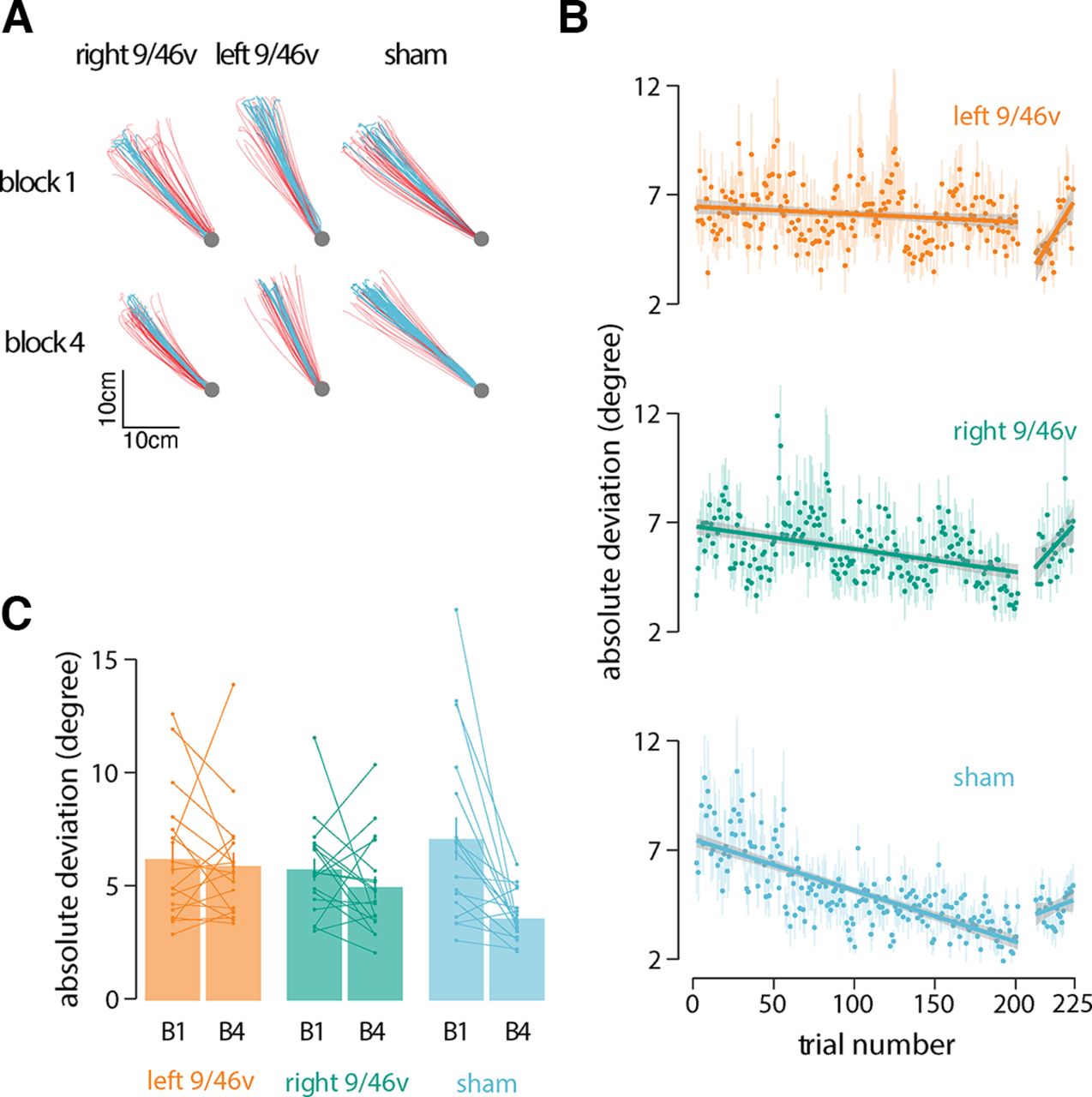
  • Figure 2.
    • Download figure
    • Open in new tab
    • Download powerpoint
    Figure 2.

    Suppression of left 9/46v using cTBS disrupts motor learning. A, Hand paths of a representative participant from each group at the start (block 1) and end of training (block 4). Hand paths shown in red are for unsuccessful movements, and those in blue are for successful movements. B, Mean absolute deviation from the center of the target zone over the course of training. The linear fit is shown across learning trials and no-feedback trials separately. The shaded region represents ±SEM. The rate of learning was less in participants who received stimulation over left 9/46v than those who received sham stimulation. C, Mean absolute deviation in the first and last block of the training. Participants in the sham stimulation condition showed a greater reduction in |AD| than participants in the left 9/46v condition.

  • Figure 3.
    • Download figure
    • Open in new tab
    • Download powerpoint
    Figure 3.

    Suppression of left 9/46v using cTBS leaves reinforcement learning intact. A, Mean percentage of rewarded trials over the course of training. A linear fit is shown across learning trials. The shaded region represents ±SEM. B, Mean percent of rewarded movements in the first and last block of the training. Participants in the sham stimulation condition received more rewards as learning progressed, whereas participants who received stimulation to left 9/46v showed no improvement at all. C, Mean absolute change in movement direction between the current trial (nth trial) and the subsequent trial (n + 1th trial) as a function of the history of rewarded movements. Reward history included three most recent movements (n, n–1, and n–2 trial), where at least one of these movements was rewarded. The left 9/46v group showed the same basic reward-history-dependent pattern as the other conditions but with greater change in direction overall. This suggests that the learning deficit after left 9/46v suppression is not because of inability to process reward but likely because of a deficit in memory for target direction.

  • Figure 4.
    • Download figure
    • Open in new tab
    • Download powerpoint
    Figure 4.

    cTBS over left or right 9/46v did not alter the excitability of motor cortex or basic movement parameters. A, Mean time series of MEPs recorded from the FDI muscle pre-cTBS (blue) and post-cTBS (red) from a representative participant in each experimental condition. The TMS pulse occurs at time = 0 ms. The shaded regions are ±SEM across 20 MEPs. B, Mean change in amplitude of MEPs measured 10 min after cTBS (computed as a percentage of pre-cTBS MEPs). Error bars give the SE across participants. C, Mean movement duration, peak velocity, and movement amplitude across experimental conditions. cTBS to either left or right 9/46v did not modify the movement parameters.

Back to top

In this issue

eneuro: 9 (5)
eNeuro
Vol. 9, Issue 5
September/October 2022
  • Table of Contents
  • Index by author
  • Ed Board (PDF)
Email

Thank you for sharing this eNeuro article.

NOTE: We request your email address only to inform the recipient that it was you who recommended this article, and that it is not junk mail. We do not retain these email addresses.

Enter multiple addresses on separate lines or separate them with commas.
Ventrolateral Prefrontal Cortex Contributes to Human Motor Learning
(Your Name) has forwarded a page to you from eNeuro
(Your Name) thought you would be interested in this article in eNeuro.
CAPTCHA
This question is for testing whether or not you are a human visitor and to prevent automated spam submissions.
Print
View Full Page PDF
Citation Tools
Ventrolateral Prefrontal Cortex Contributes to Human Motor Learning
Neeraj Kumar, Ananda Sidarta, Chelsea Smith, David J. Ostry
eNeuro 16 September 2022, 9 (5) ENEURO.0269-22.2022; DOI: 10.1523/ENEURO.0269-22.2022

Citation Manager Formats

  • BibTeX
  • Bookends
  • EasyBib
  • EndNote (tagged)
  • EndNote 8 (xml)
  • Medlars
  • Mendeley
  • Papers
  • RefWorks Tagged
  • Ref Manager
  • RIS
  • Zotero
Respond to this article
Share
Ventrolateral Prefrontal Cortex Contributes to Human Motor Learning
Neeraj Kumar, Ananda Sidarta, Chelsea Smith, David J. Ostry
eNeuro 16 September 2022, 9 (5) ENEURO.0269-22.2022; DOI: 10.1523/ENEURO.0269-22.2022
Twitter logo Facebook logo Mendeley logo
  • Tweet Widget
  • Facebook Like
  • Google Plus One

Jump to section

  • Article
    • Abstract
    • Significance Statement
    • Introduction
    • Materials and Methods
    • Results
    • Discussion
    • Footnotes
    • References
    • Synthesis
  • Figures & Data
  • Info & Metrics
  • eLetters
  • PDF

Keywords

  • motor learning
  • reinforcement
  • TMS

Responses to this article

Respond to this article

Jump to comment:

No eLetters have been published for this article.

Related Articles

Cited By...

More in this TOC Section

Research Article: New Research

  • Microglial morphological complexity in the piriform cortex is associated with olfactory aversion following chronic stress
  • Dopamine and calcium dynamics in the nucleus accumbens core during food seeking
  • Spatiotemporal Dynamics in Pre-speech Semantic Category Decoding: An intracranial EEG Study.
Show more Research Article: New Research

Sensory and Motor Systems

  • Microglial morphological complexity in the piriform cortex is associated with olfactory aversion following chronic stress
  • Neural and Behavioral Correlates of Pure Tone and Narrowband Noise Processing in Rats: A Tradeoff between Discrimination and Sensitivity
  • Cortically Mediated Muscle Responses to Balance Perturbations Increase with Perturbation Magnitude in Older Adults with and without Parkinson's Disease
Show more Sensory and Motor Systems

Subjects

  • Sensory and Motor Systems
  • Home
  • Alerts
  • Follow SFN on BlueSky
  • Visit Society for Neuroscience on Facebook
  • Follow Society for Neuroscience on Twitter
  • Follow Society for Neuroscience on LinkedIn
  • Visit Society for Neuroscience on Youtube
  • Follow our RSS feeds

Content

  • Early Release
  • Current Issue
  • Latest Articles
  • Issue Archive
  • Blog
  • Browse by Topic

Information

  • For Authors
  • For the Media

About

  • About the Journal
  • Editorial Board
  • Privacy Notice
  • Contact
  • Feedback
(eNeuro logo)
(SfN logo)

Copyright © 2026 by the Society for Neuroscience.
eNeuro eISSN: 2373-2822

The ideas and opinions expressed in eNeuro do not necessarily reflect those of SfN or the eNeuro Editorial Board. Publication of an advertisement or other product mention in eNeuro should not be construed as an endorsement of the manufacturer’s claims. SfN does not assume any responsibility for any injury and/or damage to persons or property arising from or related to any use of any material contained in eNeuro.