

Article Figures & Data
Figures
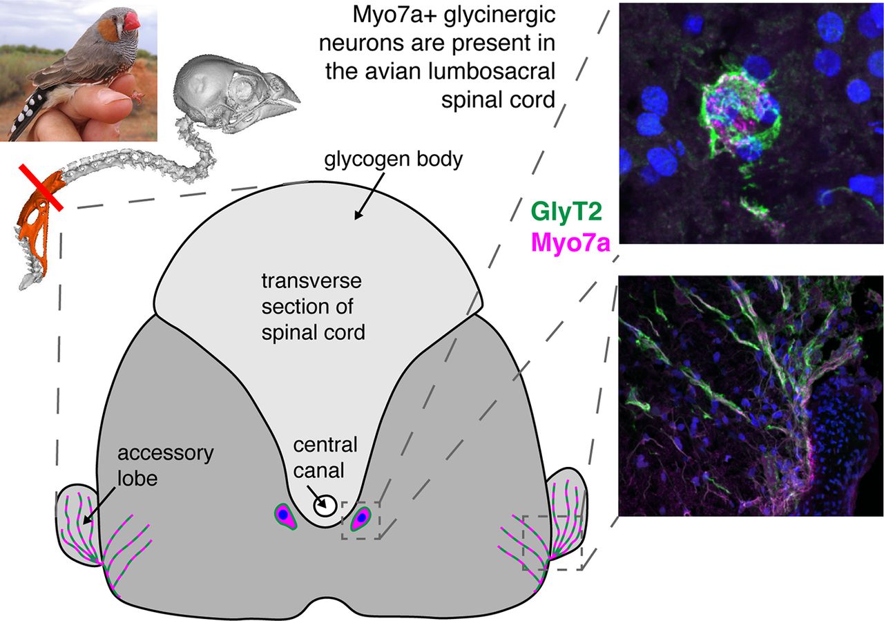
- Figure 1.
Glycinergic neuronal cell bodies near the central canal of the avian lumbosacral organ (LSO) display myosin7a immunoreactivity. A, The synsacrum (fused to the pelvic bones) is highlighted within a reconstructed CT scan of the zebra finch axial skeleton (Yale Peabody Museum specimen #125076). B, Dorsal and ventral views of the synsacrum, reconstructed from the CT scan. C, A sagittal section through the vertebral canal of the synsacrum CT scan shows the dorsal expansion of the vertebral canal that holds the glycogen body. A parasagittal section shows transverse canal-like structures on the internal dorsal side of the vertebral canal that roughly align with the accessory lobes (see Stanchak et al., 2020). D, A diagram of the neural tissue of the LSO based on dissection and histology observations and a contrast-enhanced CT scan (data from Stanchak et al., 2020). E, Cells near the central canal (middle) that are immunoreactive for both glyt2 and myo7a. F, Higher-resolution image of the right cell from E. G, Labeling with two different myo7a antibodies (mouse and rabbit). H, neun/fox3 labeling within the myo7a+ cells. White arrow in the first panel of E and G points to the ependymal layer surrounding the central canal. Upper right diagram in the first panel of E–H indicates approximately where in the transverse section the image was taken. In composite images of E–H, blue is DAPI.
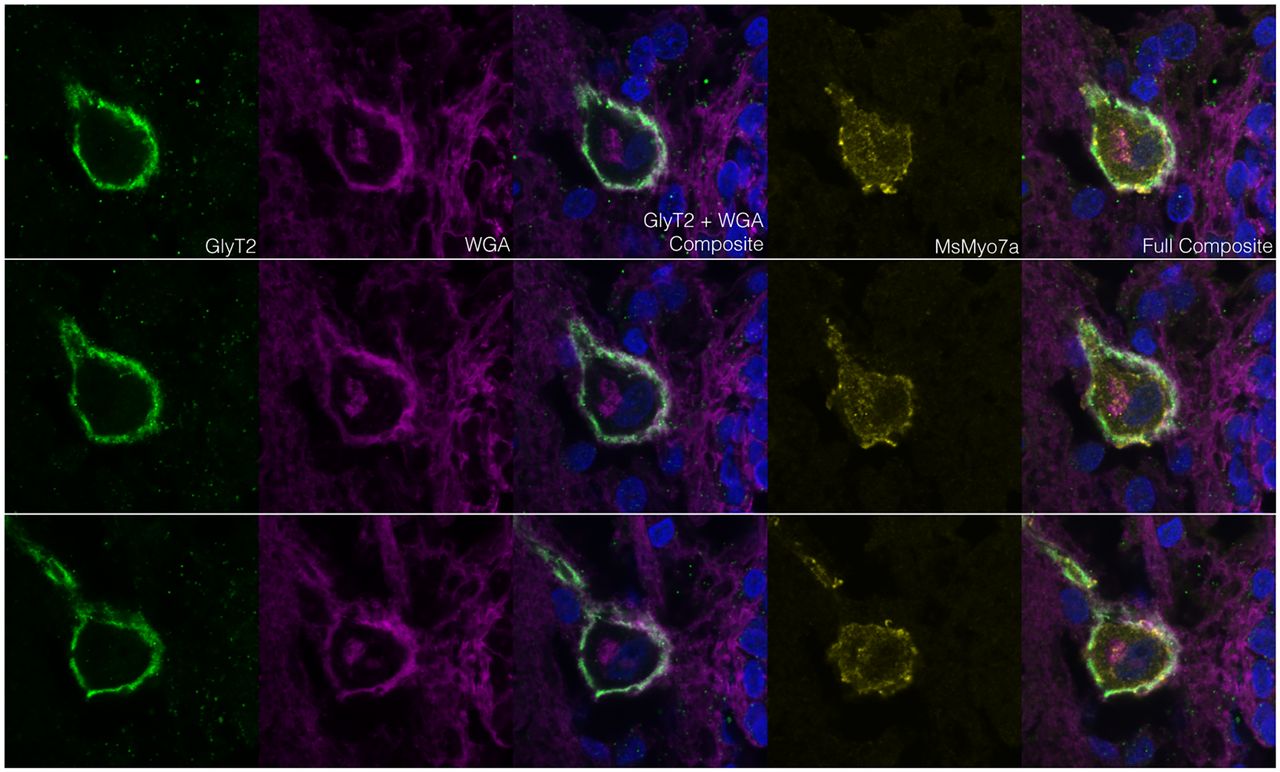
- Figure 2.
Glycine transporter 2 is located in the cell membrane of the neuronal cell bodies that display myosin7a immunoreactivity. Each horizontal row of images is a separate optical section along the z-axis of one cell. WGA is wheat germ agglutinin, a lectin that labels plasma membranes. In composite images, blue is DAPI. Nuclei of the ependymal cells of the central canal can be seen in the lower right of the composite images.
- Figure 3.
Lateral glycinergic processes are immunoreactive for proteins also present in inner ear hair cell stereocilia. A, Glyt2+ ventrolateral processes are immunoreactive for myo7a. Margin of spinal cord with dentate ligament is at lower left; upper left is accessory lobe. B, C, The accessory lobe processes (B) and terminations at the dentate ligament (C) double-label with two different antibodies for myo7a (mouse and rabbit). D, Myo7a-immunoreactive processes in the accessory lobe also react with anti-espin. A–D, In composite images, blue is DAPI. Upper right diagram in the first panel of each subfigure indicates approximately where in the transverse section the image was taken.


In this issue


