

Article Figures & Data
Figures
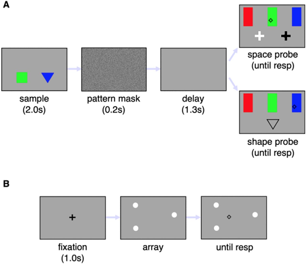
- Figure 1.
Panel A shows the event sequence in the color recall task. Participants had to remember the color of a triangle and a square displayed side by side. The sample display was followed by a pattern mask and a blank screen. The recall target was identified either by a space probe, consisting a bright cross displayed at the location previously occupied by the target, or by a shape probe, consisting of the outline of the target shape . The color was reported by placing the cursor over the corresponding rectangle and clicking the mouse button. The response (resp) initiated the next trial. Panel B shows the centroid estimation task. The visual display contained three bright dots, and the participant had to indicate the location of the center of mass of the imaginary triangle whose vertices corresponded to the dots location, by dragging the cursor and clicking.
- Figure 2.
Recall error rates. The bar graph on the left shows the group averaged proportions of swap errors, while the bar graph on the right shows the group averaged proportions of generic errors, following space and shape probes, respectively. Overall, patients made more swap errors than controls. Moreover, patients with left temporal lobectomies made more swap errors following space than shape probes, suggesting a specific impairment of spatial binding in this group only. For generic errors, group differences were marginal and were not further affected by probe dimension. For sake of clarity, the data are averaged over the two blocks. Circles are individual participants’ error rates. Continuous lines join swap error rates, following space and shape probes, respectively, of each left temporal lobectomy patient. Broken lines join the swap error rates of each right temporal lobectomy patient. Error bars are SEM.


Tables
left TLE
(n = 6)right TLE
(n = 7)controls
(n = 15)BF10 Sex (%males) 83.3 100 93.3 0.64 Age (years) 35.2 (±7.9) 33.08
(±8.9)33.3
(±7.8)0.29 Education (highest
grade)15.3
(±1.6)14.3
(±1.9)14.3
(±2.4)0.5 Full Scale IQ 92.0
(±11.7)87.3
(±9.9)99.5
(±15.5)0.9 Epilepsy onset
age (years)11.1
(±10.2)22.1
(±17.2)— 0.84 Frequency group differences were compared using a Bayesian contingency table. Continuous variables were compared with Bayesian ANOVAs or Bayesian independent samples t test. The values in parenthesis are SDs. None of the demographic or clinical variables showed appreciable group differences since the Bayes factor (BF10) was less than 1.0 for all comparisons.
Patient Gender Education (years) WAIS II HVLT BVMT CTT Block design Vocab Matrix reasoning Similar Immediate Delayed Discrimin. Immediate Delayed Discrimin. CTT1 (s) CTT2 (s) P1 M 16 16 (−0.59) 18 (−1.6) 12 (−0.41) 24 (−0.77) 19 (−1.43) 6 (−1.47) 9 (−1.9) 12 (−1.01) 4 (−1.22) 4 (−1.4) 85 (1.24) 160 (2.0) P2 M 14 18 (−0.46) 27 (−0.83) 12 (−0.41) 23 (−0.88) 26 (0.36) 9 (0.11) 12 (1.1) 10 (−1.28) 4 (−1.22) 4 (−1.4) 97 (1.74) 166 (2.22) P3 F 16 12 (−0.88) 25 (−1.0) 18 (0.53) 28 (−0.31) 24 (−0.15) 7 (−0.95) 10 (−0.9) 9 (−1.41) 6 (−0.58) 5 (−0.4) 65 (0.41) 130 (0.92) P4 M 14 16 (−0.59) 26 (−0.91) 14 (−0.09) 26 (−0.54) 22 (−0.67) 7 (−0.95) 11 (0.1) 14 (−0.75) 5 (−0.9) 4 (−1.4) 66 (0.45) 191 (3.12) P5 M 12 14 (−0.74) 28 (−0.74) 10 (−0.72) 25 (−0.66) 24 (−0.15) 8 (−0.42) 8 (−2.9) 7 (−1.68) 3 (−1.55) 3 (−2.4) 65 (0.41) 135 (1.1) P6 M 16 24 (−0.0) 20 (−1.48) 16 (0.22) 22 (−1.0) 24 (−0.15) 7 (−0.95) 11 (0.1) 21 (0.19) 7 (−0.26) 5 (−0.4) 75 (0.82) 150 (1.65) P7 M 16 14 (−0.74) 28 (−0.74) 8 (−1.03) 25 (−0.66) 19 (−1.43) 5 (−2.0) 7 (−3.9) 12 (−1.01) 5 (−0.9) 4 (−1.4) 87 (1.33) 154 (1.79) P8 M 16 39 (1.09) 45 (0.71) 21 (1.0) 30 (−0.08) 27 (0.62) 10 (0.63) 12 (1.1) 13 (−0.88) 9 (0.39) 5 (−0.4) 32 (−0.97) 85 (−0.7) P9 M 14 27 (0.21) 32 (−0.4) 18 (0.53) 26 (−0.54) 21 (−0.92) 8 (−0.42) 11 (0.1) 21 (0.19) 7 (−0.26) 5 (−0.4) 52 (−0.13) 105 (0.02) P10 M 14 19 (−0.37) 22 (−1.26) 9 (−0.88) 24 (−0.77) 18 (−1.69) 7 (−0.95) 10 (−0.9) 17 (−0.35) 7 (−0.26) 4 (−1.4) 94 (1.62) 165 (2.19) P11 M 11 26 (0.14) 22 (−1.26) 16 (0.22) 24 (−0.77) 20 (−1.18) 7 (−0.95) 10 (−0.9) 15 (−0.61) 6 (−0.58) 4 (−1.4) 80 (1.03) 151 (1.68) P12 M 12 13 (−0.81) 23 (−1.17) 12 (−0.41) 22 (−1.0) 19 (−1.43) 5 (−2.0) 8 (−2.9) 12 (−1.01) 5 (−0.9) 4 (−1.4) 86 (1.28) 153 (1.75) P13 M 16 17 (−0.52) 28 (−0.74) 11 (−0.56) 28 (−0.31) 21 (−0.92) 8 (−0.42) 10 (−0.9) 14 (−0.75) 6 (−0.58) 4 (−1.4) 83 (1.16) 158 (1.94) Raw scores are reported for each test and participant (see Materials and Methods). The corresponding normalized values are shown in parenthesis. Normalized z scores values were computed by subtracting the mean score and dividing by the SD of a reference sample (Al-Joudi et al., 2019). HVLT-R = Hopkins Verbal Learning Test–Revised; BVMT-R = Brief Visuospatial Memory Test–Revised; CTT = Color Trails Test.
Age Pathology Lesion
lateralityTemporal lobe structures HIP ERC PRC PHC ITG MTG TP STG AMG P1 25 GGs L 0 + + 0 0 0 + 0 0 P2 25 MTS R ++ 0 0 0 0 0 + 0 ++ P3 50 MG L 0 0 + 0 0 + + 0 0 P4 41 GGs R 0 + + 0 0 0 + 0 0 P5 31 MTS R ++ + 0 + 0 + + + ++ P6 27 MTS L ++ 0 0 0 0 0 0 0 0 P7 40 MG L 0 0 0 0 0 + + 0 0 P8 32 MTS R ++ 0 0 0 + 0 ++ 0 + P9 33 MTS R ++ + 0 ++ + 0 0 + + P10 22 MTS L + + 0 0 ++ 0 0 + 0 P11 25 MTS R ++ + 0 0 0 0 0 + ++ P12 47 GGs L 0 + 0 0 0 ++ ++ + 0 P13 32 MTS R + + 0 + + 0 0 + 0 The table lists the pathology and regions affected by the lobectomy for each patient. Extended Data Table 3-1 shows representative MRI slices through the MTL. GG, ganglioglioma; MG, meningioma; MTS, medial temporal sclerosis; HIP, hippocampus; ERC, entorhinal cortex; PRC, perirhinal cortex; PHC, parahippocampal cortex; ITC, inferotemporal cortex; MTG, Middle temporal gyrus; ATP, anterior temporal pole; STG, superior temporal gyrus; AMG, amygdala. 0 indicates an unaffected subregion, + a rostro-caudal lesion extent up to 20 mm, and ++ up to 40 mm.
Effects P(incl) P(incl|d) BFincl B 0.737 0.941 5.673 D 0.737 0.925 4.414 G 0.737 0.997 119.755 B·D 0.316 0.206 0.563 G·B 0.316 0.188 0.500 G·D 0.316 0.891 17.629 G·B·D 0.053 0.017 0.317 The table lists each factor and interaction for swap error rates. Extended Data Table 4-1 lists the models and associated prior and posterior probabilities from which the values in the present table are computed. P(incl) is the prior probability of the effect; P(incl|d) is the posterior probability of the effect; BFincl is Bayes factor. A BF greater than 1.0 favors the effect, a BF less than 1.0 favors a null instead. Values of the BF greater than 3.0 are in bold, to highlight those effects that have at least moderate evidence in their favor. Block, probe dimension, group, and the interaction of group by probe dimension all have at least moderately strong evidence in their favor.
Effects P(incl) P(incl|d) BFincl B 0.737 0.804 1.463 D 0.737 0.815 1.571 G 0.737 0.641 0.637 B·D 0.316 0.142 0.360 G·B 0.316 0.085 0.202 G·D 0.316 0.087 0.206 G·B·D 0.053 7.157e −4 0.013 The table lists each factor and interaction for generic error rates. Extended Data Table 5-1 lists the models and associated probabilities, used to compute the effects. P(incl) is the prior probability of the predictors; P(incl|d) is the posterior probability; BFincl is Bayes factor. A BF greater than 1.0 favors the predictor, a BF less than 1.0 favors a null effect instead. The only predictors with favorable evidence, albeit of very modest entity, are block and probe dimension.
Extended Data Table 3-1
Postsurgical MRI scans for 13 patients. The images were obtained with T1 weighted, T2 weighted, fluid-attenuated inversion recovery, and gradient recalled echo sequences. ERC, entorhinal cortex; PHC, parahippocampal cortex; PRC, perirhinal cortex; Hipp, hippocampus; AMG, amygdala; TP, temporal pole; antSTG, anterior superior temporal gyrus; antMTG, anterior middle temporal gyrus. Download Table 3-1, DOCX file.
Extended Data Table 4-1
Best model comparisons for swap errors. The table presents each of the model comparisons from the Bayesian ANOVA. The within factors are block (B) and probe dimension (D). The between factor is group G. P(M) is the a priori model probability, P(M|d) is the posterior model probability. BFM is the Bayes factor of the model, BF10 is the Bayes factor of the model relative to the best one. The best model contained all three factors and the interaction of group by probe dimension. Download Table 4-1, DOC file.
Extended Data Table 5-1
Best model comparisons for generic errors. The table presents the model comparisons obtained from a Bayesian ANOVA. The within factors are block (B) and probe dimension (D). The between factor is group G. P(M) is the a priori model probability, P(M|d) is the posterior model probability. BFM is the Bayes factor of the model, BF10 is the Bayes factor of the model relative to the best one. The best model included the three main factors, namely, block (B), probe dimension (D), and group (G). Download Table 5-1, DOC file.
In this issue


