

Article Figures & Data
Figures
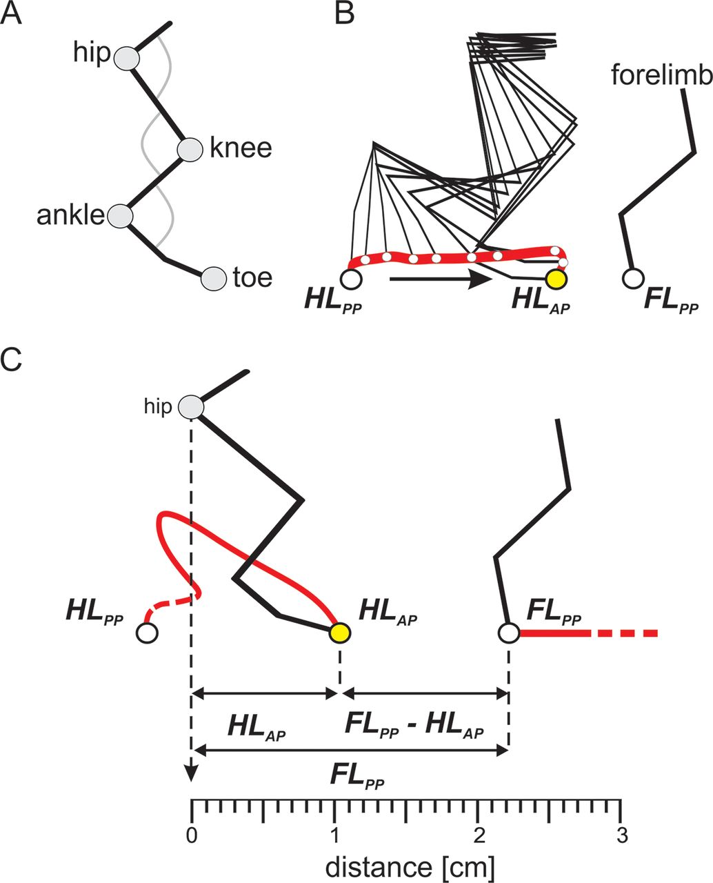
- Figure 1.
Schematic representation of precise foot placement assessment during locomotion on the treadmill. A, Kinematic data obtained by reconstruction of the hindlimb by means of detecting the coordinates of markers attached to the skin over imposed hindlimb segments. B, Stick diagrams reconstructed by connecting marker coordinates showing the kinematics of a swing phase while the hindlimb is traveling from a posterior position (HLPP) to an anterior position (HLAP), alongside coordinates of the posterior position of the forelimb (FLPP). C, Hindlimb targeting obtained by measurements of distances between toe to the hip joint (HLAP), forelimb paw to hip joint (FLPP), and (FLPP – HLAP) along the horizontal x-axis. The swing phase diagram represents measurements taken from an SCR event.
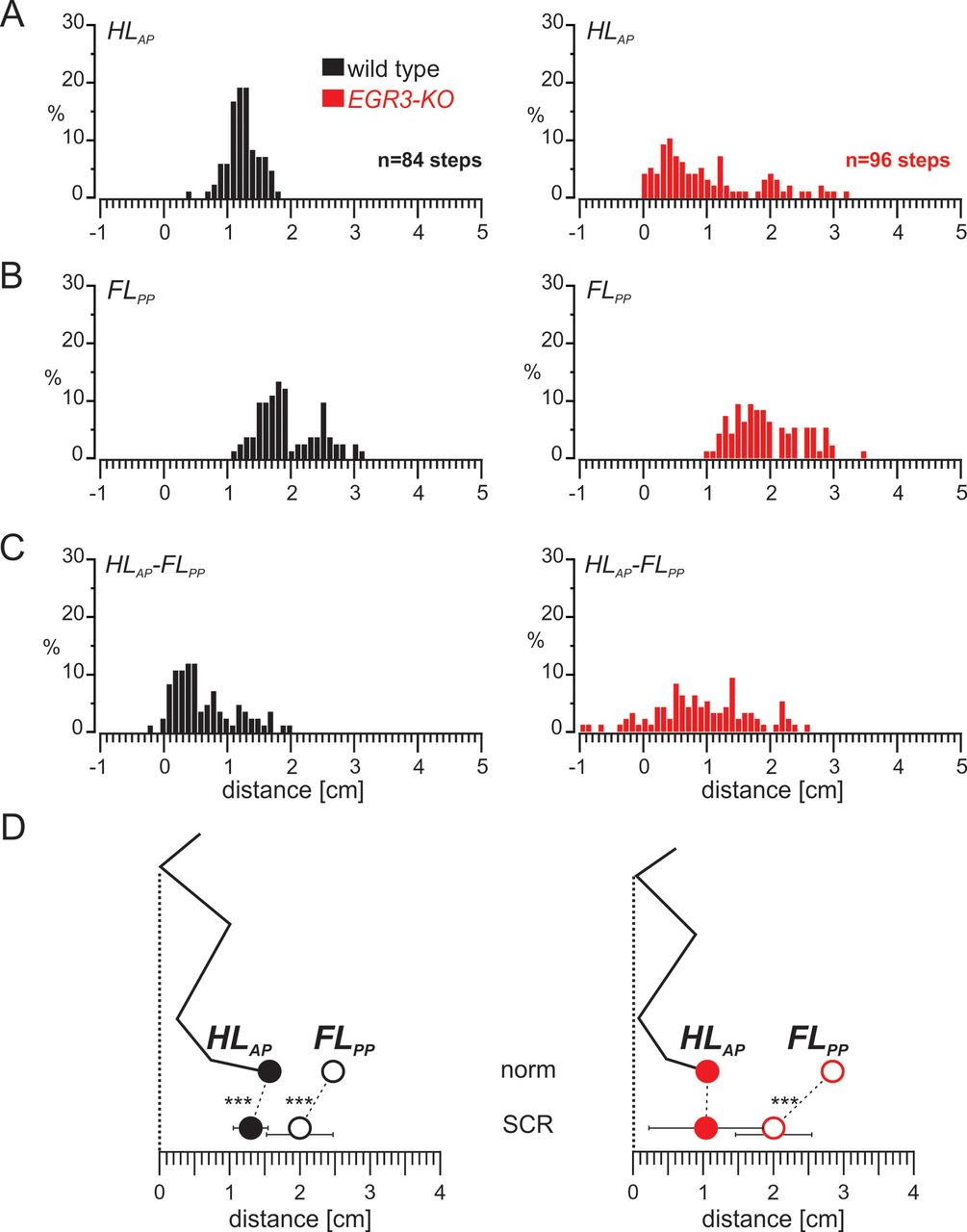
- Figure 2.
The well organized swing movement in mice requires proprioceptive sensory feedback from the muscle spindles. A, Trajectories of the hindlimb paw of wild-type mice (left) are very stereotyped across animals, whereas more variability in movement is observed in Egr3-KO mice (right). The colored lines are swings from an individual animal, and the bold black line is the pooled average. The average posterior paw position at swing onset and the anterior paw position at swing offset are illustrated as open and closed circles, respectively. B, Histogram representing the distribution of hindlimb paw positions relative to the hip joint at the end of hindlimb swing movement (HLAP) in wild-type (left) and Egr3-KO (right) mice. C, Histogram showing the distribution of forelimb paw positions relative to hip joint at the end of forelimb stance movement (FLPP) in wild-type (left) and Egr3-KO (right) mice. D, Histogram presenting the distribution of the distance between HLAP and FLPP in wild-type (left) and Egr3-KO (right) mice. E, Diagram illustrating the relationship between the average (±SD) of the HLAP and FLPP in wild-type (left) and Egr3-KO (right) mice during unperturbed hindlimb swing movements.
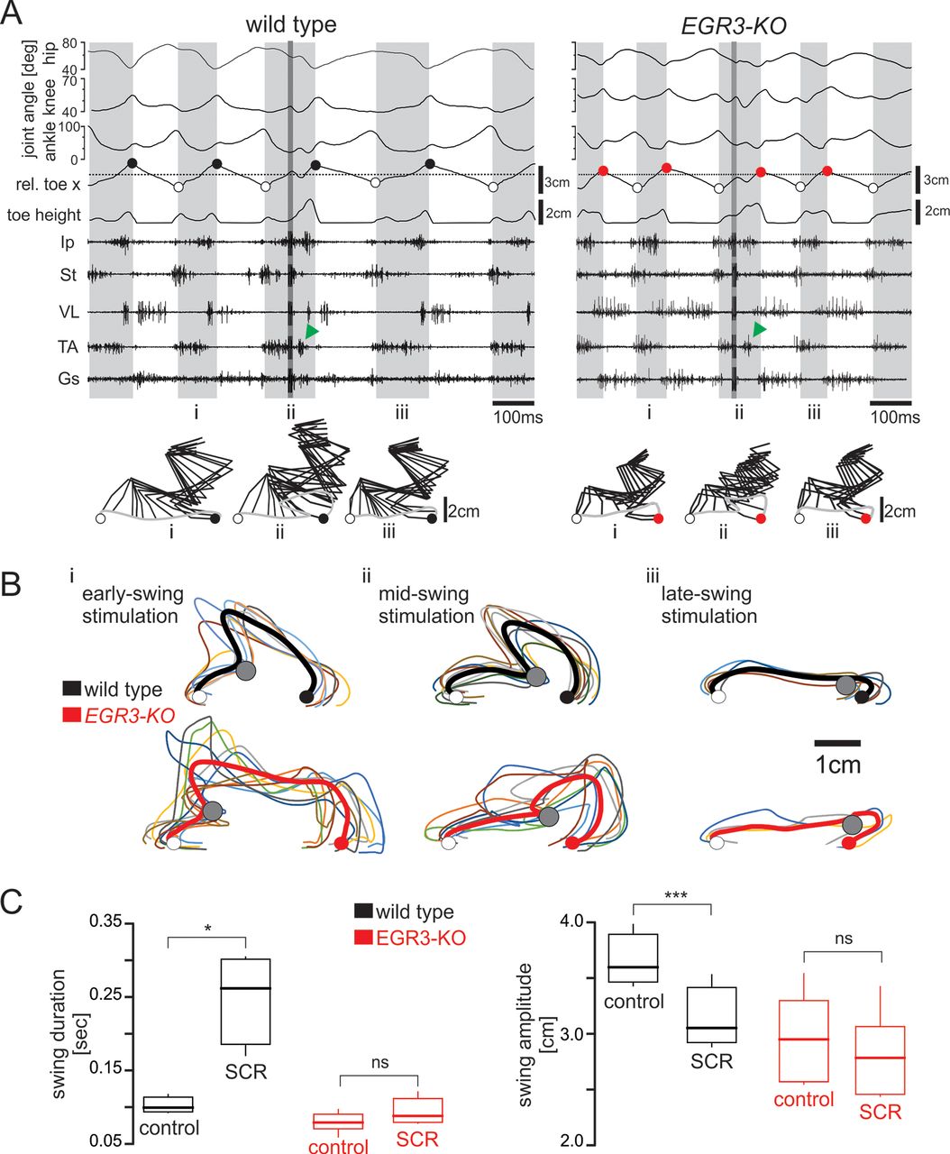
- Figure 3.
Induction of a stumbling corrective reaction is independent of proprioceptive sensory feedback from the muscle spindles. A, Hip, knee, and ankle joint angles, relative toe x- and y-coordinates (toe height) synchronized with raw EMG activity of flexor (Ip, St, TA) and extensor (VL, Gs) muscles. Electrical stimulation of the saphenous nerve during swing phase is indicated by the darker gray region inside the third swing phase. Green arrows point to the activity of the flexor muscle elicited by the stimulation. Stick diagram reconstruction of a swing phase before SCR (i), an SCR (ii), and a swing phase after SCR (iii) are illustrated below. B, Trajectories of the hindlimb paw during SCR are very stereotyped in wild-type animals (top) but are disorganized in Egr3-KO mice (bottom). The colored lines are individual swings, and the bold line is the pooled average from one animal. The average posterior paw position at swing onset and the anterior paw position at swing offset are illustrated as open and closed circles, respectively. The point of nerve stimulation is indicated by the gray circle. C, Changes in control swing and SCR durations (p = 0.016) and distances between the paw position from liftoff to touchdown (swing amplitude) in SCR events in wild-type mice (p < 0.001). No changes were observed in swing duration (p = 0.074) and swing amplitude (p = 0.534) during SCR events in Egr3-KO mice.
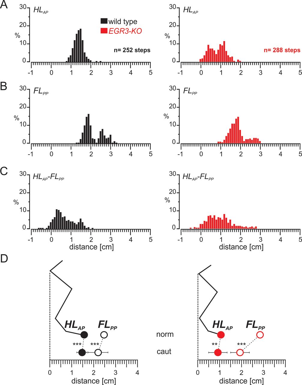
- Figure 4.
Limb movement during SCR remains well organized in wild-type mice but is more variable in the absence of proprioceptive sensory feedback from the muscle spindles. A, Histogram representing the distribution of HLAP during SCR in wild-type (left) and Egr3-KO (right) mice. B, Histogram showing the distribution of FLPP during the SCRs in wild-type (left) and Egr3-KO (right) mice. C, Histogram presenting the distribution of the distance between the HLAP and FLPP during SCRs in wild-type (left) and Egr3-KO (right) mice. D, Diagram illustrating the relationship between the average (±SD) of the HLAP and FLPP during normal swing movements (norm) and SCRs in wild-type (left) and Egr3-KO (right) mice during SCRs. ***p < 0.001. Histograms illustrating the distribution of FLPP during SCR in individual WT and Egr3-KO mice are shown in Extended Data Figure 4-1.
- Figure 5.
The swing movement during cautious locomotion is well organized in wild-type mice but is more variable in the absence of proprioceptive sensory feedback from the muscle spindles. A, Histogram representing the distribution of HLAP in swing movements during cautious locomotion in wild-type (left) and Egr3-KO (right) mice. B, Histogram showing the distribution of FLPP in swing movements during cautious locomotion in wild-type (left) and Egr3-KO (right) mice. C, Histogram showing the distribution of the distance between the HLAP and FLPP in swing movements during cautious locomotion in wild-type (left) and Egr3-KO (right) mice. D, Diagram illustrating the relationship between the average (±SD) of the HLAP and FLPP during normal swing movements (norm) and swing movements during cautious locomotion (caut) in wild-type (left) and Egr3-KO (right) mice during SCRs. **p < 0.01, ***p < 0.001. Histograms illustrating the distribution of FLPP during cautious locomotion in individual WT and Egr3-KO mice are shown in Extended Data Figure 5-1.
- Figure 6.
The distance between HLAP and FLPP is relatively constant in wild-type mice but is more variable in the absence of proprioceptive feedback from muscle spindles. A, Diagram illustrating the relationship between the average (±SD) of the HLAP and FLPP in wild-type (left) and Egr3-KO (right) mice during control swing movements (norm), SCRs, and swing movements during cautious locomotion (caut). B, Graphs illustrating the average (±SD) of the distance between the HLAP and FLPP in wild-type (left) and Egr3-KO (right) mice during control swing movements (norm), SCRs, and swing movements during cautious locomotion (caut). **p < 0.01, ***p < 0.001.


Movies
- Movie 1.
Stumbling corrective reaction in a wild-type mouse elicited by stimulating the saphenous nerve, indicated by the red circle.
- Movie 2.
Stumbling corrective reaction in an Egr3-KO mouse elicited by stimulating the saphenous nerve, indicated by the red circle.
Figure 4-1
Histograms of forelimb paw distance from the hip joint of individual wild-type and Egr3-KO animals during SCRs. Histograms of FLPP from individual wild-type (left) and Egr3-KO (right) animals. In the last histograms on the bottom, all animals are superimposed in a single histogram. Download Figure 4-1, TIF file.
Figure 5-1
Histograms of forelimb paw distance from the hip joint of individual wild-type and Egr3-KO animals during swing movements of cautious locomotion. Histograms of FLPP from individual wild-type (left) and Egr3-KO (right) animals. In the last histogram on the bottom, all animals are superimposed in a single histogram. Download Figure 5-1, TIF file.
In this issue


