
Main menu

  • HOME
  • CONTENT
    • Early Release
    • Featured
    • Current Issue
    • Issue Archive
    • Blog
    • Collections
    • Podcast
  • TOPICS
    • Cognition and Behavior
    • Development
    • Disorders of the Nervous System
    • History, Teaching and Public Awareness
    • Integrative Systems
    • Neuronal Excitability
    • Novel Tools and Methods
    • Sensory and Motor Systems
  • ALERTS
  • FOR AUTHORS
  • ABOUT
    • Overview
    • Editorial Board
    • For the Media
    • Privacy Policy
    • Contact Us
    • Feedback
  • SUBMIT

User menu

Search

  • Advanced search
eNeuro
eNeuro

Advanced Search


  • HOME
  • CONTENT
    • Early Release
    • Featured
    • Current Issue
    • Issue Archive
    • Blog
    • Collections
    • Podcast
  • TOPICS
    • Cognition and Behavior
    • Development
    • Disorders of the Nervous System
    • History, Teaching and Public Awareness
    • Integrative Systems
    • Neuronal Excitability
    • Novel Tools and Methods
    • Sensory and Motor Systems
  • ALERTS
  • FOR AUTHORS
  • ABOUT
    • Overview
    • Editorial Board
    • For the Media
    • Privacy Policy
    • Contact Us
    • Feedback
  • SUBMIT
PreviousNext
Research ArticleResearch Article: New Research, Cognition and Behavior

FKBP51 in the Oval Bed Nucleus of the Stria Terminalis Regulates Anxiety-Like Behavior

Clara Engelhardt, Fiona Tang, Radwa Elkhateib, Joeri Bordes, Lea Maria Brix, Lotte van Doeselaar, Alexander S. Häusl, Max L. Pöhlmann, Karla Schraut, Huanqing Yang, Alon Chen, Jan M. Deussing and Mathias V. Schmidt
eNeuro 6 December 2021, 8 (6) ENEURO.0425-21.2021; https://doi.org/10.1523/ENEURO.0425-21.2021
Clara Engelhardt
1Research Group Neurobiology of Stress Resilience, Max Planck Institute of Psychiatry, Munich 80804, Germany
  • Find this author on Google Scholar
  • Find this author on PubMed
  • Search for this author on this site
Fiona Tang
1Research Group Neurobiology of Stress Resilience, Max Planck Institute of Psychiatry, Munich 80804, Germany
  • Find this author on Google Scholar
  • Find this author on PubMed
  • Search for this author on this site
Radwa Elkhateib
1Research Group Neurobiology of Stress Resilience, Max Planck Institute of Psychiatry, Munich 80804, Germany
  • Find this author on Google Scholar
  • Find this author on PubMed
  • Search for this author on this site
Joeri Bordes
1Research Group Neurobiology of Stress Resilience, Max Planck Institute of Psychiatry, Munich 80804, Germany
  • Find this author on Google Scholar
  • Find this author on PubMed
  • Search for this author on this site
Lea Maria Brix
1Research Group Neurobiology of Stress Resilience, Max Planck Institute of Psychiatry, Munich 80804, Germany
2International Max Planck Research School for Translational Psychiatry (IMPRS-TP), Munich 80804, Germany
  • Find this author on Google Scholar
  • Find this author on PubMed
  • Search for this author on this site
  • ORCID record for Lea Maria Brix
Lotte van Doeselaar
1Research Group Neurobiology of Stress Resilience, Max Planck Institute of Psychiatry, Munich 80804, Germany
2International Max Planck Research School for Translational Psychiatry (IMPRS-TP), Munich 80804, Germany
  • Find this author on Google Scholar
  • Find this author on PubMed
  • Search for this author on this site
Alexander S. Häusl
1Research Group Neurobiology of Stress Resilience, Max Planck Institute of Psychiatry, Munich 80804, Germany
  • Find this author on Google Scholar
  • Find this author on PubMed
  • Search for this author on this site
Max L. Pöhlmann
1Research Group Neurobiology of Stress Resilience, Max Planck Institute of Psychiatry, Munich 80804, Germany
  • Find this author on Google Scholar
  • Find this author on PubMed
  • Search for this author on this site
Karla Schraut
1Research Group Neurobiology of Stress Resilience, Max Planck Institute of Psychiatry, Munich 80804, Germany
  • Find this author on Google Scholar
  • Find this author on PubMed
  • Search for this author on this site
Huanqing Yang
1Research Group Neurobiology of Stress Resilience, Max Planck Institute of Psychiatry, Munich 80804, Germany
  • Find this author on Google Scholar
  • Find this author on PubMed
  • Search for this author on this site
Alon Chen
3Department of Stress Neurobiology and Neurogenetics, Max Planck Institute of Psychiatry, Munich 80804, Germany
4Department of Neurobiology, Weizmann Institute of Science, Rehovot 76100, Israel
  • Find this author on Google Scholar
  • Find this author on PubMed
  • Search for this author on this site
Jan M. Deussing
5Research Group Molecular Neurogenetics, Max Planck Institute of Psychiatry, Munich 80804, Germany
  • Find this author on Google Scholar
  • Find this author on PubMed
  • Search for this author on this site
Mathias V. Schmidt
1Research Group Neurobiology of Stress Resilience, Max Planck Institute of Psychiatry, Munich 80804, Germany
  • Find this author on Google Scholar
  • Find this author on PubMed
  • Search for this author on this site
  • Article
  • Figures & Data
  • Info & Metrics
  • eLetters
  • PDF
Loading

Article Figures & Data

Figures

  • Figure
    • Download figure
    • Open in new tab
    • Download powerpoint
  • Figure 1.
    • Download figure
    • Open in new tab
    • Download powerpoint
    Figure 1.

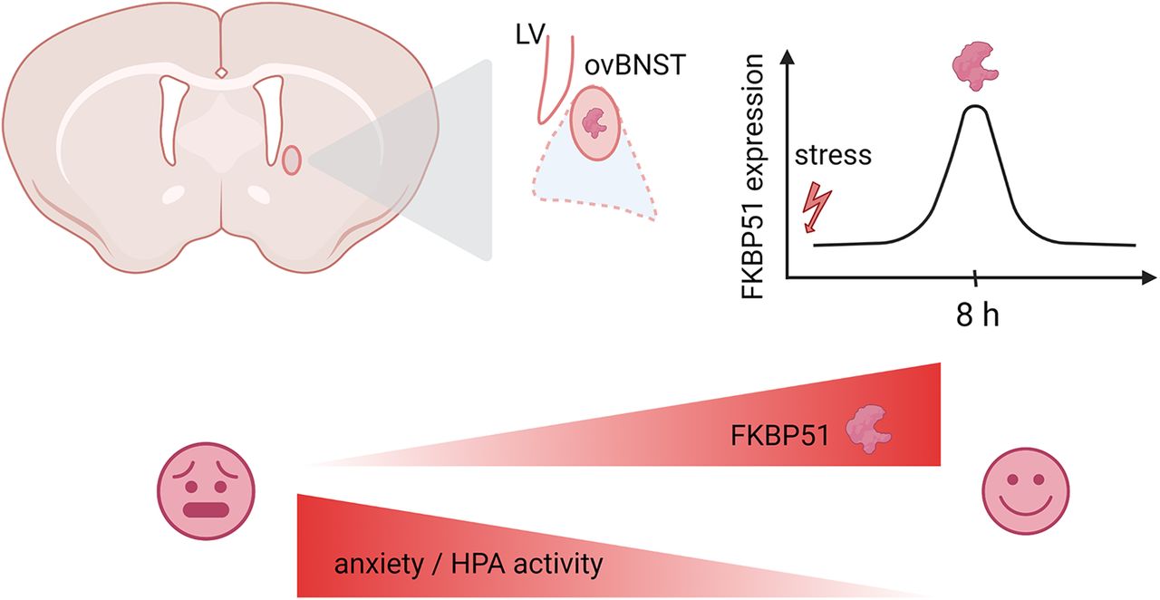
    Expression and regulation of Fkbp5 in the ovBNST. A, Fkbp5 is among other regions expressed in the ovBNST at basal level and highly upregulated after exposure to acute stress. Stress regulation of FKBP51 is shown through β-gal upregulation (B) and on mRNA level (C). Furthermore, Fkbp5 is expressed and regulated in the majority of GABAergic neurons (D). Black arrow, GABAergic cell expressing Fkbp5; white arrow, non-GABAergic cell expressing Fkbp5. This was further confirmed by RNAscope (E), demonstrating a clear coexpression of Fkbp5 and GAD65. F, Fkbp5 was not significantly upregulated after exposure to CSDS. However, exposure to a different type of acute stress, acute fear conditioning, reliably resulted in a significant upregulation of Fkbp5 in the ovBNST (G). A time course of Fkbp5 revealed a significant upregulation, with Fkbp5 regulation peaking at its highest after 4 h (H). Data are mean ± SEM; *p < 0.05, **p < 0.01, #p < 0.001.

  • Figure 2.
    • Download figure
    • Open in new tab
    • Download powerpoint
    Figure 2.

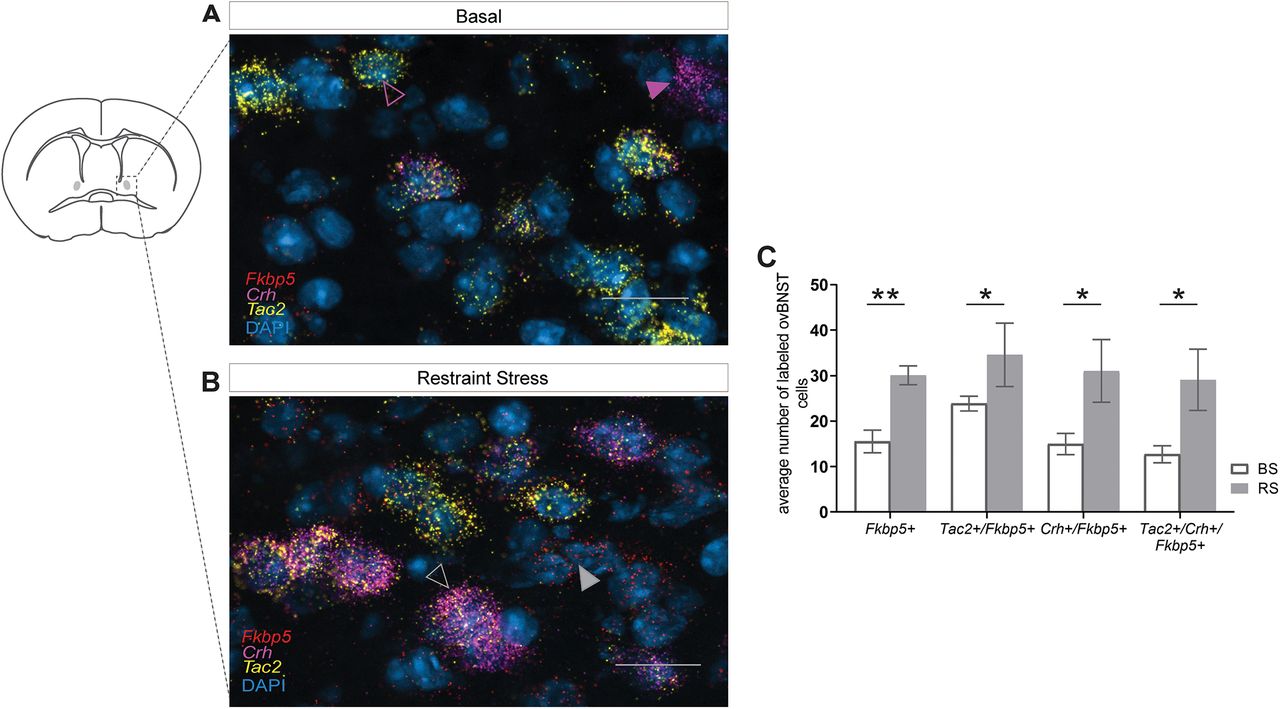
    Fkbp5+ cells in the ovBNST coexpress with the neuropeptides Crh and Tac2 and their number is significantly increased after exposure to ASR. A, Fkbp5 and Tac2 are coexpressed in the ovBNST, as can be seen in detail (violet outline arrow). Fkbp5 and Crh are also coexpressed in the ovBNST as shown in detail (violet arrow). B, Expression patterns of Fkbp5 with Tac2 and Crh in the ovBNST also strongly overlapped (gray outline arrow). In addition, there were some cells that expressed Fkbp5 only (gray arrow). C, Quantification of the number of cells expressing Fkbp5 only, coexpressing Fkbp5 and Tac2, coexpressing Fkbp5 and Crh, and coexpressing Fkbp5, Tac2, and Crh after exposure to ASR resulted in significant upregulation across all cell types. Scale bar (A, B): 25 μm. Data are mean ± SEM; *p < 0.05, **p < 0.01.

  • Figure 3.
    • Download figure
    • Open in new tab
    • Download powerpoint
    Figure 3.

    OE of Fkbp5 in the BNST. A, Schematic representation of viral manipulation and experimental timeline including testing battery. B, Fkbp5 ISH demonstrating correct viral expression (scale bar: 250 μm). C, Quantification of Fkbp5 OE. The right panel illustrates the virus injection sites. D, There were no significant differences in open-arm time spent and number of open-arm entries in the EPM. E, FKBP51BNST-OE and control animals also did not differ in the total distance covered during the OF test. F, FKBP51BNST-OE animals entered the lit compartment of the DALI more frequently than control animals, but there was no significant difference in the distance covered within the light zone. G, FKBP51BNST-OE animals demonstrated significantly lower corticosterone levels after a 15-min ARS. H, Both the coexpressed neuropeptides Tac2 and Crh were significantly upregulated in FKBP51BNST-OE animals. Data are mean ± SEM; *p < 0.05, **p < 0.01.

  • Figure 4.
    • Download figure
    • Open in new tab
    • Download powerpoint
    Figure 4.

    KO of Fkbp5 in the BNST. A, Schematic of viral manipulation as well as experimental timeline and testing battery. B, Example of virus expression and correct targeting through Cre and Fkp5 ISH. Scale bar: 250 μm. C, Quantification of Fkbp5 knock-down in the ovBNST. The right panel illustrates the virus injection sites. D, In the EPM, FKBP51BNST-KO animals (n = 13) spent significantly less time in the open arms and entered open arms less frequently than control animals (n = 10). E, FKBP51BNST-KO animals covered significantly less distance within the light area of the dark-light box (DALI) test. F, Total distance covered within the OF test was not significantly different. G, FKBP51BNST-KO animals demonstrated a matching neuroendocrine phenotype, exposing significantly higher corticosterone levels after a 15-min ASR. H, In line with previous results, the coexpressed neuropeptides Tac2 and Crh were significantly downregulated in FKBP51BNST-KO animals. Data are mean ± SEM; *p < 0.05, **p < 0.01, #p < 0.001.

  • Figure 5.
    • Download figure
    • Open in new tab
    • Download powerpoint
    Figure 5.

    Activity-dependent KO of Fkbp5 in the BNST. A, Schematic representation of ESARE promoter driven and 4OHT-dependent conditional KO of previously activated neurons in the ovBNST, as well as subsequent experimental timeline. B, Fkbp5 ISH to validate correct viral manipulation. Scale bar: 250 μm (C–F). Activity-dependent KO of Fkpb5 in the ovBNST indicated an anxiogenic phenotype. C, FKBP51ovBNST-KO (KO) animals spent significantly less time in the open arms of the EPM. In addition, they showed a tendency to travel less distance within the light zone of the dark-light box (DALI). The OF test (E) and the ASR response (F) did not show any significant differences between the two groups. Data are mean ± SEM; *p < 0.05, **p < 0.01.

  • Figure 6.
    • Download figure
    • Open in new tab
    • Download powerpoint
    Figure 6.

    Timeframe of anxiety-like behavior after exposure to ASR. Exposure to ASR caused an anxiolytic-like phenotype. A, Schematic representation of experimental timeline. B, In the EPM, restrained animals (rs) spent significantly more time in the open arms. However, there was no difference regarding the number of entries to the open arms between the two groups. C, The OF test did not show any significant differences between the two groups. Data are mean ± SEM; *p < 0.05, **p < 0.01.

Back to top

In this issue

eneuro: 8 (6)
eNeuro
Vol. 8, Issue 6
November/December 2021
  • Table of Contents
  • Index by author
  • Ed Board (PDF)
Email

Thank you for sharing this eNeuro article.

NOTE: We request your email address only to inform the recipient that it was you who recommended this article, and that it is not junk mail. We do not retain these email addresses.

Enter multiple addresses on separate lines or separate them with commas.
FKBP51 in the Oval Bed Nucleus of the Stria Terminalis Regulates Anxiety-Like Behavior
(Your Name) has forwarded a page to you from eNeuro
(Your Name) thought you would be interested in this article in eNeuro.
CAPTCHA
This question is for testing whether or not you are a human visitor and to prevent automated spam submissions.
Print
View Full Page PDF
Citation Tools
FKBP51 in the Oval Bed Nucleus of the Stria Terminalis Regulates Anxiety-Like Behavior
Clara Engelhardt, Fiona Tang, Radwa Elkhateib, Joeri Bordes, Lea Maria Brix, Lotte van Doeselaar, Alexander S. Häusl, Max L. Pöhlmann, Karla Schraut, Huanqing Yang, Alon Chen, Jan M. Deussing, Mathias V. Schmidt
eNeuro 6 December 2021, 8 (6) ENEURO.0425-21.2021; DOI: 10.1523/ENEURO.0425-21.2021

Citation Manager Formats

  • BibTeX
  • Bookends
  • EasyBib
  • EndNote (tagged)
  • EndNote 8 (xml)
  • Medlars
  • Mendeley
  • Papers
  • RefWorks Tagged
  • Ref Manager
  • RIS
  • Zotero
Respond to this article
Share
FKBP51 in the Oval Bed Nucleus of the Stria Terminalis Regulates Anxiety-Like Behavior
Clara Engelhardt, Fiona Tang, Radwa Elkhateib, Joeri Bordes, Lea Maria Brix, Lotte van Doeselaar, Alexander S. Häusl, Max L. Pöhlmann, Karla Schraut, Huanqing Yang, Alon Chen, Jan M. Deussing, Mathias V. Schmidt
eNeuro 6 December 2021, 8 (6) ENEURO.0425-21.2021; DOI: 10.1523/ENEURO.0425-21.2021
Twitter logo Facebook logo Mendeley logo
  • Tweet Widget
  • Facebook Like
  • Google Plus One

Jump to section

  • Article
    • Visual Abstract
    • Abstract
    • Significance Statement
    • Introduction
    • Materials and Methods
    • Results
    • Discussion
    • Acknowledgments
    • Footnotes
    • References
    • Synthesis
    • Author Response
  • Figures & Data
  • Info & Metrics
  • eLetters
  • PDF

Keywords

  • anxiety
  • BNST
  • FKBP5
  • mouse
  • stress

Responses to this article

Respond to this article

Jump to comment:

No eLetters have been published for this article.

Related Articles

Cited By...

More in this TOC Section

Research Article: New Research

  • Robust representation and nonlinear spectral integration of harmonic stacks in layer 4 of mouse primary auditory cortex
  • Changes in palatability processing across the estrous cycle are modulated by hypothalamic estradiol signaling
  • Automatic, but not autonomous: Implicit adaptation is modulated by goal-directed attentional demands
Show more Research Article: New Research

Cognition and Behavior

  • Robust representation and nonlinear spectral integration of harmonic stacks in layer 4 of mouse primary auditory cortex
  • Changes in palatability processing across the estrous cycle are modulated by hypothalamic estradiol signaling
  • Automatic, but not autonomous: Implicit adaptation is modulated by goal-directed attentional demands
Show more Cognition and Behavior

Subjects

  • Cognition and Behavior
  • Home
  • Alerts
  • Follow SFN on BlueSky
  • Visit Society for Neuroscience on Facebook
  • Follow Society for Neuroscience on Twitter
  • Follow Society for Neuroscience on LinkedIn
  • Visit Society for Neuroscience on Youtube
  • Follow our RSS feeds

Content

  • Early Release
  • Current Issue
  • Latest Articles
  • Issue Archive
  • Blog
  • Browse by Topic

Information

  • For Authors
  • For the Media

About

  • About the Journal
  • Editorial Board
  • Privacy Notice
  • Contact
  • Feedback
(eNeuro logo)
(SfN logo)

Copyright © 2026 by the Society for Neuroscience.
eNeuro eISSN: 2373-2822

The ideas and opinions expressed in eNeuro do not necessarily reflect those of SfN or the eNeuro Editorial Board. Publication of an advertisement or other product mention in eNeuro should not be construed as an endorsement of the manufacturer’s claims. SfN does not assume any responsibility for any injury and/or damage to persons or property arising from or related to any use of any material contained in eNeuro.