

Article Figures & Data
Figures
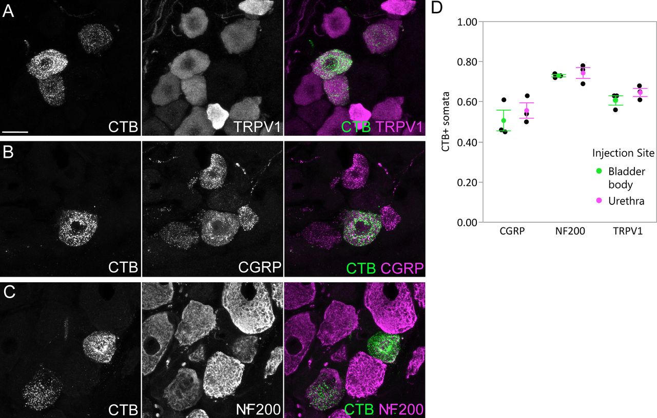
- Figure 1.
Uptake of CTB by afferents following intramural injection into the bladder body or proximal urethra. Confocal micrographs of L6 DRG cryosections immunolabelled for CTB and (A) TRPV1, (B) CGRP, or (C) NF200, following injection of the bladder body or urethra of male rats. D, Proportion (mean ± SE) of CTB-labeled somata immunoreactive for TRPV1, CGRP, or NF200, following CTB injection into the bladder body or urethra. CGRP, calcitonin gene-related peptide; CTB, cholera toxin B subunit; NF200, neurofilament 200 kDa; TRPV1, transient receptor potential vanilloid 1. Scale bar: 20 μm.
- Figure 2.
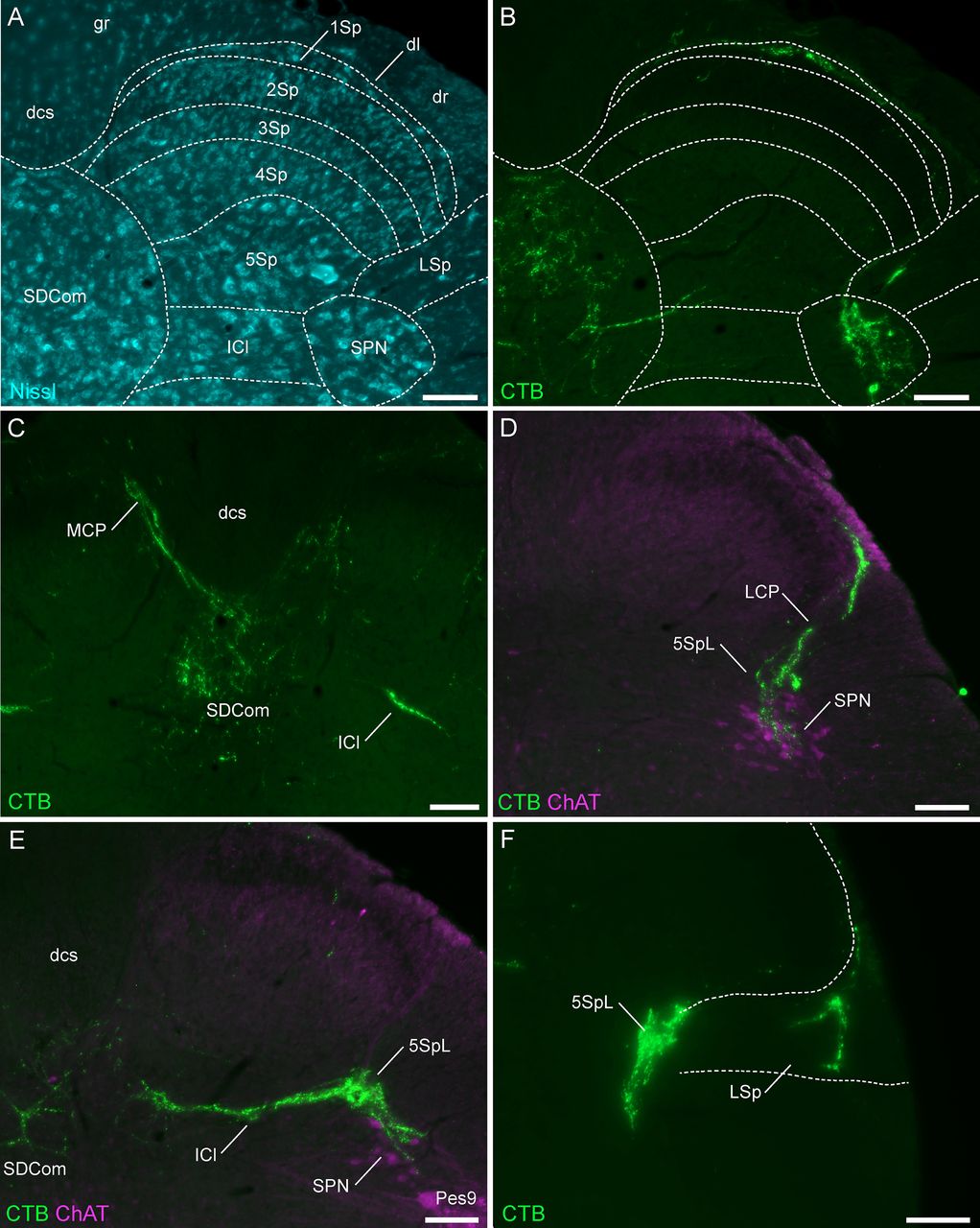
Major projections of CTB-labeled LUT afferents in the lumbosacral (L6-S1) spinal cord. A, Cytoarchitecture shown by Nissl staining in an L6 spinal cord section and corresponding spinal cord regions from the atlas of Watson et al. (2009). B, Urethra afferents (male) of the same section as A, showing afferents in the SPN, SDCom, lateral spinal nucleus, and Lamina I of the dorsal horn. C, Bladder afferents (male) in the medial collateral pathway, SDCom, and intercalated nucleus of the L6 spinal cord. D, Urethra afferents (male) projecting in the lateral collateral pathway to lateral Lamina V of the dorsal horn and the SPN (co-labeled for ChAT) in the S1 spinal cord. E, Bladder afferents (female) in the SPN, SDCom, and intercalated nucleus of the L6 spinal cord. F, Bladder afferents (female) innervating lateral Lamina V of the dorsal horn and the lateral spinal nucleus in the L6 spinal cord. CC, central canal; ChAT, choline acetyltransferase; CTB, cholera toxin B subunit; dl, dorsolateral fasciculus; dcs, dorsal corticospinal tract; g, gracile fasciculus; ICl, intercalated nucleus; LCP, lateral collateral pathway; LSp, lateral spinal nucleus; LUT, lower urinary tract; MCP, medial collateral pathway; 1–10Sp, Laminae I–X of the spinal gray (dorsal horn); SDCom, sacral dorsal commissural nucleus; SPN, sacral preganglionic nucleus. Each image is oriented with dorsal at the top of the micrograph; the lateral dorsal horn is on the right for all panels except C, midline view. Scale bars: 100 μm.
- Figure 3.
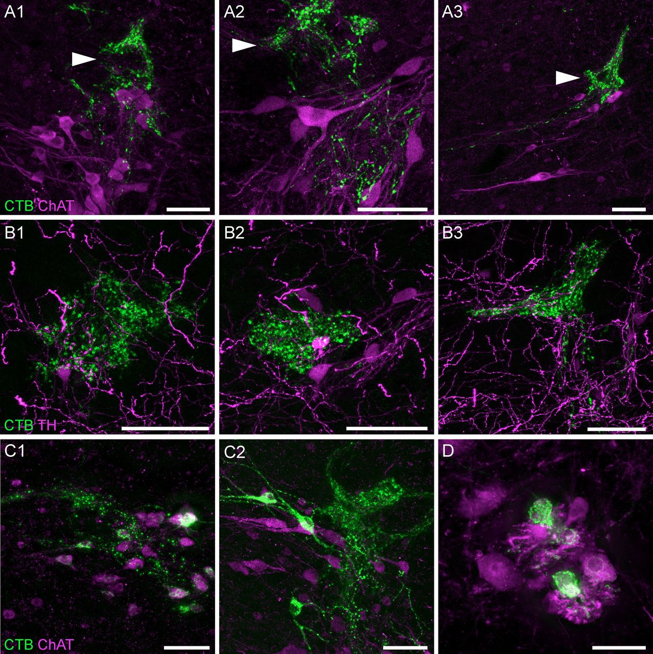
LUT afferent innervation of the SPN and surrounding regions. A, Confocal micrographs demonstrate the close apposition of CTB-labeled bladder afferents to cholinergic (ChAT) neurons in the SPN of a male rat; most of the afferent innervation in this region is located immediately dorsal to the SPN, in lateral Lamina V (arrowheads). B, Confocal micrographs of bladder afferent innervation of TH-positive neurons in lateral Lamina V of a male rat. C, CTB-labeled cholinergic neurons of the SPN in (C1) male and (C2) female rats following urethra injections of CTB. D, CTB-labeled neurons in the dorsolateral motor nucleus of a female rat after CTB injection into the urethra. ChAT, choline acetyltransferase; CTB, cholera toxin B subunit; LUT, lower urinary tract; TH, tyrosine hydroxylase. Scale bars: 50 μm.
- Figure 4.
The SPN location within the lumbosacral spinal cord. A, A 3D reconstruction was created from the digital alignment of a 1:2 series of 121 cryosections of the lumbosacral spinal cord (female rat). A1, Transverse view of the aligned L6 and S1 segments showing motoneuron nuclei and preganglionic autonomic neurons identified via ChAT immunolabelling. Preganglionic neurons are less strongly immunolabelled than motoneurons. A2, The boundary of the SPN in each section was identified at higher magnification and delineated manually on each section. B, The full 3D reconstruction is shown here in the horizontal plane, demonstrating that the SPN is present in both L6 and S1 segments but does not extend for the full length of L6. Examples of individual sections from four locations along the cord are indicated by arrowheads in C–F. C–F, Individual cryosections demonstrating that preganglionic neurons are absent in rostral L6 (C) but present in (D) caudal L6, (E) rostral S1, and (F) caudal S1. Specific motoneuron nuclei confirm segment position; these nuclei reduce in cell number in the caudal direction until they end at the L6-S1 junction. The rostral boundary of the SPN in L6 coincides with the narrowing of the Pes9 nucleus, approximately halfway along the L6 segment, as also observed in (G) light sheet image stacks of iDISCO-cleared intact lumbosacral spinal cord (male rat). Motoneurons of Lamina IX: Ax9 (axial), ExU9* (external urethral sphincter), Gl9 (gluteal), Hm9 (hamstring), Pes9 (pes), Tail9 (tail); CC, central canal; ChAT, choline acetyltransferase; SPN sacral preganglionic nucleus. *, the location of ExU9 is indicated according to Schrøder (1980), McKenna and Nadelhaft (1986), Nadelhaft and Vera (1996, 2001), and Xu et al. (2007). Scale bars: 200 μm.
- Figure 5.
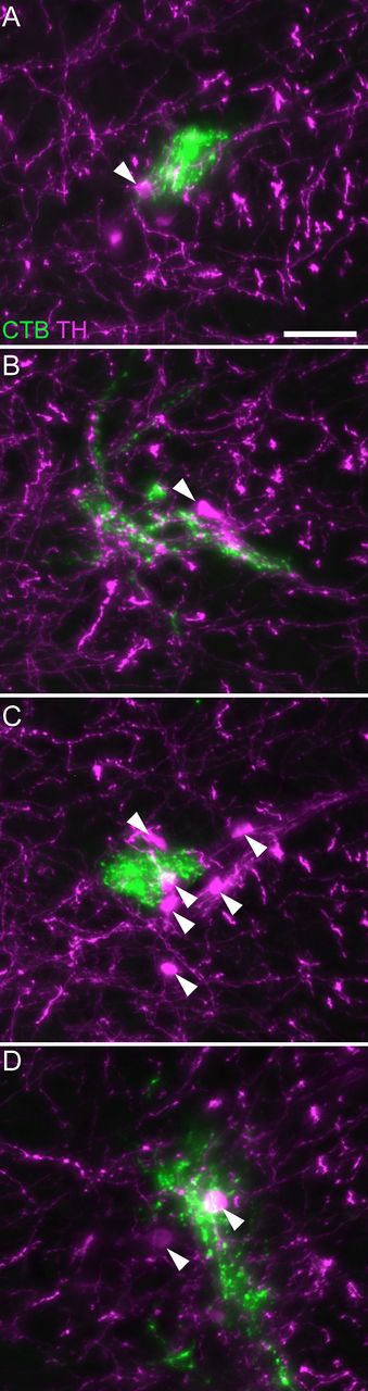
Association of LUT afferents with TH neurons in lateral Lamina V. CTB-labeled bladder afferents innervating lateral Lamina V in (A) rostral L6, (B) mid-L6, (C) caudal L6, and (D) rostral S1 of a female rat. Many but not all TH neurons show close associations with LUT afferents. Arrowheads indicate examples of TH-positive neurons. CTB, cholera toxin subunit B; LUT, lower urinary tract; TH, tyrosine hydroxylase. Scale bar: 50 μm.
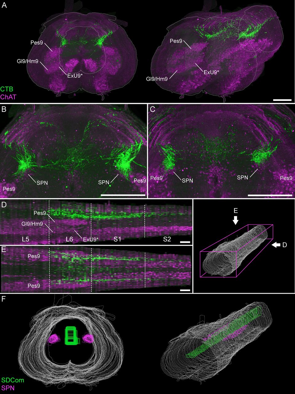
- Figure 6.
Visualizing the rostrocaudal distribution of bladder afferents in 3D reconstructions of lumbosacral spinal cord. A, Visualization of 100 ordered and aligned sections in a single view along the rostrocaudal axis of the spinal cord (female rat), oriented with the rostral boundary at the front of the field of view in transverse (A1) and oblique (A2) views. Motor and preganglionic autonomic neurons were visualized by ChAT. B, C, Bladder afferents in a female (B) and male (C) rat, shown in a maximum intensity projection of the 3D reconstructed lumbosacral spinal cord. Sagittal (D) and horizontal (E) views of bladder afferent innervation (male rat) each demonstrate the rostrocaudal limits of LUT afferent innervation, indicated by the segment boundaries and the location of the lumbosacral motor nuclei. Schematic on the right shows the viewing orientation of D, E. F, For further visualization and quantitative studies, the perimeters of individual sections were outlined (white) and specific ROIs defined, such as the SPN (magenta) and the SDCom (green). Motoneurons of Lamina IX: ExU9* (external urethral sphincter), Gl9 (gluteal), Hm9 (hamstring), Pes9 (pes). *, the location of ExU9 indicated according to Schrøder (1980), McKenna and Nadelhaft (1986), Nadelhaft and Vera (1996, 2001), and Xu et al. (2007). ChAT, choline acetyltransferase; CTB, cholera toxin B subunit; LUT, lower urinary tract; SDCom, sacral dorsal commissural nucleus; SPN, sacral preganglionic nucleus. Scale bars: 500 μm.
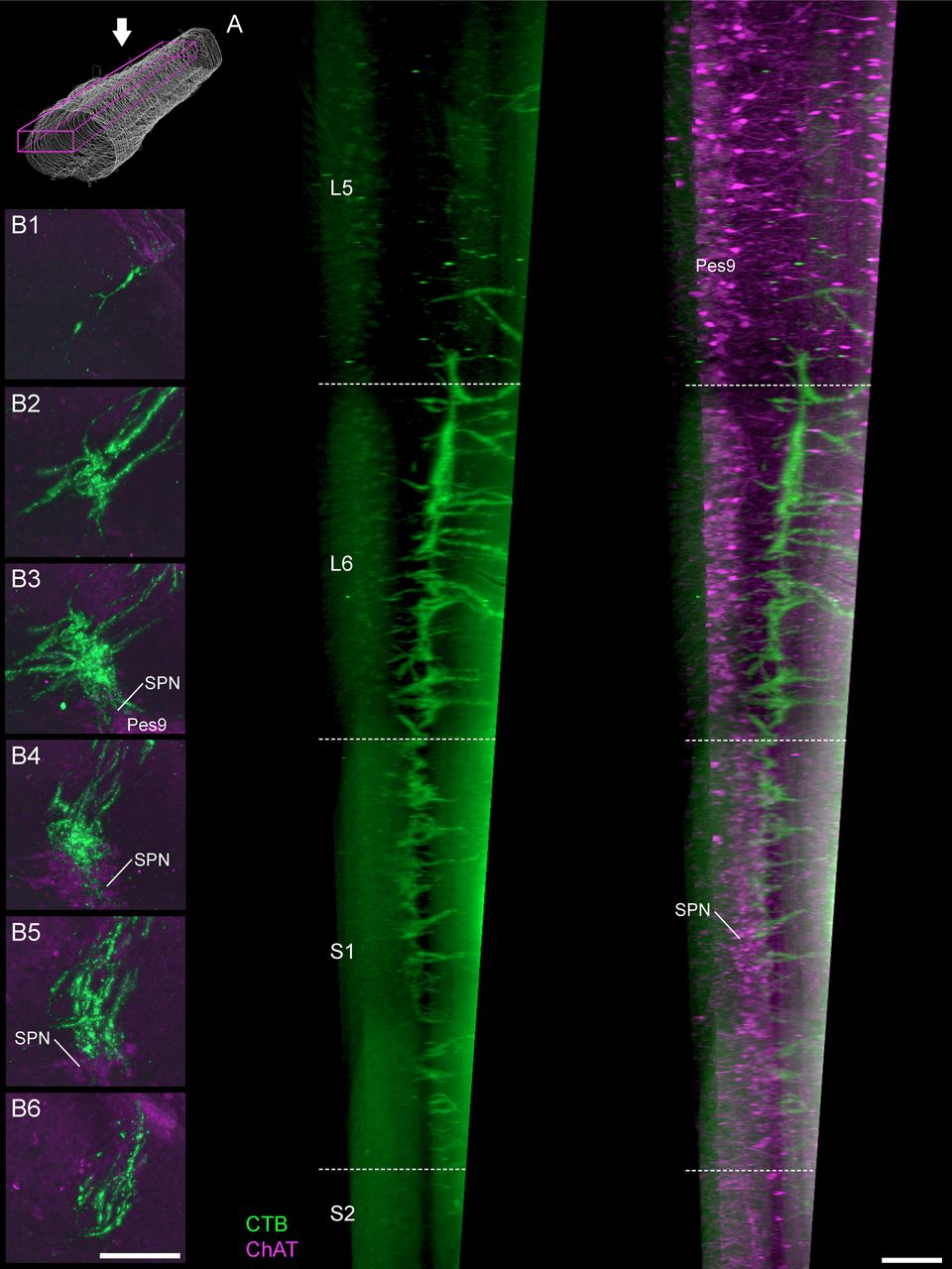
- Figure 7.
Projections of LUT afferents in the lateral collateral pathway. A, In a parasagittal view of the cleared lumbosacral spinal cord, bladder afferents (male rat) show distinct gradations in density of the lateral collateral pathway projection. This extends from caudal L5 to rostral S2 but is most dense across the length of the L6 segment. Schematic shows viewing orientation. B, Maximum intensity projections of discrete portions of the reconstructed spinal cord datasets reveal the distribution of bladder afferents (female rat) at different levels of the spinal cord: (B1) caudal L5, (B2) rostral L6, (B3) caudal L6, (B4) rostral S1, (B5) caudal S1, and (B6) rostral S2. This includes regions more proximal (B1, B2) and more caudal (B6) to the SPN, as demonstrated by immunoreactivity for ChAT; ChAT, choline acetyltransferase; CTB, cholera toxin B subunit; LUT, lower urinary tract; Pes9, Pes motoneurons of lamina IX; SPN, sacral preganglionic nucleus. Scale bars: 200 μm.
- Figure 8.
Projections of LUT afferents in the medial collateral pathway. A, Discrete portions of the reconstructed spinal cord datasets were visualized with maximum intensity projections of bladder afferents (female rat), focusing on the medial collateral pathway, SDCom, and intercalated nucleus: (A1) caudal L5, (A2) rostral L6, (A3) caudal L6, (A4) rostral S1, (A5) caudal S1, and (A6) rostral S2. B, In cleared lumbosacral spinal cord, individual tracts of the medial collateral pathway are most prominently observed in the parasagittal view. In the horizontal (C) and transverse (D) views, the LUT afferent innervation of the intercalated nucleus between the SDCom and the lateral collateral pathway are revealed. Schematic on B, left, shows the viewing orientation of B-D. ICl, intercalated nucleus; LCP, lateral collateral pathway; MCP, medial collateral pathway; ChAT, choline acetyltransferase; CTB, cholera toxin B subunit; LUT, lower urinary tract; SDCom, sacral dorsal commissural nucleus. Scale bars: 100 μm.
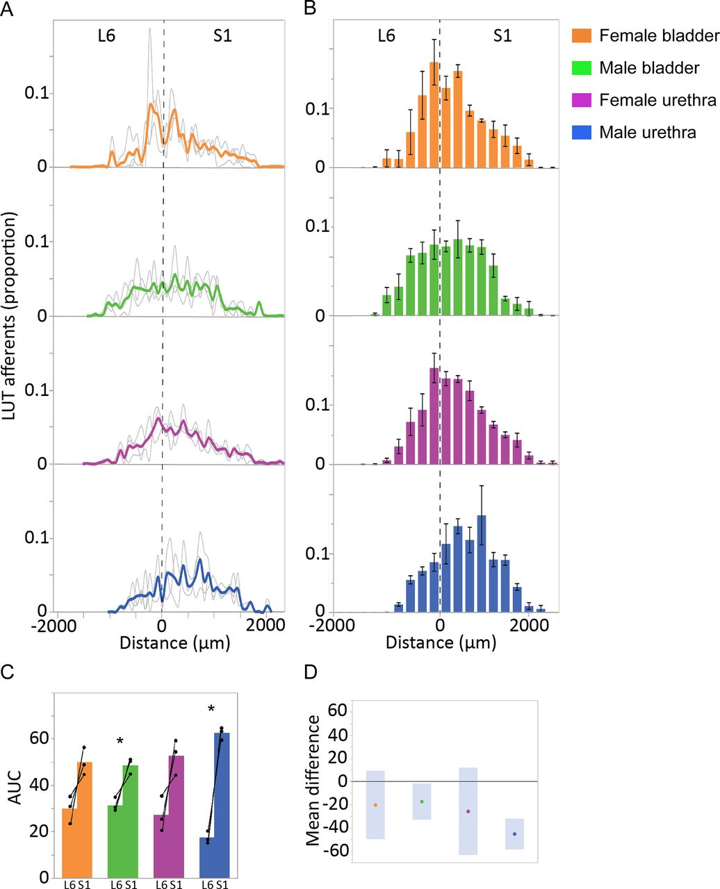
- Figure 9.
Quantitation of LUT afferent innervation of the SPN. A, CTB-labeled afferents in each section, segmented and quantified as puncta, are expressed as the proportion of the total number of CTB puncta in the SPN of the same section. Data shown for individual animals (gray lines) and group mean (bold colors) and plotted as distance (μm) from the L6-S1 junction (0 μm, vertical broken line). B, Data from individual sections was summed (three sections per bin) to better visualize overall distribution patterns (n = 3 rats per group, mean ± SE). C, Total afferent innervation of the SPN in L6 and S1 segments determined by AUC, shown for individual animals and group data (mean ± SE) for each of the four experimental groups. In each animal, regardless of LUT region and sex, a larger number of afferent structures were found in S1 than L6. This segmental difference was also detected in analysis of the grouped data for the male bladder and male urethra groups [p = 0.042 (male bladder) and p = 0.005 (male urethra), paired two-tailed t test] but could not be detected for either of the female groups (female bladder, female urethra) where there was more intersubject variation in AUC measurements. D, Plot of difference in group means (dots) and 95% confidence intervals (gray bars) for AUC measurements in SPN of L6 and S1 spinal cord. LUT, lower urinary tract; SPN, sacral preganglionic nucleus.
- Figure 10.
Quantitation of LUT afferent innervation of the SDCom. A, The innervation of the SDCom extends beyond the L6 and S1 spinal cord segments, as exemplified in a dataset (bladder afferents, female rat) in which aligned cryosections were collected from caudal L4 until the caudal limit of S2. This also demonstrates the strong innervation of this nucleus in the rostral half of L6, a spinal level where preganglionic neurons are absent. B, CTB-labeled afferents in each section, segmented and quantified as puncta, are expressed as the proportion of the total number of CTB puncta in the SDCom of the same section. Data shown for individual animals (gray lines) and group mean (bold colors) and plotted as distance (μm) from the L6-S1 junction (0 μm, vertical broken line). C, Data from individual sections was summed (three sections per bin) to better visualize overall distribution patterns (n = 3 rats per group, mean ± SE). D, Total afferent innervation of the SDCom in L6 and S1 segments determined by AUC, shown for individual animals and group data (mean ± SE) for each of the four experimental groups. No significant difference between L6 and S1 segments were detected in any of the four groups (paired, two-tailed t test). E, Plot of difference in group means (dots) and 95% confidence intervals (gray bars) for AUC measurements in the SDCom of the L6 and S1 spinal cord. LUT, lower urinary tract; SDCom, sacral dorsal commissural nucleus.


Tables
Primary antibodies RRID Antigen Host Dilution Supplier Catalog number AB_1658411 CGRP Mouse 1:500 Abcam AB81887 AB_11214092 ChAT Goat 1:500 Millipore AB144P AB_258833 CTB Rabbit 1:30,000 (sections);
1:3000 (iDisco clearing)Sigma-Aldrich C3062 AB_477257 NF200 Mouse 1:4000 Sigma-Aldrich NO142 AB_90755 TH Sheep 1:1000 Millipore AB1542 AB_1624144 TRPV1 Goat 1:1000 Neuromics GT15129 Secondary antibodies RRID Antigen Tag Host Dilution Supplier Catalog number AB_2307351 Goat IgG Cy3 Donkey 1:2000 Jackson ImmunoResearch 705-165-147 AB_2340433 Goat IgG AF594 Donkey 1:500 Jackson ImmunoResearch 705-585-147 AB_2340846 Mouse IgG AF488 Donkey 1:2000 Jackson ImmunoResearch 711-545-150 AB_2535789 Mouse IgG AF594 Donkey 1:1000 Molecular Probes A-21203 AB_2313584 Rabbit IgG AF488 Donkey 1:1000 Jackson ImmunoResearch 711-545-152 AB_2307443 Rabbit IgG Cy3 Donkey 1:3000 Jackson ImmunoResearch 711-165-152 AB_2536183 Rabbit IgG AF647 Donkey 1:1000 Molecular Probes A-31573 AB_10374882 Sheep IgG AF647 Donkey 1:500 Molecular Probes A-21448 CGRP, calcitonin gene-related peptide; ChAT, choline acetyltransferase; CTB, cholera toxin B subunit; NF200, neurofilament 200 kDa; TH, tyrosine hydroxylase; TRPV1, transient receptor potential vanilloid 1.
In this issue


