

Article Figures & Data
Figures
- Figure 1.
Opn3-eGFP immunodetection (red) at E10.5 in sagittal (A–E) and coronal (F–K) serial sections counterstained with DAPI (blue). On the left, schematics of E10.5 embryos including planes of sections shown in A–K. A, Di: diencephalic vesicle, Mes: mesencephalic vesicle, opv: optic vesicle, Rh: rhombencephalic vesicle, v.g: trigeminal ganglion. B, ix.g: glossopharyngeal ganglion, Mes: mesencephalic vesicle, o: otic vesicle, oe: olfactory epithelium, Rh: rhombencephalic vesicle, vii-viii.g: facio-acoustic ganglia, x.g: vagus ganglion. A, B, Parts of the amnion surrounding the embryo were digitally removed (raw data in Extended Data Fig. 1-3). C, ix.g: glossopharyngeal ganglion, o: otic vesicle, Rh: rhombencephalic vesicle, v.g: trigeminal ganglion, vii-viii.g: facio-acoustic ganglia, x.g: vagus ganglion, x.n: vagal nerve. D, drg: dorsal root ganglia, drg.x: dorsal root afferent processes. E, ap: alar plate, bp: basal plate, drg: dorsal root ganglia, drg.x: dorsal root afferent processes, Rh: rhombencephalic vesicle. F, oe: olfactory epithelium. G, Di: diencephalic vesicle, opv: optic vesicle, os: optic stalk. H, ix.n: glossopharyngeal nerve, o: otic vesicle, v.g: trigeminal ganglion, vii.g: geniculate ganglion, viii.g: vestibulocochlear ganglion, x.n: vagal nerve. I, rs: rootlets of the v/vii/viii.g, v.g: trigeminal ganglion, vii.g: geniculate ganglion, viii.g: vestibulocochlear ganglion. J, bp: basal plate, drg: dorsal root ganglion, SC: spinal cord. K, mnl: mantle layer, s.n: spinal nerves. Scale bars: 100 μm. Extended Data Figures 1-1, 1-2 are supporting this figure.
- Figure 2.
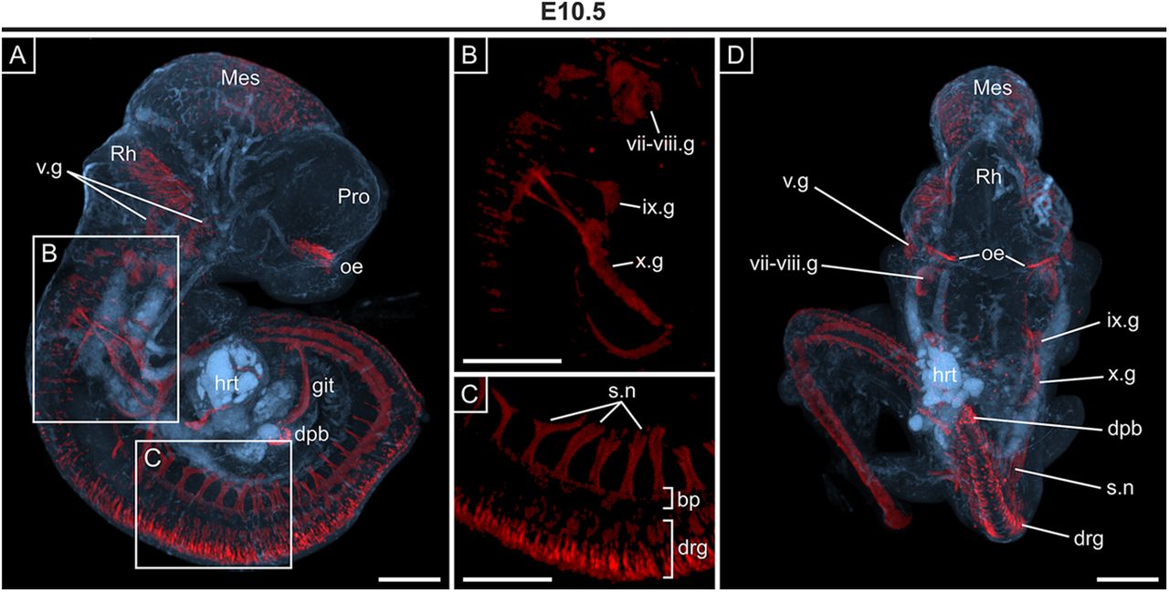
3D OPT imaging of whole E10.5 embryo showing Opn3-eGFP immunodetection. A, Sagittal view showing detection of Opn3-eGFP (red) against a background of auto-fluorescing anatomical structures (blue). Developing ganglia that form the sensory branches of CNs V, CN VII, and CN VIII CNs are shown in B, and spinal nerves highlighted in C, D. D, Dorsal view of embryo shown in A. bp: basal plate, dpb: dorsal pancreatic bud, drg: dorsal root ganglia, git: gastrointestinal tract, hrt: heart; ix.g: glossopharyngeal ganglion, Mes: mesencephalic vesicle, oe: olfactory epithelium, Pro: prosencephalic vesicle, Rh: rhombencephalic vesicle, s.n: spinal nerves, v.g: trigeminal ganglion, vii-viii.g: facio-acoustic ganglia (comprising the vii.g geniculate ganglion and viii.g: vestibulocochlear ganglion), x.g: vagus ganglion. Scale bars: 250 μm.
- Figure 3.
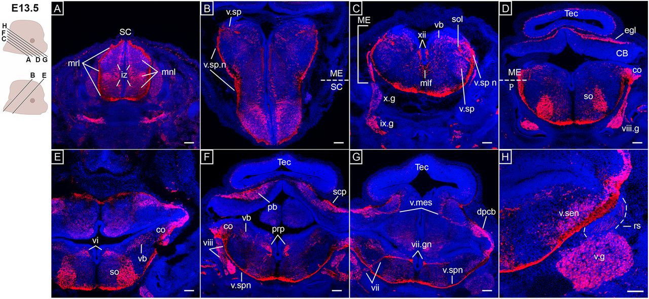
Opn3-eGFP immunodetection (red) at E13.5 in horizontal (A, C, D, F–H) and coronal (B, E) serial sections in caudo-rostral direction from the spinal cord to hindbrain areas counterstained with DAPI (blue). Left, Schematics of E13.5 embryonic heads including planes of sections shown in A–H. A, iz: intermediate zone, mnl: mantle layers, mrl: marginal layers, SC: spinal cord. B, ME: medulla, SC: spinal cord, v.sp n: spinal trigeminal nerve, v.sp: spinal trigeminal nucleus. C, ix.g: glossopharyngeal ganglion, ME: medulla, mlf: medial longitudinal fasciculus, sol: solitary tract, v.sp: spinal trigeminal nucleus, vb: vestibular nuclei, x.g: vagus ganglion, xii: hypoglossal nucleus. D, CB: cerebellum, co: cochlear nucleus, egl: external granular layer of cerebellum, ME: medulla; P: pons, so: superior olivary complex, Tec: tectum of midbrain, vii.g: geniculate (facial) ganglion. E, co: cochlear nucleus, so: superior olivary complex, vb: vestibular nuclei, vi: abducens nucleus. F, pb: parabrachial nuclei, prp: nucleus prepositus, scp: superior cerebellar peduncle, Tec: tectum of midbrain, v.sp.n: spinal trigeminal nerve, viii: vestibular nerve. G, dpcb: deep cerebellar nuclei, Tec: tectum of midbrain, v.mes: mesencephalic trigeminal nucleus, v.sp.n: spinal trigeminal nerve, vii.gn: genu of facial nerve, vii: sensory/parasympathetic facial nerve. H, rs: rootlets of the v.g, v.g: trigeminal ganglion, v.sen: sensory trigeminal nucleus. Scale bars: 100 μm.
- Figure 4.
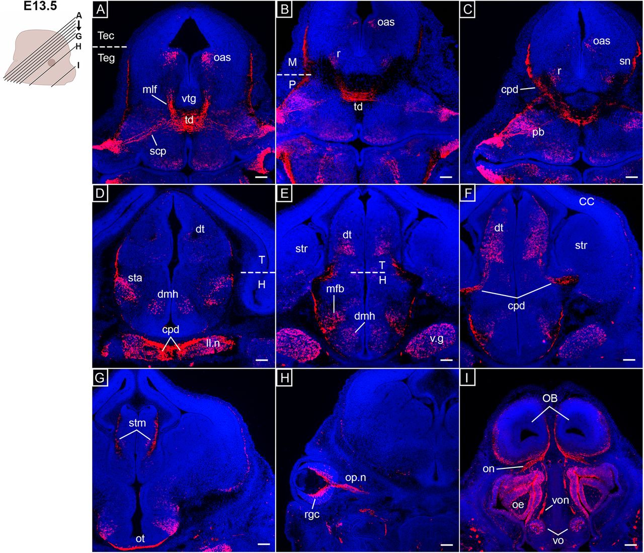
Opn3-eGFP immunodetection at E13.5 (red) in coronal serial sections caudo-rostral direction from midbrain to forebrain areas counterstained with DAPI (blue). On the left, a schematic of an E13.5 embryonic head including planes of sections shown in A–I. A, mlf: medial longitudinal fasciculus, oas: oculomotor associated subnuclei, scp: superior cerebellar peduncle, td: tegmental decussations, Tec: tectum of midbrain, Teg: midbrain and pontine tegmentum, vtg: ventral tegmentum. B, M: midbrain, oas: oculomotor associated subnuclei, P: pons, r: red nucleus, td: tegmental decussations. C, cpd: cerebral peduncle, oas: oculomotor associated subnuclei, pb: parabrachial nuclei, r: red nucleus, sn: substantia nigra. D, cpd: cerebral peduncle, dmh: dorsomedial hypothalamic nucleus, dt: dorsal thalamus, H: hypothalamus, ll.n: nucleus of lateral lemniscus, sta: subthalamic area, T: thalamus, v.g: trigeminal ganglion. E, dt: dorsal thalamus, H: hypothalamus, mfb: medial forebrain bundles, str: striatum, T: thalamus, dmh: dorsomedial hypothalamic nucleus. F, CC: cerebral cortex, cpd: cerebral peduncle, dt: dorsal thalamus, str: striatum. G, stm: stria medullaris, ot: optic tract. H, op.n: optic nerve, rgc: retinal ganglion cells. I, OB: olfactory bulb, oe: olfactory epithelia, on: olfactory nerve, vo: vomeronasal organ, von: vomeronasal nerve. Scale bars: 100 μm.
- Figure 5.
Opn3-eGFP immunodetection (red) at E15.5 in horizontal serial sections in caudo-rostral direction from the medulla to pons areas counterstained with DAPI (blue). On the left, a schematic of an E15.5 embryonic head including planes of sections shown in A–H. A, ME, medulla, P: pons, so: superior olivary complex, sol.n: solitary nucleus, xii: hypoglossal nucleus. B, marn: magnocellular reticular nucleus, mern: medullary reticular nuclei, ro: nucleus raphe obscurus. C, mcp: medial cerebellar peduncle, pn: pontine nuclei. D, cf: cuneate fasciculus, gf: gracile fasciculus, ll.n: nucleus of lateral lemniscus, ME: medulla, P: pons, sol: solitary tract, v.sen: sensory trigeminal nucleus, v.sp: spinal trigeminal nucleus. E, mlf: medial longitudinal fasciculus, prn: pontine reticular nuclei, vii.gn: genu of facial nerve. F, ME: medulla, P: pons, prp: nucleus prepositus, sol.n: solitary nucleus. G, lav: lateral vestibular nucleus, ll.n: nucleus of lateral lemniscus, prn: pontine reticular nuclei, suv: superior vestibular nucleus, v.m: motor trigeminal nucleus, v.sen: sensory trigeminal nucleus, v.sp: spinal trigeminal nucleus. H, CB: cerebellum, dn: dentate nucleus, fn: fastigial nucleus, IntA: interposed nucleus, scp: superior cerebellar peduncle. Scale bars: 100 μm.
- Figure 6.
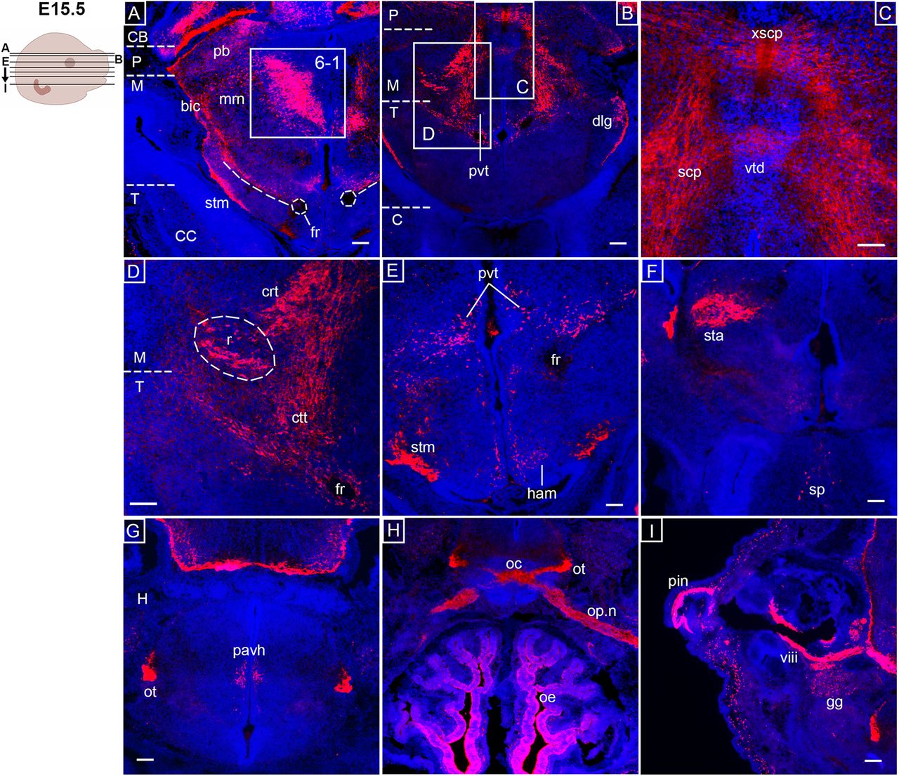
Opn3-eGFP immunodetection (red) at E15.5 in horizontal serial sections in caudo-rostral direction from the midbrain to forebrain areas counterstained with DAPI (blue). Left, Schematic of an E15.5 embryonic head including planes of sections shown in A–I. A, bic: brachium of inferior colliculus, CB: cerebellum, CC: cerebral cortex, fr: fasciculus retroflexus, M: midbrain, mrn: midbrain reticular nuclei, P: pons, pb: parabrachial nuclei, stm: stria medullaris, T: thalamus. The inserted box indicates area shown in Extended Data Figure 6-1. B, C: cerebrum, dlg: dorsal lateral geniculate nucleus, M: midbrain, P: pons, pvt: pvt: periventricular thalamic nucleus, T: thalamus. C, scp: superior cerebellar peduncle, vtd: ventral tegmental decussation, xscp: decussation of superior cerebellar peduncle. D, crt: cerebellorubral tract, ctt: cerebellothalamic tract, fr: fasciculus retroflexus, M: midbrain, r: red nucleus, T: thalamus. E, fr: fasciculus retroflexus, ham: medial habenula, pvt: periventricular thalamic nucleus, stm: stria medullaris. F, sp: septal nuclei, sta: subthalamic area. G, H: hypothalamus, ot: optic tract, pavh: paraventricular hypothalamus nucleus. H, oc: optic chiasm, oe: olfactory epithelium, op.n: optic nerve, ot: optic tract. I, gg: geniculate ganglion, pin: pinna of the ear, viii: vestibulocochlear nerve. Scale bars: 100 μm.
- Figure 7.
Opn3-eGFP immunodetection (red) at E17.5 in horizontal (B) and coronal (A, C–P) serial sections from the hindbrain to forebrain areas counterstained with DAPI (blue). Left, Schematic of E17.5 embryonic heads including planes of sections shown in A–P. A, ic: inferior colliculus. B, CA: cerebral aqueduct, CB: cerebellum, dnsc: deep nuclei of superior colliculus, fn: fastigial nucleus, hbr: hooked bundle of Russel, M: midbrain, pag: periaqueductal gray. C, CB: cerebellum, cun: cuneiform nucleus, dncb: deep nuclei of cerebellum, ll.d dorsal component of lateral lemniscus, P: pons, Tec: tectum of midbrain, Teg: tegmentum of midbrain. D, CB: cerebellum, ME: medulla, mern: medullary reticular nuclei, P: pons, pb: parabrachial nuclei, prn: pontine reticular nuclei. E, dnsc: deep nuclei of superior colliculus dtd: dorsal tegmental decussation, mlf: medial longitudinal fasciculus, mrn: midbrain reticular nuclei, oas: oculomotor associated subnuclei. F, M: midbrain, oas: oculomotor associated subnuclei, r: red nucleus, rst: rubrospinal tract, Tec: tectum of midbrain, Teg: tegmentum of midbrain, vtd: ventral tegmental decussation. G, sn: substantia nigra, pf: parafascicular nucleus. H, ll: lateral lemniscus, MB: mamillary body, mcp: medial cerebellar peduncle, pn: pontine nuclei (gray), prf: pontine reticular formation. I, fh: field H, pvt: paraventricular thalamic nucleus, sta: subthalamic area, stm: stria medullaris, sut: subthalamic nucleus. J, CC: cerebral cortex, cpd: cerebral peduncle, fh: field H, H: hypothalamus, lf: lenticular fasciculus, pf: parafascicular nucleus, π: pineal gland, sta: subthalamic area, T: thalamus, vmt: ventromedial thalamic nucleus. K, fh: field H, H: hypothalamus, hal lateral habenula, ot: optic tract, stm: stria medullaris, T: thalamus, thf: thalamic fasciculus, dmh: dorsomedial hypothalamic nucleus, vmt: ventromedial thalamic nucleus. L, hip: hippocampus, H: hypothalamus, pavh: paraventricular hypothalamic nucleus, T: thalamus. M, H: hypothalamus, ot: optic tract, pavh: paraventricular hypothalamic nucleus, pvh periventricular hypothalamic nucleus, T: thalamus. N, H: hypothalamus, ot: optic tract, pvh periventricular hypothalamic nucleus, T: thalamus. O, C: cerebrum, sfo: subfornical organ, sp: septum of brain, T: thalamus. P, nac: nucleus accumbens, cpf: pyriform (olfactory) cortex, mpn: medial preoptic nucleus, sp: septum of brain, str: striatum. Scale bar: 100 μm. Extended Data Figure 7-1 is supporting this figure.
- Figure 8.
Opn3-eGFP immunodetection (red) at P0.5 in horizontal sections of forebrain areas counterstained with DAPI (blue). On the left, a schematic of a P0.5 brain including planes of sections shown in A–D. A, CC: cerebral cortex, Hip: hippocampus, p: posterior thalamus, rt: thalamic reticular nucleus, str: striatum, vpl: ventral posterolateral nucleus, vpm: ventral posteromedial nucleus. B, C: cerebrum, dlg: dorsal lateral geniculate nucleus, ld: lateral dorsal nucleus, sfo: subfornical organ, sp: septal nuclei, T: thalamus, Tec: tectum of midbrain. C, dg: dentate gyrus, str: striatum. D, acc: anterior cingulate cortex, str: striatum. Scale bars: 100 μm. Extended Data Figure 8-1 is supporting this figure.
- Figure 9.
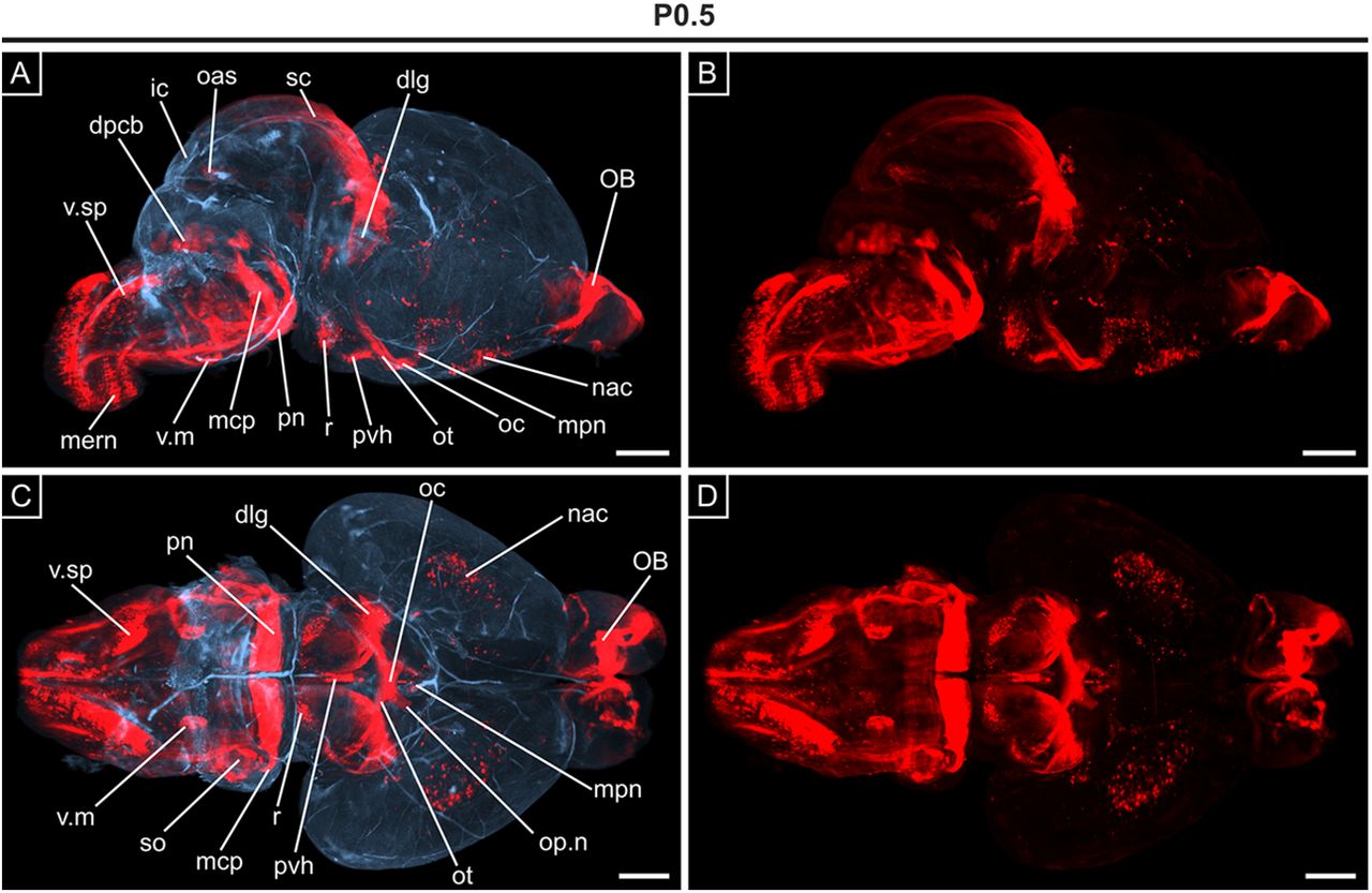
3D OPT imaging of Opn3-eGFP immunodetection in the brain at P0.5. A, Sagittal view showing the presence of Opn3-eGFP (red) against a background of auto-fluorescing anatomic structures (blue). B, Repeat of (A) showing unmasked Opn3-eGFP (red) staining only. C, D, Ventral views of the brain shown in A, B, respectively. dlg: dorsal lateral geniculate nucleus, dpcb: deep nuclei of cerebellum, ic: inferior colliculus, mcp: medial cerebellar peduncle, mern: medullary reticular nuclei, mpn: medial preoptic nucleus, nac: nucleus accumbens, oas: oculomotor associated subnuclei, OB: olfactory bulb, oc: optic chiasm, op.n: optic nerve, ot: optic tract, pn: pontine nuclei (also pontine nuclei gray), pvh: periventricular hypothalamic nucleus, r: red nucleus, sc: superior colliculus, so: superior olivary complex, v.m: motor trigeminal nucleus, v.sp: spinal trigeminal nucleus. Scale bars: 1 mm.


Tables
- Table 1.
A Summary outlining the onset of Opn3-eGFP expression in various structures of the CNS and PNS, indicating novel Opn3-eGFP+ structures identified in this study (•)
PNS and sensory organs CNS Age Trigeminal ganglion • Neural tube Basal plate E9.5 Facio-acoustic complexganglion • Brainvesicles Basal plate of diencephalon Olfactory placode Dorsal outer cells of mesencephalon Dorsal root ganglion Spinal cord Motor neurons E10.5 Inferior vagus ganglion • Glossopharyngealganglion • Brainvesicles Optic stalk Migratory olfactoryneurons Retinal ganglion cells Spinal cord Dorsal, lateral and ventral mantleand marginal layers E13.5 Optic nerve Olfactory epithelium • Hindbrain Spinal trigeminal nucleus • Olfactory nerve • Principal sensory trigeminal nucleus • Vomeronasal organ • Mesencephalic trigeminal nucleus • Vomeronasal nerve • Genu of facial nerve • Solitary tract Parabrachial nuclei • Hypoglossal nucleus • Nucleus prepositus • Vestibular nuclei • Medial longitudinal fasciculus Abducens nucleus • Cochlear nuclei • Superior olivary complex External granular layers of cerebellum Deep cerebellar nuclei Superior cerebellar peduncle Midbrain Red nucleus Oculomotor associated sub nuclei • Tegmental fibres Forebrain Subthalamic area Dorsal thalamus Dorsal medial hypothalamic nucleus Stria medullaris Optic tract Olfactory bulb Pinna of the ear Hindbrain Medullary reticular nuclei E15.5 Magnocellular reticular nucleus Raphe obscurus • Cuneate and gracile nuclei • Pontine nuclei Medial cerebellar peduncle • Motor trigeminal nucleus Nucleus of lateral lemniscus Brachium of inferior colliculus Midbrain Midbrain reticular nuclei Inferior colliculus Brachium of inferior colliculus Forebrain Periventricular thalamic nucleus Dorsal lateral geniculate nucleus Septal nuclei • Paraventricular hypothalamic nucleus Cerebellum Uncinate fasciculus of cerebellum E17.5 Midbrain Periaqueductal gray • Cuneiform nucleus Rubrospinal tract Substantia nigra Epithalamus Pineal gland Thalamus Parafascicular nucleus • Ventromedial thalamic nucleus Subthalamus Subthalamic nucleus Hypothalamus Periventricular hypothalamic nucleus Medial hypothalamic nucleus Cerebrum Subfornical organ Nucleus accumbens Pyriform cortex Thalamus Posterior thalamus P0.5 Lateral dorsal nucleus Ventral posteromedial nucleus Ventral posterolateral nucleus Subpallium Dentate gyrus of hippocampus Pallium Anterior cingulate cortex Frontal cortex Hypothalamus Mammillary nuclei P10 Posterior nucleus Subpallium Fornix Subiculum Corticostrial circuitry Meninges Outer membrane Hypothalamus Perifornical nuclei Adult Tanycytes Supraoptic nucleus Bed nucleus of stria terminalis
Movies
- Movie 1.
Video of 3D imaging of a whole E10.5 embryo (along the y-axis) showing Opn3-eGFP immunodetection (red) against a background of auto-fluorescing anatomical structures (blue).
- Movie 2.
Video of 3D imaging of a P0.5 brain (along the x-axis) showing Opn3-eGFP immunodetection (red) against a background of auto-fluorescing anatomical structures (blue).
Extended Data Figure 1-1
Opn3-eGFP immunodetection (red) at E9.5 in sagittal (A–D) and coronal (E–L) serial sections counterstained with DAPI (blue). On the left, schematics of E9.5 embryos including planes of sections shown in A–L. A, bp: basal plate, dpb: dorsal pancreatic bud, nt: neural tube, php: pharyngeal pouches. B, dpb: dorsal pancreatic bud, nt: neural tube, php: pharyngeal pouches, v.g: trigeminal ganglion. C, bp: basal plate, nt: neural tube, o: otic vesicle, php: pharyngeal pouches, vii-viii.g: facio-acoustic ganglia. D, git: gastrointestinal tract, Mes: mesencephalic vesicle, Pro: prosencephalic vesicle, Rh: rhombencephalic vesicle. E, Te: telencephalic vesicle. F, op: olfactory placode, Te: telencephalic vesicle. G, Di: diencephalic vesicle, inf: infundibulum. H, Di: diencephalic vesicle. I, Mes: mesencephalic vesicle. J, Mes: mesencephalic vesicle. K, o: otic vesicle, Rh: rhombencephalic vesicle, v.g: trigeminal ganglion, vii-viii.g: facio-acoustic ganglia. L, bp: basal plate, nt: neural tube. Scale bars: 100 μm. Download Figure 1-1, TIF file.
Extended Data Figure 1-2
No DAPI labelled nuclei were detected in Opn3-eGFP-positive projection areas. A–F, Opn3-eGFP immunodetection (red) at E10.5 in (A–C) and at E13.5 (D–F) horizontal sections counterstained with DAPI (blue). A, bp: basal plate, drg: dorsal root ganglion, SC: spinal cord. B, s.n: spinal nerves, mnl: mantle layer. C, s.n: spinal nerves. D, dpcb: deep nuclei of cerebellum, Tec: tectum of midbrain, v.mes: mesencephalic trigeminal nucleus. E, v.spn: spinal trigeminal nerve, vii: facial nerve, vii.gn: genu of facial nerve. F, v.spn: spinal trigeminal nerve, vii: facial nerve, vii.gn: genu of facial nerve. Scale bars: 100 μm. Download Figure 1-2, TIF file.
Extended Data Figure 1-3
Raw data of the main images in Figure 1A,B. Opn3-eGFP immunodetection (red) at E10.5 in horizontal serial sections counterstained with DAPI (blue) showing the (in Fig. 1A,B) digitally removed Opn3-eGFP-positive amnion surrounding the embryo (arrowheads). Scale bar: 100 μm. Download Figure 1-3, TIF file.
Extended Data Figure 6-1
Confirmation of DAPI labelled nuclei in the region of the oculomotor associated subnuclei. A–C, Opn3-eGFP immunodetection (red) at E15.5 in horizontal (A–C) sections counterstained with DAPI (blue). A, bic: brachium of inferior colliculus, CB: cerebellum, CC: cerebral cortex, fr: fasciculus retroflexus, M: midbrain, mrn: midbrain reticular nuclei, P: pons, pb: parabrachial nuclei, stm: stria medullaris, T: thalamus. B, oas: oculomotor associated subnuclei. C, 3dv: third ventricle. Scale bars: 100 μm. Download Figure 6-1, TIF file.
Extended Data Figure 7-1
Opn3-eGFP expression was not observed in GFAP+ astrocytes. On the left, a schematic of an E17.5 head including planes of sections shown in A–D. A–D, Opn3-eGFP (red) and GFAP (green) immunodetection at E17.5 in coronal sections counterstained with DAPI (blue). Opn3-eGFP and GFAP was not co-expressed in GFAP+ astrocytes in the hippocampal, thalamic or hypothalamic regions. C, D, Minimal co-localization, but not co-expression, of Opn3-eGFP and GFAP was observed in parts of the optic tract. A, gl: glia limitans, Hip: hippocampus, stm: stria medullaris. B, dg: dentate gyrus, fim: fimbria, stm: stria medullaris. C, 3dv: third ventricle, dg: dentate gyrus, fim: fimbria, ot: optic tract, pvt: paraventricular thalamic nucleus, stm: stria medullaris, vmt: ventromedial thalamic nucleus. D, 3dv: third ventricle, mpn: medial preoptic nucleus, ot: optic tract, pavh: paraventricular hypothalamic nucleus, pvh periventricular hypothalamic nucleus. Scale bars: 100 μm. Download Figure 7-1, TIF file.
Extended Data Figure 8-1
Opn3-eGFP immunodetection (red) at P10 in horizontal (A–D) and at adult stage in coronal (E–H) serial sections of forebrain areas counterstained with DAPI (blue). On the left, schematics of P10 and adult brains including planes of sections shown in A-H. A, MB: mammillary bodies, mh: medial hypothalamic nuclei. B, fx: posterior fibers of the fornix, pavh: paraventricular hypothalamic nucleus, ph: posterior hypothalamus, sn: substantia nigra. C, dg: dentate gyrus, dlg: dorsal lateral geniculate nucleus, EC: entorhinal cortex, mg: medial geniculate nucleus, om: outer membrane, sub: subiculum. D, acc: anterior cingulate cortex, CC: cerebral cortex, fx: fibers of fornix, hc: hippocampal commissure, ld: lateral dorsal thalamic nucleus, sfo: subfornical organ, str: striatum, sp: septal nuclei. E-H, Opn3-eGFP immunodetection using an anti-rabbit 488 antibody, here pseudo-colored (from green to red). E, dmh: dorsomedial hypothalamic nucleus, fx: fibers of the fornix, pfn: perifornical lateral hypothalamic nuclei, tn: tanycytes. F, pavh: paraventricular hypothalamic nucleus, pvh: periventricular hypothalamic nucleus. G, son.rx: retrochiasmatic supraoptic nucleus. H, bnst: bed nucleus of stria terminalis, mepn: median preoptic nucleus, mpn: medial preoptic nucleus, son.p: supraoptic nucleus proper. Scale bars: 100 μm. Download Figure 8-1, TIF file.
In this issue


