

Article Figures & Data
Figures
- Figure 1.
Sex differences in astrocyte-mediated synaptogenesis. A, Representative ICC images of cortical neurons isolated from either male (top row) or female (bottom row) pups and treated with either NGM only or cultured with sex-matched astrocyte inserts (Astro) that allow for the continuous exchange of media/factors between cell types. Co-localized presynaptic (Bassoon; magenta) and postsynaptic (Homer1; green) puncta reveal sites of excitatory synapses (white arrowheads). B, Exposure to astrocyte-secreted factors via culture inserts significantly increased excitatory synapse number per ROI in both sexes. B’, Fold-change of synapse number induced by male or female-derived astrocyte inserts (shown as percent of sex-matched NGM control); ****p < 0.0001; n.s., not significant (Kruskal–Wallis test with Dunn’s multiple comparisons post hoc analysis; K-W statistic = 65.23). C, Representative ICC images of male (top row) and female (bottom row)-derived cortical neurons treated with ACM previously purified from either males or females (M-ACM and F-ACM, respectively), stained as in A. D, ACM derived from either sex promoted excitatory synapses in male neurons. In females, this increase was limited to F-ACM. D’, Fold-change of synapse number induced by male or female-derived ACM (shown as percent of sex-matched NGM control); *p < 0.05, **p < 0.01, ****p < 0.0001 (Kruskal–Wallis test with Dunn’s multiple comparisons post hoc analysis; K-W statistic = 77.03). In B, D, the mean difference for each comparison is shown in the Cumming estimation plot. The raw data are plotted on the upper axes; each mean difference (within sex) is plotted on the lower axes as a bootstrap sampling distribution. Mean differences are depicted as dots; 95% CIs are indicated by the ends of the vertical error bars.
- Figure 2.
A, Brightfield image of a cortical neuron in whole cell patch clamp configuration for the recording of mEPSCs. B, Sample mEPSC traces from male and female-derived cortical neurons treated with either standard NGM or NGM with the addition of sex-matched ACM. C and D, Sex-matched ACM increased both frequency (C) and amplitude (D) of mEPSCs recorded from either male or female-derived cortical neurons. C’, Fold-change of mEPSC frequency values (in Hz) induced by male or female-derived ACM (shown as percent of sex-matched NGM control); *p < 0.05, ***p < 0.001 (one-way ANOVA with Tukey’s multiple comparisons post hoc analysis; F = 10.29). D’, Fold-change of mEPSC amplitude values (in pA) induced by male or female-derived ACM (shown as percent of sex-matched NGM control); *p < 0.05, **p < 0.01; n.s., not significant (Kruskal–Wallis test with Dunn’s multiple comparisons post hoc analysis; K-W statistic = 22.61).
- Figure 3.
Increased synaptic response to astrocytic factor TSP2 by male but not female-derived cortical neurons. A, Representative ICC images of male and female-derived cortical neurons. Co-localized presynaptic (Bassoon; magenta) and postsynaptic (Homer1; green) puncta reveal sites of excitatory synapses (white arrowheads). B, No baseline difference in the number of co-localized excitatory synaptic puncta between male and female-derived neurons with standard NGM treatment. The mean difference between M and F is shown in the Gardner–Altman estimation plot. Both groups are plotted on the left axes; the mean difference is plotted on a floating axis on the right as a bootstrap sampling distribution. The mean difference is depicted as a dot; the 95% CI is indicated by the ends of the vertical error bar. C, Sample mEPSC traces from male and female-derived cortical neurons treated with either standard NGM or NGM with the addition of purified TSP2. D, E, TSP2 treatment increased frequency (D) but not amplitude (E) of mEPSCs recorded from male-derived cortical neurons. Female-derived cortical neurons showed no change in mEPSC frequency but had a small decrease in amplitude after TSP2.
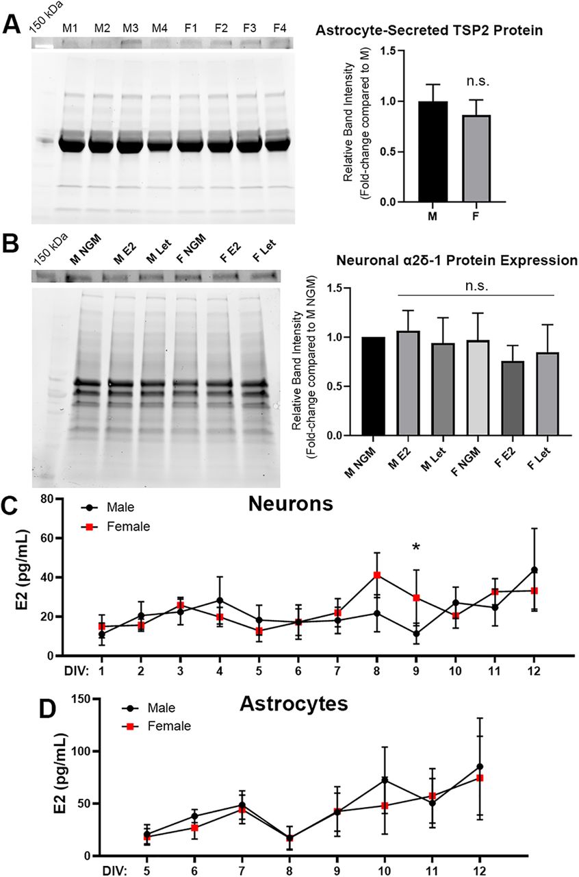
- Figure 4.
Transient increase in neuronally-derived estradiol in cultured female cortical neurons. A, left, Western blotting for TSP2 [top, ∼160-kDa observed band (∼130 kDa expected); bottom, total protein blot for normalization] from astrocyte media collected on DIV10. Right, No difference was found in secreted TSP2 levels between male and female astrocytes; p = 0.568 [unpaired Student’s t test; n = 4 independent experimental replicates (M1-4, F1-4)]. B, left, Western blotting for α2δ−1 (top, 143-kDa observed band; bottom, total protein blot for normalization) from purified male and female cortical neuron lysates. Lysates were collected on DIV13 following treatment with NGM only, 100 nm E2, or 100 nm letrozole (Let) on DIV7 and DIV10. Right, No difference in total α2δ−1 protein expression was observed between male or female-derived neurons at baseline or following treatment with either E2 or Let; p = 0.805 (two-way ANOVA, interaction: F(2,12) = 0.220; n = 3 independent experimental replicates). C, Immunoassay results for levels of E2 detected in male versus female neuron-conditioned media. An increase in secreted E2 was detected in female cultures at the start of the second week, reaching significance at DIV9; *p < 0.05 [linear mixed-effects model (REML) with Holm–Sidak’s post hoc analysis, sex: F(1,55) = 7.00; n = 4 independent experimental replicates]. D, Estradiol measurements from male versus female cortical ACM. No significant differences were observed (linear mixed-effects model, sex: F(1,29) = 3.80; n = 3 independent experimental replicates).
- Figure 5.
Inhibition of neuronal estrogen production modulates TSP/astrocyte-induced synaptogenesis. A, ICC image of a male-derived cortical neuron fixed and stained on DIV13 with presynaptic (Bassoon; magenta) and postsynaptic (Homer1; green) markers. Dotted box indicates example dendritic ROI sampled for images in B, which shows co-localized excitatory synaptic puncta (white, arrowheads) along male and female-derived cortical neurites treated with NGM only or NGM plus TSP2, 100 nm letrozole (Let), or TSP2/letrozole on DIV7 and DIV10. C, left, Letrozole had a mild synaptogenic effect on male-derived cortical neurons but attenuated the synapse-promoting ability of TSP2. Right, Female-derived cortical neurons showed no synaptogenic response to TSP2 unless treatment was combined with Let. D, Delayed treatment (DIV13 and DIV16, fixed and stained DIV19) revealed no differences in synapse number from NGM-only condition following TSP2 and/or Let treatment in neurons from either sex. E, Quantification of Bassoon/Homer1 co-localized synaptic puncta following DIV7/DIV10 treatment schedule showed that Let abolished the synaptogenic effects of sex-matched astrocyte inserts (Astro) and ACM in male-derived neurons as well as ACM in female-derived neurons. No attenuation was observed with Let and Astro in females; *p < 0.05, ***p < 0.001, ****p < 0.0001 [Kruskal–Wallis test with Dunn’s multiple comparisons post hoc analysis; K-W statistic = 52.57 (male), 66.18 (female); n = 30 cells per condition per experimental replicate, 3 independent experimental replicates].
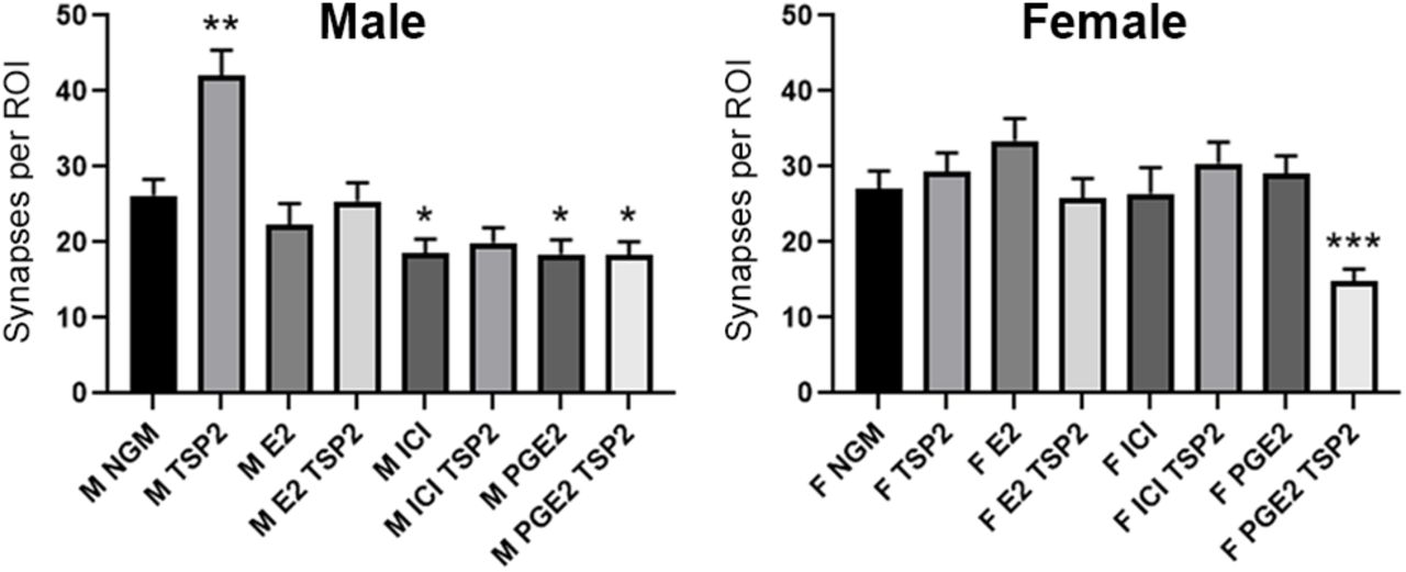
- Figure 6.
Endogenous estrogen signaling permits TSP2-induced synapse formation in male but not female neurons. Quantification of Bassoon/Homer1 co-localized synaptic puncta following DIV7/DIV10 treatment schedule showed that modulating endogenous estrogen signaling, either with exogenous 100 nm E2, 100 nm ICI 182780 (ER antagonist), or 100 nm PGE2 (aromatase agonist), precluded the ability of TSP2 to promote synapse formation in either male (left)-derived or female (right)-derived cortical neurons. PGE2, in particular, was notable in that it lowered synapse number compared with NGM alone in males and when combined with TSP2 in both sexes; *p < 0.05, **p < 0.01, ***p < 0.001 [Kruskal–Wallis test with Dunn’s multiple comparisons post hoc analysis; K-W statistic = 64.21 (male), 44.75 (female); n = 30 cells per condition per experimental replicate, 3 independent experimental replicates].


Tables
- Table 1
Statistical results for Western blottings, estradiol immunoassay, and ICC (fold-change and multiple group comparisons)
Figure Figure panel Statistical test Conditions Results 1 B' Kruskal–Wallis test with Dunn’s post hoc comparison M NGM vs M Astro K-W = 65.23 p < 0.0001 1 B' Kruskal–Wallis test with Dunn’s post hoc comparison F NGM vs F Astro K-W = 65.23 p < 0.0001 1 B' Kruskal–Wallis test with Dunn’s post hoc comparison M Astro vs F Astro K-W = 65.23 p > 0.9999 1 D' Kruskal–Wallis test with Dunn’s post hoc comparison M NGM vs M M-ACM K-W = 77.03 p < 0.0001 1 D' Kruskal–Wallis test with Dunn’s post hoc comparison M NGM vs M F-ACM K-W = 77.03 p < 0.0001 1 D' Kruskal–Wallis test with Dunn’s post hoc comparison M M-ACM vs M F-ACM K-W = 77.03 p > 0.9999 1 D' Kruskal–Wallis test with Dunn’s post hoc comparison M M-ACM vs F M-ACM K-W = 77.03 p = 0.0142 1 D' Kruskal–Wallis test with Dunn’s post hoc comparison M F-ACM vs F F-ACM K-W = 77.03 p = 0.1693 1 D' Kruskal–Wallis test with Dunn’s post hoc comparison F NGM vs F M-ACM K-W = 77.03 p = 0.2781 1 D' Kruskal–Wallis test with Dunn’s post hoc comparison F NGM vs F F-ACM K-W = 77.03 p = 0.0036 2 C' One-way ANOVA with Tukey's post hoc comparison M NGM vs M ACM F(3,75) = 10.29 p = 0.0002 2 C' One-way ANOVA with Tukey's post hoc comparison F NGM vs F ACM F(3,75) = 10.29 p = 0.0195 2 C' One-way ANOVA with Tukey's post hoc comparison M ACM vs F ACM F(3,75) = 10.29 p = 0.2119 2 D' Kruskal–Wallis test with Dunn’s post hoc comparison M NGM vs M ACM K-W = 22.61 p = 0.0165 2 D' Kruskal–Wallis test with Dunn’s post hoc comparison F NGM vs F ACM K-W = 22.61 p = 0.0018 2 D' Kruskal–Wallis test with Dunn’s post hoc comparison M ACM vs F ACM K-W = 22.61 p > 0.9999 4 A Unpaired Student's t test TSP2 relative band intensity,
male vs femalep = 0.568 4 B Two-way ANOVA (interaction) α2δ−1 relative band intensity F(2,12) = 0.220 p = 0.805 4 B Two-way ANOVA (sex) α2δ−1 relative band intensity F(1,12) = 0.647 p = 0.437 4 B Two-way ANOVA (treatment) α2δ−1 relative band intensity F(2,12) = 0.093 p = 0.912 4 C Linear mixed-effects model (REML) with Holm–Sidak’s
post hoc comparison (sex)E2 (pg/ml) F(1,55) = 6.999 p = 0.011 4 D Linear mixed-effects model (REML) with Holm–Sidak’s
post hoc comparison (sex)E2 (pg/ml) F(1,29) = 3.799 p = ns 5 E, left Kruskal–Wallis test with Dunn’s post hoc comparison NGM vs Astro K-W = 52.57 p = 0.0002 5 E, left Kruskal–Wallis test with Dunn’s post hoc comparison NGM vs Astro + Let K-W = 52.57 p > 0.9999 5 E, left Kruskal–Wallis test with Dunn’s post hoc comparison NGM vs ACM K-W = 52.57 p = 0.0002 5 E, left Kruskal–Wallis test with Dunn’s post hoc comparison NGM vs ACM + Let K-W = 52.57 p > 0.9999 5 E, left Kruskal–Wallis test with Dunn’s post hoc comparison Astro vs Astro + Let K-W = 52.57 p = 0.0198 5 E, left Kruskal–Wallis test with Dunn’s post hoc comparison ACM vs ACM + Let K-W = 52.57 p < 0.0001 5 E, right Kruskal–Wallis test with Dunn’s post hoc comparison NGM vs Astro K-W = 66.18 p < 0.0001 5 E, right Kruskal–Wallis test with Dunn’s post hoc comparison NGM vs Astro + Let K-W = 66.18 p = 0.0123 5 E, right Kruskal–Wallis test with Dunn’s post hoc comparison NGM vs ACM K-W = 66.18 p < 0.0001 5 E, right Kruskal–Wallis test with Dunn’s post hoc comparison NGM vs ACM + Let K-W = 66.18 p > 0.9999 5 E, right Kruskal-Wallis test with Dunn’s post hoc comparison Astro vs Astro + Let K-W = 66.18 p = 0.2655 5 E, right Kruskal-Wallis test with Dunn’s post hoc comparison ACM vs ACM + Let K-W = 66.18 p < 0.0001 6 left Kruskal–Wallis test with Dunn’s post hoc comparison NGM vs TSP2 K-W = 64.21 p = 0.0039 6 left Kruskal–Wallis test with Dunn’s post hoc comparison NGM vs E2 K-W = 64.21 p = 0.2220 6 left Kruskal–Wallis test with Dunn’s post hoc comparison NGM vs E2 TSP2 K-W = 64.21 p > 0.9999 6 left Kruskal–Wallis test with Dunn’s post hoc comparison NGM vs ICI K-W = 64.21 p = 0.0452 6 left Kruskal–Wallis test with Dunn’s post hoc comparison NGM vs ICI TSP2 K-W = 64.21 p = 0.2420 6 left Kruskal–Wallis test with Dunn’s post hoc comparison NGM vs PGE2 K-W = 64.21 p = 0.0377 6 left Kruskal–Wallis test with Dunn’s post hoc comparison NGM vs PGE2 TSP2 K-W = 64.21 p = 0.0467 6 left Kruskal–Wallis test with Dunn’s post hoc comparison E2 vs E2 TSP2 K-W = 64.21 p > 0.9999 6 left Kruskal–Wallis test with Dunn’s post hoc comparison ICI vs ICI TSP2 K-W = 64.21 p > 0.9999 6 left Kruskal–Wallis test with Dunn’s post hoc comparison PGE2 vs PGE2 TSP2 K-W = 64.21 p > 0.9999 6 right Kruskal–Wallis test with Dunn’s post hoc comparison NGM vs TSP2 K-W = 44.75 p > 0.9999 6 right Kruskal–Wallis test with Dunn’s post hoc comparison NGM vs E2 K-W = 44.75 p > 0.9999 6 right Kruskal–Wallis test with Dunn’s post hoc comparison NGM vs E2 TSP2 K-W = 44.75 p > 0.9999 6 right Kruskal–Wallis test with Dunn’s post hoc comparison NGM vs ICI K-W = 44.75 p = 0.8729 6 right Kruskal–Wallis test with Dunn’s post hoc comparison NGM vs ICI TSP2 K-W = 44.75 p > 0.9999 6 right Kruskal–Wallis test with Dunn’s post hoc comparison NGM vs PGE2 K-W = 44.75 p > 0.9999 6 right Kruskal–Wallis test with Dunn’s post hoc comparison NGM vs PGE2 TSP2 K-W = 44.75 p = 0.0002 6 right Kruskal–Wallis test with Dunn’s post hoc comparison E2 vs E2 TSP2 K-W = 44.75 p > 0.9999 6 right Kruskal–Wallis test with Dunn’s post hoc comparison ICI vs ICI TSP2 K-W = 44.75 p = 0.6315 6 right Kruskal–Wallis test with Dunn’s post hoc comparison PGE2 vs PGE2 TSP2 K-W = 44.75 p < 0.0001
In this issue


