
Main menu

  • HOME
  • CONTENT
    • Early Release
    • Featured
    • Current Issue
    • Issue Archive
    • Blog
    • Collections
    • Podcast
  • TOPICS
    • Cognition and Behavior
    • Development
    • Disorders of the Nervous System
    • History, Teaching and Public Awareness
    • Integrative Systems
    • Neuronal Excitability
    • Novel Tools and Methods
    • Sensory and Motor Systems
  • ALERTS
  • FOR AUTHORS
  • ABOUT
    • Overview
    • Editorial Board
    • For the Media
    • Privacy Policy
    • Contact Us
    • Feedback
  • SUBMIT

User menu

Search

  • Advanced search
eNeuro
eNeuro

Advanced Search


  • HOME
  • CONTENT
    • Early Release
    • Featured
    • Current Issue
    • Issue Archive
    • Blog
    • Collections
    • Podcast
  • TOPICS
    • Cognition and Behavior
    • Development
    • Disorders of the Nervous System
    • History, Teaching and Public Awareness
    • Integrative Systems
    • Neuronal Excitability
    • Novel Tools and Methods
    • Sensory and Motor Systems
  • ALERTS
  • FOR AUTHORS
  • ABOUT
    • Overview
    • Editorial Board
    • For the Media
    • Privacy Policy
    • Contact Us
    • Feedback
  • SUBMIT
PreviousNext
Review, Sensory and Motor Systems

Vertebrate Evolution Conserves Hindbrain Circuits despite Diverse Feeding and Breathing Modes

Shun Li and Fan Wang
eNeuro 11 March 2021, 8 (2) ENEURO.0435-20.2021; https://doi.org/10.1523/ENEURO.0435-20.2021
Shun Li
Department of Neurobiology, Duke University, Durham, NC 27710
  • Find this author on Google Scholar
  • Find this author on PubMed
  • Search for this author on this site
  • ORCID record for Shun Li
Fan Wang
Department of Neurobiology, Duke University, Durham, NC 27710
  • Find this author on Google Scholar
  • Find this author on PubMed
  • Search for this author on this site
  • ORCID record for Fan Wang
  • Article
  • Figures & Data
  • Info & Metrics
  • eLetters
  • PDF
Loading

Abstract

Feeding and breathing are two functions vital to the survival of all vertebrate species. Throughout the evolution, vertebrates living in different environments have evolved drastically different modes of feeding and breathing through using diversified orofacial and pharyngeal (oropharyngeal) muscles. The oropharyngeal structures are controlled by hindbrain neural circuits. The developing hindbrain shares strikingly conserved organizations and gene expression patterns across vertebrates, thus begs the question of how a highly conserved hindbrain generates circuits subserving diverse feeding/breathing patterns. In this review, we summarize major modes of feeding and breathing and principles underlying their coordination in many vertebrate species. We provide a hypothesis for the existence of a common hindbrain circuit at the phylotypic embryonic stage controlling oropharyngeal movements that is shared across vertebrate species; and reconfiguration and repurposing of this conserved circuit give rise to more complex behaviors in adult higher vertebrates.

  • breathing
  • central rhythm generator
  • evolution
  • feeding
  • hindbrain

Significance Statement

Understanding how a highly conserved hindbrain generates diverse feeding/breathing patterns is important for elucidating neural mechanisms underlying the execution and coordination of these two vital behaviors. Here, we first briefly summarize key modes of vertebrates feeding/breathing, discuss main principles coordinating feeding/breathing, and provide a unifying hypothesis for the existence of a shared oropharyngeal movement control circuit across species. By synthesizing behavior, structural and neural mechanisms for feeding/breathing functions across evolution, we believe that this review and our hypothesis can open new research avenues for elucidating the precise hindbrain circuits controlling feeding, breathing, and other oropharyngeal functions.

Introduction

The orofacial and pharyngeal regions of vertebrates, derived from pharyngeal arches (PAs) in the embryo, are critical for executing the two key survival functions, breathing and feeding. Vertebrates share a highly conserved embryonic hindbrain organization, both in terms of gene expression profiles and stereotyped rhombomere arrangement (Kiecker and Lumsden, 2005; Parker and Krumlauf, 2020). Yet, they use widely different approaches for feeding and breathing, that rely on a diversity of oropharyngeal structures and muscles (Bels and Whishaw, 2019). In this review, we first summarize major modes of feeding and breathing and then examine different manners through which these two actions are coordinated throughout vertebrate evolution. Finally, we provide a hypothesis for how a highly conserved embryonic hindbrain can assemble neural circuits that control drastically different feeding and breathing apparatuses and behaviors across species. Important terminologies are listed and defined in Table 1.

View this table:
  • View inline
  • View popup
Table 1

Glossary

Feeding

Since animals are unable to directly exploit solar energy, they must obtain energy from resources available in their habitat. Feeding is thus one of the primary and essential behaviors of survival. Vertebrate feeding is commonly separated into four principle stages, namely ingestion, intraoral transport, processing, and swallowing (Schwenk, 2000a). These feeding stages can be viewed as solving two major subtasks, transportation and reduction of food. The transportation process includes stages of ingestion, intraoral transport, and swallowing that move food (in original form or after reduction) from the outside environment to the esophagus, while reduction is the process of mechanically breaking down food (and this step is not needed for ingesting liquid). Vertebrates living in different environment (e.g., aquatic vs terrestrial) develop distinct oropharyngeal musculoskeletal structures and feeding mechanisms (Fig. 1; Table 2; for more details, see Bels and Whishaw, 2019).

View this table:
  • View inline
  • View popup
Table 2

Summary of major feeding and breathing modes and their respective oropharyngeal apparatus, controller muscle groups, and putative innervating nerves

Figure 1.
  • Download figure
  • Open in new tab
  • Download powerpoint
Figure 1.

Anatomy of the oropharyngeal region involved in feeding and breathing in major vertebrate species. Schematic representation of gross oropharyngeal anatomy of different vertebrates in sagittal section. Major structures color-coded as indicated. Blue arrows indicate pathways for breathing, while brown arrows indicate pathways for feeding. A, Sagittal section of a hagfish. Different gill pouches structures of Atlantic and Pacific hagfish are depicted. During breathing, the tooth plate is in its retracted position, while the velum repeatedly scrolls and unscrolls to allow water flowing in from the nostril. The water then flows through the gills and exits either at the common PCD or at individual gill slit. B, Schematic sagittal view of fish oral cavity with gills depicted. C, Schematic sagittal view of a frog oral cavity. D, Position of a feeding snake in sagittal plane (prey is not depicted for simplicity) showing the protruded glottis. E, Schematic sagittal view of a bird’s partially separated oral and nasal cavities. F, Sagittal representation of an adult pilot whale (Globicephala melaena). The intranasal position of the larynx allows simultaneous feeding and vocalization. G, Sagittal view of a human infant. H, Sagittal view of a human adult. The larynx in adult is descended compared with the elevated position in infant. I, Oropharyngeal structure of human adult during swallowing. The food bolus (brown) pushes the epiglottis down, allowing it to contact with the elevated larynx to assist airway protection during swallowing in adult humans. Drawings are based on or modified from Eom and Wood (2019), Ding et al. (2019), Mason et al. (2020), Cundall et al. (2014), Brown and Stallknecht (2008), Laitman and Reidenberg (1997), and Arvedson and Lefton-Greif (1998).

Aquatic food transportation

Food transportation in aquatic vertebrates is medium-dependent, operated mainly by producing an inward water current that pulls the food into the mouth. The most primitive form of current generation is through directional beating of cilia (no muscles involved) to produce a slow inward flow and the use of a mucus filter to capture suspended food particles (Clark and Uyeno, 2019). The evolution of branchial muscles enabled the production of larger current flows. For example, lampreys use branchial muscles to contract the branchial basket (the wall of the buccal cavity), thereby squeezing the water rapidly out of the mouth (Rovainen, 1977; Missaghi et al., 2016). The subsequent elastic (but passive) recoil of the buccal cavity brings food-bearing water into the mouth. Then water flows out through the pharyngeal slits, while food is retained and swallowed.

This primitive form of compression-followed-by-passive expansion suction evolved into a more complex sequence of active expansion-suction phases. Briefly, in most sharks and fish, feeding starts with a sudden expansion of the oropharyngeal cavity produced by rapid and nearly synchronous activation of jaw opening and branchial muscles, capturing passing preys and moving the intake caudally at high velocity (Grubich, 2001). After ingestion, through rounds of sequential moderate cavity expansions and relaxations, food is slowly transported toward the esophagus in a step-like pattern (van Meer et al., 2019; Weller et al., 2020). In addition to fish, aquatic mammals (e.g., seals and beaked whales) also secondarily evolved suction using tongue retraction and depression (Marshall et al., 2014; Kienle and Berta, 2016; Kienle et al., 2017), pointing to suction as an optimal solution for transporting prey in the aquatic environment.

Besides suction, other solutions of food transportation in the aquatic environment emerge at various stages of vertebrate evolution. For example, hagfish, which will be discussed more thoroughly in later section, repeatedly protracts and retracts the tooth plate to rip off and move the food to the esophagus (Clark et al., 2010; Zintzen et al., 2011). This cyclic pattern of protraction and retraction is aided by the posteriorly curved palatal tooth, which acts as a rachet to prevent leakage of food (Clark and Summers, 2007). On the other hand, balaenid whales constantly keep their mouths open and “swim over” dense patches of food such as copepod. The prey-laden water then flows through the baleen system, formed of structures inside the mouth that uses close-knit hair fringes called baleen hairs to filter out water and collect preys in front of the esophageal opening (Werth and Potvin, 2016; Goldbogen et al., 2017). Moreover, rorqual whales (e.g., blue whales) lunge toward and engulf a huge volume of prey-laden water during feeding. Prey items are then being filtered and transported to the esophagus mainly through hydraulic forces (Arnold et al., 2005; Werth, 2007; Werth and Ito, 2017).

Terrestrial food transportation

In terrestrial habitats, the air’s lower density means that food transportation is no longer achievable through suction. Instead, most tetrapods (amphibians, reptiles, birds, mammals) possess a hyolingual apparatus (hyoid and tongue) that is fundamental to food transportation for these terrestrial animals. Evolution of these complex oral structures allow the animal to push or squeeze the food to the esophagus in lieu of suction.

Intraoral transport in most lizards and mammals involve repetitive cycles of tongue protrusion and retraction. Importantly, these tongue movement cycles are also tightly integrated with the cyclic jaw movements (i.e., gape cycles). Based on the velocity and direction of jaw movements, gape cycles can be divided into slow opening of the jaw, fast opening, fast closing, and slow closing-power stroke phases (Bramble and Wake, 1985; Schwenk, 2000a). During slow opening, the tongue moves anteriorly and slides under the food. The tongue and hyoid then retract back along with the food at the end of the fast opening while the jaw rapidly closes (fast closing) to prevent escape of the prey. The gape cycle then enters the last phase (slow closing-power stroke) during which food items are crushed by the teeth, which slows the jaw closing motion. Underlying mechanism of such tongue-jaw coordination is still under extensive investigations, and current results indicate contributions from both neural mechanism via common premotor neurons (Stanek et al., 2014, 2016) and mechanical linkages (for more detail, see Matsuo and Palmer, 2010).

After the coordinated actions of tongue and jaw that move the food through the oral cavity, swallowing is the final step that pushes food into the esophagus via a posteriorly-directed muscle activation sequence. Reptiles and mammals have different swallowing procedures. In many reptile species like lizards and turtles, swallowing consists of two discrete stages. First, the jaw closes and food items are being pushed to and accumulate in the pharynx in the pharyngeal packing stage by posterior and dorsal movements of the hyolingual apparatus (Schwenk and Rubega, 2005). This is then followed by the pharyngeal emptying stage that squeezes the food bolus into the esophagus by constricting the pharynx through the constrictor colli muscle (Schwenk, 2000b). In mammals, swallowing is rapidly executed within a process called deglutition because of the evolutionarily more recent invention of internal pharyngeal muscles (Schwenk, 2000a). In deglutition, the jaw is closed when the food bolus is formed. The closing of the jaw stabilizes the mandible, and the hyoid is elevated, which is accompanied by posterior tongue retraction that thrusts the food bolus posteriorly into the pharynx (Fig. 1I). Hyoid elevation also leads to closure of the glottis (the opening of trachea) for airway protection, i.e., preventing food from entering into the airway. As the bolus enter the pharynx, the internal pharyngeal muscles initiate a powerful descending contraction wave of the pharyngeal wall, pushing the bolus into the esophagus in a peristaltic motion (Fig. 1I; Ertekin and Aydogdu, 2003; Thexton et al., 2007; Miller, 2008; Lang, 2009). After the passing of food, the hyoid resumes its normal position. Together, this highly choreographed muscle activation sequence allows mammals to integrate deglutition into food reduction (called mastication, see Food Reduction) and intraoral transport cycles rather than being a discrete and prolonged kinematic stage like that in non-mammalian species (Schwenk, 2000a). Major oropharyngeal muscles, their functions, and neuronal innervations are further explained in Table 3.

View this table:
  • View inline
  • View popup
Table 3

Major action and cranial motor nerve supply of oropharyngeal muscles in human

In addition to this hyolingual apparatus-powered mode of intraoral food transport, other land-dwelling species also evolved a diversity of methods for moving food from mouth to esophagus. For example, most frogs, crocodiles, and birds do not develop tongue and hyoid structures complicated enough to be fully responsible for hyolingual transportation. In crocodiles, the tongue lacks intrinsic musculature and is entirely connected to the mandible (Iwasaki et al., 2019). Therefore, crocodiles elevate and protrude their heads while their jaws release the food, allowing gravity along with the retracting hyoid to drop the food toward the esophagus (Cleuren and de Vree, 1992). In frogs, the root of the tongue is usually attached at the anterior portion of the mouth’s floor (Fig. 1C). When feeding, frogs flip out their tongues, stick to the prey, and flip back along with the prey into the mouth (Nishikawa, 2000; Herrel et al., 2019; Iwasaki et al., 2019). The subsequent swallowing is believed to be primarily driven by tongue retraction, but also substantially aided by head elevations and even retraction of eyeballs, which help push the food into the esophagus (Regal and Gans, 1976; Ritter and Nishikawa, 1995; Tso et al., 1995; Levine et al., 2004). In snakes, the jaw muscles and body both contribute to eating and swallowing preys, without assistance from the tongue, which is completely reserved for chemosensory functions (Fig. 1D). Using highly kinetic joints, snakes can achieve extremely large gap and cranial kinesis, providing substantial degree of freedom to the upper jaw to move relatively independently of the head. Therefore, jaws of the snake can be opened and advanced unilaterally in an alternating pattern of protraction of the upper and lower jaw on either side, functioning as the primary vector of transportation/swallowing (Moon et al., 2019). This motion is aptly termed the “pterygoid walk,” since the snake basically “walks over” the prey through a combination of ratchet-like jaw movements and concertina-like body undulation (Boltt and Ewer, 1964; Kley and Brainerd, 2002).

Food reduction

Evolution of feeding motor programs is also dependent on food size. While animals like lampreys and balaenid whales are microphagous that transport food items in bulk, wide variety of vertebrates are macrophagous, posing the need of reduce the food into manipulatable sizes. Food reduction strategies of vertebrates, from agnathans (e.g., hagfish) to mammals, all predominantly rely on the use of teeth in both aquatic and terrestrial environment.

Macrophagy perhaps first evolved in hagfish, one of the most primitive members of existing vertebrates (Miyashita et al., 2019). The feeding apparatus of a hagfish consists of feeding musculature, a basal plate, and dentitions (tooth plates; Clark and Uyeno, 2019). The feeding musculature of hagfish are all innervated by the trigeminal nerve (Lindström, 1949) and can be divided into anterior hard component for protraction and a posterior soft component for retraction (Clark et al., 2010; Clark and Uyeno, 2019). During feeding, a hagfish repeatedly protracts tooth plates to press into the food and retracts them into the mouth along with the torn food (Clark et al., 2010). After retraction, the oral mucosa and palatal tooth dislodge the food from the teeth and transport it to the esophagus via additional protraction-retraction cycles (Clark and Summers, 2007). Because hagfish lacks the opposing jaw, to generate enough force to tear off pieces of food, the soft retractor musculature is arranged as a muscular hydrostat, which stiffens to provide a compression-resistant support (Uyeno and Clark, 2015; Clubb et al., 2019). Additionally, hagfish can twist its boneless body into a knot (a movement called body knotting) to provide further leverage (for details, see Haney et al., 2020). Thus, although hagfish is jawless, the turgid muscular hydrostat together with the body knot functions like a jaw that generates forceful bites similar to other jawed vertebrates (Clark and Summers, 2007).

In contrast (to jawless fish), mammals possess well-developed jaw apparatus. The upper and lower teeth in most mammals occlude such that they fit nicely together (Schwenk, 2000a). Occlusion within an advanced jaw enables a highly efficient form of food chewing called mastication, which allows food items to be fully comminuted because of the precise fit of the teeth. Chewing occurs mostly during the ingestion and intraoral transport stage before swallowing. The masticatory muscles for rhythmic jaw opening and closing are described in Table 3. In mammalian (e.g., sea otters) and non-mammalian gnathostomes whose teeth do not occlude, macrophagic methods like crushing and tearing become the common solution for reduction (Kienle et al., 2017). In this case, food items, usually hard-shell invertebrates, are punctured and crushed in the oral cavity so that salivary enzymes are able to further soften the food. In addition to crushing, tearing is used by carnivorous species like crocodiles, orcas, or sharks. Similar to hagfish feeding, during tearing the jaw together with the teeth grasp and rip off small pieces of food suitable for swallowing (for more details, see Bels and Whishaw, 2019).

Breathing

Respiration/breathing is also indispensable for vertebrate survival as cells within the body need O2 replenishment and CO2 excretion to produce energy and properly metabolize. Although breathing behaviors in vertebrates all includes active muscle contractions to move O2 and CO2 in or out of the body, respiration per se is based on passive gas diffusion (Kardong, 2018). O2 is normally subject to a higher partial pressure in the external environment, allowing it to naturally diffuse into the blood while CO2 tends to diffuse out from the blood. The passive nature of gas exchanges means that all movements associated with breathing have for purpose to maintain the partial pressure gradients across the exchange surfaces, by moving gas-containing medium (water or air). Throughout evolution, many solutions for this task have emerged across different families of vertebrates, depending on the specific properties of the respiratory organ as well as the metabolic demand of the animal.

Aquatic breathing

The emergence of active aquatic breathing involves parallel evolution of water-transporting motor programs and specialized respiration structures. Active breathing in early vertebrates is believed to be originated from feeding, partially driven by the continued evolution of pharyngeal slits (Alheid et al., 2004; Graham and Richardson, 2012; Gillis and Tidswell, 2017). In primitive chordates like amphioxus, breathing occurs mostly through the skin (cutaneous breathing; Schmitz et al., 2000; Milsom et al., 2004), and their pharyngeal slits, alongside the body, function to assist filter feeding (Graham and Richardson, 2012). As the efficiency of feeding increased because of the innovation of the muscular suction pump, the pharyngeal slits that were previously dedicated to feeding became free to take on respiratory functions. In some agnathans such as Pacific hagfish, each pharyngeal slit, also referred to as gill pouch, is separated and has its own opening to the outside, whereas the gill pouches in Atlantic hagfish are connected to a common opening called pharyngo-cutaneous duct (PCD; Fig. 1A). During hagfish aquatic breathing, rhythmic contraction of velum brings water into mouth through the nostril (inhalation), followed by active contractions of gill pouches to expel water out either through individual gill slits or via the common PCD (Fig. 1A; Malte and Lomholt, 1998; Eom and Wood, 2019). Similar motor program is also observed in premetamorphic larval lampreys (ammocoetes), in which breathing and feeding are accomplished within the same action: water enters from the mouth and exits through the pharyngeal slits by the scrolling and unscrolling action of a velum (a muscular structure that is located at the midline of pharynx), plus the compression and recoil of the branchial basket (Rovainen, 1996).

Eventually, gills are evolved to replace the pharyngeal slits in cartilaginous and teleost fish. In these species, breathing happens through gill ventilation, which uses a dual pump mechanism, namely the successive expansion and compression of the buccal and opercular cavity controlled by branchial muscles (Fig. 1B; Kardong, 2018; Milsom, 2018). This dual pump mechanism, generated by rostral-caudally propagating wave of brainstem motor activity (Taylor, 1992; Taylor et al., 2009b), sequentially drives the water first into the buccal cavity (buccal expansion), then across the gill curtain (buccal compression along with opercular expansion) where O2 diffuses passively into the gill vasculature. Finally, water containing CO2 to be excreted exits from the opercular valve (through opercular compression). In principle, the hydraulic nature of both fish breathing and feeding means that the same subset of cranial muscles can be used for both processes, although muscular activity during gill ventilation alone (no feeding) is slower and less powerful (Taylor et al., 2010; Milsom, 2018). Under hypoxic conditions, fish recruit additional feeding muscles to generate higher suction power to increase water flow (Taylor et al., 2006).

An important exception to the common dual pump ventilation is ram ventilation. Here, the swimming motion itself generates respiration-needed water flow across the gills, thereby saving energy costs associated to activating cranial muscles during normal gill ventilation (Roberts and Rowell, 1988). Many elasmobranchs, such as great white sharks or whale sharks, do not possess an operculum as an expiratory flap and are obligate ram ventilators that need to swim continuously for breathing (Mallatt, 1997). Other fish (e.g., nurse shark, trout) transition between gill and ram ventilation depending on swimming speed, water velocity, and/or water O2 tension, displaying what might be the earliest form of breathing-locomotion coordination (Randall, 1982; Steffensen, 1985).

Air breathing

Air breathing possibly first evolved in certain bony fish species that dwell in O2-depleted aquatic habitats, by modifying the existing suction-based feeding-breathing mechanism. Two of the most common air-breathing organs (ABOs) are the lung and the gas bladder. The opening of ABOs to the oral cavity is gated by the glottis near the floor of digestive tract. Air-breathing fish only come out of water to breathe intermittently when they need to. During water immersion, the glottis is closed, and the cycles of buccal expansion and compression are used to move water in and out of mouth for feeding as mentioned above. During air breathing, the fish swims to the surface of the water, where the same cycles of buccal expansion and compression coupled with the opening and closing of the glottis are used for drawing air into the buccal cavity and exchanging the “old” air in the ABO with fresh air (Brainerd, 1994; Brainerd and Ferry‐Graham, 2005; Milsom, 2018). This buccal pump mechanism for breathing is retained in anuran species like frogs and toads (Fig. 1C; Taylor et al., 2010).

Subsequently during evolution, aspiration breathing substituted this primitive form of air breathing, with the recruitment of axial muscles for higher volume and a more efficient gas exchange. A first instance of aspiration breathing is exemplified by the traverse abdominal muscle that some salamanders use during active expiration (Brainerd, 1999). Then both inspiration and expiration became powered by axial muscles, thus drastically increasing the volume of gas exchange. For example, in lepidosaurs such as lizards and snakes, intercostal muscles are used to create a rotation of the ribs that expands the thoracoabdominal cavity and the lung (Carrier, 1989). Similar aspiration pump mechanism has been documented in birds, during which air sacs are inflated by rib rotation and caudal sternum depression (Claessens, 2004). A unique aspect of avian breathing is that within the air sac system airflow is unidirectional (Farmer, 2015). Using two aerodynamic valves, inspiring air can only enter through one valve into one set of chambers, subsequently the air moves in one direction, and expiring air leaves from the other valve back into the trachea (for more details, see Cieri and Farmer, 2016). This aerodynamic valving system maximizes breathing efficiency.

However, the reliance on axial muscles has its downsides. For instance, intercostal and abdominal muscles in lizards are also needed for locomotion. While breathing requires bilateral and synchronous activation of both muscle groups, locomotion unilaterally and alternatively activates these muscles to bend the body or to stiffen the trunk (Carrier, 1990, 1991). This functional conflict is especially evident during high-speed sprinting, during which axial muscles are solely recruited for locomotion and thus reduce the lizard’s tidal volume to near zero (Carrier, 1987). To solve this problem, many lizards, such as savannah monitor lizards, use gular (throat) pumping to circumvent the breathing-locomotion conflict. In a highly similar fashion to fish and amphibian’s buccal pumping for breathing, the gular cavity expands by retracting and depressing the hyobranchial skeleton to assist in inspiration. The addition of this non-conflicting motor program allows these lizards to draw more than two times the air volume than available through costal inspiration alone (Brainerd and Owerkowicz, 2006).

Mammals solved the respiration-locomotion conflict by evolving a novel respiratory muscle, the diaphragm, a dome-shaped muscle. Contraction of the diaphragm flattens the curvature of the dome, leading to expansion of the thoracic cavity and air influx. The diaphragm allows mammals to possess much greater stamina during locomotion, such that ventilation volume can increase along with moving speed during locomotion. Testudines (turtles and tortoises) and crocodilians also adopted similar strategies of using a dome-shaped muscle for aspiration breathing. In turtles, the internal oblique muscle contracts during inspiration, flattening the dome and increasing the thoracoabdominal cavity volume to draw air into the lung (Gaunt and Gans, 1969; Landberg et al., 2003). In crocodiles, the diaphragmaticus muscle retracts the liver that divides the thoracoabdominal cavity, thereby expanding the cavity and bringing a large volume of air into the lungs (Naifeh et al., 1970; Gans and Clark, 1976; Kardong, 2018).

Coordination between Feeding and Breathing

Both feeding and breathing are vital to survival, and both food and O2 enter into the body from orofacial openings. The diverse modes of feeding and breathing through evolution also involve various mechanisms for coordinating these two behaviors.

Aquatic suction feeding and aquatic ventilation (through pharyngeal slit or gill) use many of the same muscles to bring water into the oral cavity, to push water out through pharyngeal pore/gill for gas exchange while retaining food. Thus, aquatic feeding and breathing can occur simultaneously and harmoniously as part of the same motor program. For species carrying out aquatic feeding and air-breathing, feeding (in water) and breathing (on surface of water) never overlap in time, and the glottis is normally closed to guard the lung until the time of breathing. Again, the same sets of muscles that draw water into buccal cavity are used to draw air into the mouth and the lung.

In contrast, for most tetrapods, the combination of terrestrial feeding and air breathing poses two main challenges. (1) As feeding typically takes substantial time for intraoral transportation and reduction of food, for species that depend on continuous air breathing (such as most mammals), mechanisms are needed to allow concurrent respiration and food processing (before swallowing) to occur. (2) While both air and food enter via orofacial openings, air goes to the lung, whereas food needs to enter the esophagus. Thus, air-breathing tetrapod needs to prevent food from being swallowed into the lung. Different species evolved distinct musculoskeletal elements to solve these two problems.

First, to allow concurrent breathing and food processing, structural innovations were implemented during evolution to separate the transportation pathway of air and food. For example, many snakes swallow the prey whole regardless of the size, and thus require a prolonged period of intraoral transport and slow swallowing. To enable simultaneous breathing and feeding, the snake’s oropharyngeal region has undergone significant rearrangements (Cundall et al., 2014). First, the glottis of snakes is situated at the anterior region of the oral cavity and is able to protrude outside the oral cavity during feeding to maintain breathing (Fig. 1D). Furthermore, the opening of the esophagus in snakes is distinctively anterior and thus effectively shortens the length of the oral cavity, such that the prey is essentially transported directly into the esophagus (Fig. 1D).

Another seminal structural innovation in tetropods regarding the oropharyngeal region is the development of the secondary palate (the posterior portion of hard palate and all the soft palate) that enabled the formation of a separated nasal cavity dedicated to breathing, while food only enters into the oral cavity. The secondary palatal shelves in birds or lizards are not fully fused at the midline leaving a midline cleft (Fig. 1E). By contrast, in mammals, crocodilians, and sea turtles, the secondary palate is completely fused at the midline, thereby fully isolating the nasal cavity from the oral cavity (Fig. 1G; Abramyan and Richman, 2015). This allows breathing to remain uninterrupted during the majority of food processing steps before swallowing, such as chewing and suckling in mammals or ingestion in crocodilians (who immerse their whole head except the nostrils into water during lurking).

Second, to prevent the aspiration of food into the lung during swallowing, either unique structural changes or new motor coordination programs were added. In terms of structural changes, cetaceans (whales, dolphins, and porpoises) further developed a complete segregation of their airways from the feeding passage. In these species, the larynx (entry point to trachea and lung) inserts into and completely interlocks with the nasal cavity via the palatopharyngeal sphincter (Fig. 1F; Reidenberg and Laitman, 1987, 1994). Food enters the oral cavity and bypasses the airway to be swallowed. Thus, cetaceans are obligatory nasal breathers and can carry out breathing/vocalization completely independent of feeding. A less extreme situation is observed in most mammals (including human infants), in which the larynx is positioned above the pharyngeal floor where food will be passed into esophagus (Negus, 1949; Laitman et al., 1977; Wolfson and Laitman, 1990; Harrison, 1995; Crompton et al., 1997; Laitman and Reidenberg, 1997). In this configuration, the epiglottis that cover the entrance of the larynx is in contact with the posterior end of the soft palate, thereby preventing the food bolus from entering the glottis (Fig. 1G), akin to extending a snorkeling tube above the water. This elevated position of epiglottis/larynx allows human infants to breathe and suckle milk simultaneously, and also means that most mammals are essentially nasal breathers (Moss, 1965; Laitman and Reidenberg, 1993; Maynard et al., 2020).

As human infants develop, the larynx gradually descends to the adult position (below the level of pharyngeal floor) around the second or third year (Fig. 1H; Lieberman et al., 2001). The descend of larynx acts as a double-edged sword: it enables a much richer vocalization repertoire, removes the obligation for nasal breathing, but also greatly increases chances of food getting into the trachea, making humans the most susceptible for food aspiration among mammalian species (Laitman and Reidenberg, 1997). To solve this problem, a reflex/motor program evolved in which swallowing temporarily suppresses breathing, with concurrent activation of muscles that close the glottis, block the nasal cavity via the soft palate, and elevate the larynx to contact the epiglottis (Fig. 1I).

A hypothesis: how conserved embryonic hindbrain generates circuits for diverse modes of feeding and breathing

Ultraconserved hindbrain and PAs during development

Despite the diverse modes of feeding and breathing, these two vital behaviors are controlled by neural circuits originated in the hindbrain in all vertebrates (see Fig. 2; Table 2). Importantly, all vertebrates share highly conserved embryonic developmental programs during the phylotypic period of pharyngula stage, characterized by conserved segmented structures of the embryonic hindbrain and peripheral tissues called rhombomeres (r) and PAs, respectively (Fig. 2; Irie and Kuratani, 2011; Sugahara et al., 2017; Parker et al., 2019). Each rhombomere is characterized by its unique but conserved expression of genes and transcription factors (in particular Hox and Hox-regulating genes), where specific types of motor neurons, interneurons, and neural crest cells (precursors to sensory neurons) are generated (Fig. 2; Lumsden and Keynes, 1989; Kiecker and Lumsden, 2005; Parker and Krumlauf, 2020). Each PA receives distinct yet stereotyped sensorimotor innervation and forms arch-specific musculoskeletal elements (Fig. 2). In most vertebrates, the first PA forms the jaw and is innervated by motoneurons of the Vth nerve derived from r2/r3. The second arch forms the hyoid apparatus and facial muscles and is innervated by r4/r5-derived VIIth motoneurons. The more posterior PAs either develop into gill in fish or pharyngeal and laryngeal apparatuses in tetrapods and are innervated by IXth, Xth, and XIIth motoneurons derived from r6/r7/r8 (Graham and Smith, 2001; Graham et al., 2019; Maynard et al., 2020). In other words, there is a roughly ordered anterior-to-posterior relationship between the rhombomere origins of the motor nerves and the PAs that the nerves innervate in all vertebrates. To generate diverse feeding/breathing modes based on highly conserved embryonic hindbrain and PAs, we hypothesize that initially during development, the hindbrain produces a basic rhythmic motor program common to all vertebrates, and that this early conserved circuit can be repurposed, reconfigured, or replaced during later developmental stages to generate distinct patterns of feeding and breathing. Below we consider some evidence for this hypothesis.

Figure 2.
  • Download figure
  • Open in new tab
  • Download powerpoint
Figure 2.

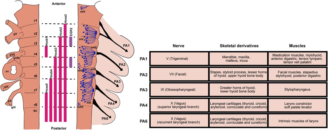
The highly conserved embryonic hindbrain and PAs and their associated circuits and peripheral structures. Left panel, Schematic representation of the vertebrate embryonic hindbrain. The developing hindbrain is segmented into rhombomeres (r1–r8), which is defined by combinatorial expression of different genes (e.g., Hox genes) and transcription factors (e.g., EGR2, also called Krox-20), depicted in the middle. Locations of cranial sensory ganglia (gV, gVII–gXI) and otic vesicles (ov) are shown on the left side of the hindbrain. The right side of the hindbrain shows motoneuron distribution of major cranial motor nuclei and their respective exit points. Neural crest cells form migratory streams (black arrows) that originate from rhombomeres to their respective PA. Right panel, Each PA is characterized by distinct nerve innervation, skeletal and muscular derivatives (table on the right). Drawings of rhombomeres and PAs and their derivatives are based on Kiecker and Lumsden (2005) and Maynard et al. (2020).

A common vertebrate embryonic hindbrain circuit for rhythmic ingestion of water/amniotic fluid

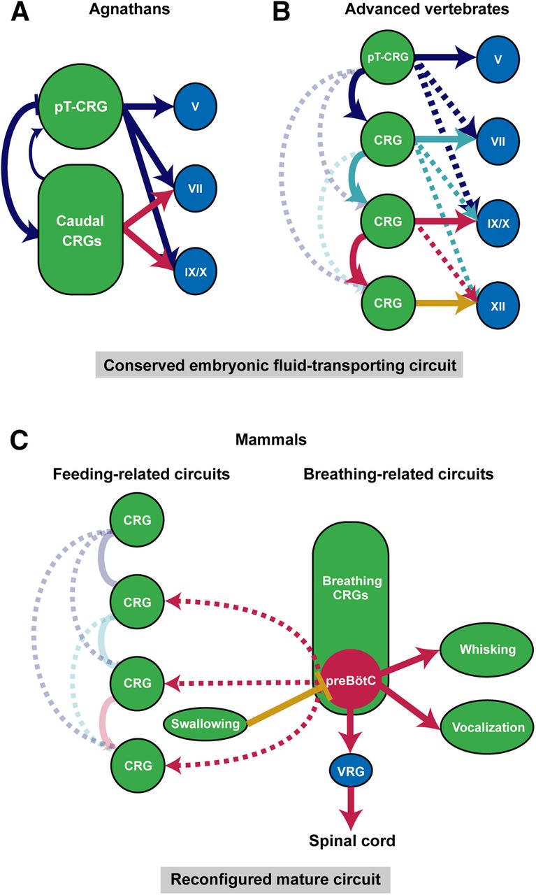
As described above, aquatic breathing and aquatic feeding share the same sets of cranial muscles and motor programs. Notably, while fish and amphibians are born directly into water, amniotes like reptiles, birds, and mammals go through embryonic development encircled by amniotic fluid. Therefore, bringing fluid into the mouth to be swallowed is likely the fundamental process common to all developing vertebrates. For example, it is known that fetal swallowing of amniotic fluid emerges right after major organogenesis around 10–14 weeks of gestation in human and is observed in early development of chicks, sheep, rodents, and monkeys (Ross and Nijland, 1998; Delaney and Arvedson, 2008; Mashayekhi et al., 2011; Gross and Trapani-Hanasewych, 2017; Maynard et al., 2020). This process of transporting fluid into the body is repeatedly executed and requires coordinated activation of oropharyngeal muscles (Sherman et al., 1990). Therefore, the developing hindbrain in all vertebrates must contains a basic circuit that produces rhythmic expansion and compression (or opening and closing) of the oral cavity for moving fluid into the mouth. We propose that this basic circuit contains either one dominant central rhythm generator (CRG) in primitive vertebrates, or a series of sequentially coupled CRGs that each has its own intrinsic rhythm in more evolved vertebrates, such that fluid is periodically transported into the mouth to be swallowed in a unidirectional manner (Fig. 3A,B).

Figure 3.
  • Download figure
  • Open in new tab
  • Download powerpoint
Figure 3.

Hypothesized conserved vertebrate embryonic hindbrain circuit for intraoral fluid transportation and its later stage reconfigurations in mammals. The fluid-surrounding environment and fluid ingestion behavior in aquatic and in embryonic terrestrial vertebrates suggest the existence of a conserved embryonic circuit that generates the fluid-transporting behavior in all vertebrates (A, B). A, In primitive vertebrates like lampreys, this fluid-transporting circuit consists of two CRGs, with pT-CRG as the dominant CRG, that drive rhythmic and sequential activations of downstream motor nuclei. B, In advanced vertebrates, a series of CRGs for each pair of rhombomeres work together to support the directional fluid-transporting behavior. Hypothesized serial CRGs are sequentially coupled such that an anterior CRG preferentially drives the activity of posterior CRGs, thereby produces sequential activation of oropharyngeal muscles and directional movement of fluid. C, Splitting, reconfiguration and repurposing of the embryonic fluid-transporting CRGs result in separated feeding-related and breathing-related CRGs in adult mammalian hindbrain. Only preBötC is highlighted among the breathing CRGs for simplicity. The need of continuous breathing makes preBötC the dominant CRG, which broadcasts its rhythm to other CRGs or motoneurons, ventral respiratory group (VRG). Feeding-related CRGs control chewing and licking largely function independent of breathing CRGs because of structural segregations of food and air intake pathways. Swallowing inhibits breathing to prevent aspiration.

Existence and locations of putative fluid-transporting CRGs in developing hindbrain

Where in embryonic hindbrain might the fluid-transporting CRGs reside? Let us first consider the larval lampreys, which use rhythmic movement of the velum to pump water into the mouth for feeding and for breathing. The velum located at the midline of pharynx is innervated by the Vth nerve. During larval lamprey aquatic breathing, muscles of velum and branchial basket (innervated by VIIth, IXth, Xth motoneurons) contract repeatedly in a fixed sequence (Russell, 1986; Rovainen, 1996). This then led to the discovery of a CRG adjacent to the Vth motor nucleus termed paratrigeminal respiratory group and a distributed CRG network around VIIth, IXth, Xth motor nuclei (Fig. 3A; Martel et al., 2007; Missaghi et al., 2016). It is postulated that these two CRG networks are coupled such that their sequential outputs drive the repeated velar and basket pumping (and relaxation) actions to bring in water (Fig. 3A). Since all later jawed vertebrates (Gnathostomes) use trigeminal (V)-innervated jaw opening and closing to bring water/amniotic fluid into the oral cavity, it is likely that the CRG located initially adjacent to the Vth motor nucleus [peritrigeminal CRG (pT-CRG)] is conserved in all vertebrates (at least in developmental stages; Fig. 3A,B).

As vertebrates evolved more muscles, the lamprey-type CRG configuration may be insufficient to drive coordinated movements of many muscles used for the repeated and directional fluid transportation and swallowing processes. Unlike agnathans such as lampreys and hagfish that predominantly relies on trigeminal-innervated velum for breathing, feeding/gill ventilation in fish starts from the opening of the jaw (V), followed by the activation of branchial muscles (VII) that results in sequential expansion and compression of first the buccal and then the opercular cavity. To swallow the food into the mouth, the sequence starts after jaw closure, followed by the activation of branchial basket muscles (IX, X) that transports the food to the esophagus through rounds of oropharyngeal expansions and compression. This sequential motor pattern likely requires serially coupled-CRGs in fish (Hyde, 1904; Sundin et al., 2000; Taylor et al., 2009a). This hypothesis is further supported by “sheep-dip” experiments, in which isolated fish brainstems are gradually submerged into high magnesium solution that blocks excitatory transmission necessary for rhythmogenesis (see next section). These experiments show that rhythmic outputs of VII nerve degrade gradually rather than abruptly, indicating that the gill ventilation rhythm in fish is generated by several mutually-coupled segmental CRGs distributed throughout the hindbrain (Duchcherer et al., 2010). In the mammalian fetal swallowing process, the fluid transportation also similarly follows the sequential activation of jaw, hyolingual, and pharyngeal muscles (Sherman et al., 1990; Thexton et al., 2007).

Furthermore, evidence suggests that in higher vertebrates, each pair of rhombomeres contains its own CRG. In chick embryos, isolated hindbrain segment preparations containing either V, VII, or XII motor nuclei all exhibit spontaneous rhythmic activities that drive their respective segmental motor nerves (Fig. 3B; Fortin et al., 1995; Borday et al., 2003). It was also revealed that odd-even pairs of rhombomeres generate higher frequencies of rhythmic bursts (Coutinho et al., 2004). In rodent preparations, similar conclusion was drawn that rhythmic firings in V, VII, and XII motoneurons representing ingestive movements are induced by their respective segmental CRG (Nakamura et al., 1999). We further speculate that the serial CRGs in pairs of rhombomeres form directionally biased connections such that the more anteriorly located CRG preferentially drives the activity of the posteriorly located CRG, thereby facilitating the propagating wave of oropharyngeal muscle activities that moves fluid from outside of the mouth to the esophagus (Fig. 3B). Notably, the rhombomeres form and differentiate in a rostral-to-caudal (from r1 to r8) order (Gavalas, 2002). Thus, the serial CRGs may also follow a developmental sequential manner of maturation thereby allowing the anterior CRGs to preferentially drive the posterior CRGs (Fig. 3B). The sequential coupling of CRGs is also likely dependent on and facilitated by the developing sensory neurons that form feedback circuits in favor of sequential activation. In isolated whole embryonic hindbrain preparations, it was shown that all cranial motor nerves exhibit synchronized rhythmic activities (Fortin et al., 1995; Abadie et al., 2000), suggesting that in the absence of sensory feedback, a single CRG (likely the most anterior pT-CRG) dominates.

Evolutionary conserved characteristics of rhythm-generating mechanism

Investigations of rhythm-generating mechanisms behind oropharyngeal behaviors in vertebrates have largely focused on breathing, especially in lampreys, frogs, and rodents. These studies primarily examined the roles of excitatory glutamatergic mechanisms, inhibitory GABAergic and glycinergic mechanisms, putative pacemaker properties, and neuromodulators in rhythm generations. Decades of studies suggest that some rhythmogenic mechanisms are evolutionary conserved.

First, excitatory glutamatergic system is essential for rhythm generation in nearly all vertebrate breathing CRGs. Both lamprey’s pT-CRG and mammalian’s inspiration CRG, preBötzinger complex (preBötC), contain large ensembles of glutamatergic neurons and are sensitive to modulations by opioids and substance P (Mutolo et al., 2010; Del Negro et al., 2018). In lampreys, microinjections of AMPA and NMDA blockers in pT-CRG temporary abolish its rhythm (Martel et al., 2007; Cinelli et al., 2013). On the other hand, manipulations that facilitate glutamatergic transmission (e.g., application of AMPA or perfusion of mGluRs I and II agonists) accelerate pT-CRG rhythm (Bongianni et al., 2002; Martel et al., 2007; Cinelli et al., 2013). In mammals, many neurons that express transcription factor developing brain homeobox protein 1 (DBX1) during the pharyngula stage later become glutamatergic neurons, including neurons in preBötC. Dbx1-knock-out mice do not breathe and die at birth and optogenetic inhibition of DBX1 lineage cells slows and stops breathing rhythm (for more detail, see Del Negro et al., 2018). Glutamatergic transmission is also found to be essential in CRGs responsible for lung ventilation in turtles (Johnson and Mitchell, 1998) and frogs (Chen and Hedrick, 2008; Baghdadwala et al., 2016). Interestingly, recent results from Ashhad and Feldman (2020) suggest that inspiratory rhythmogenesis in neonatal mammalian preBötC is critically driven by emergent network properties, where strong synchronization among excitatory rhythmogenic neurons within preBötC produces inspiratory bursts.

Second, inhibitory systems heavily modulate the frequency and amplitude of rhythm but may not be essential for rhythm generation per se. For example, respiratory rhythm still persists after blockade of GABAergic and glycinergic transmission in lampreys (Bongianni et al., 2006; Cinelli et al., 2014, 2016), in frogs (lung ventilation; Leclère et al., 2012; Baghdadwala et al., 2016), turtles (Johnson et al., 2002, 2007), and in mammals (Janczewski et al., 2013; Sherman et al., 2015; Baertsch et al., 2018). However, in different species or different brainstem nuclei, inhibition has different effects. Blocking GABAA in lamprey produce significant increases in frequency and amplitude of breathing rhythm, while injection of GABAA agonist suppresses pT-CRG rhythm (Bongianni et al., 2006; Cinelli et al., 2014). In rodents, it is proposed that GABAergic and glycinergic transmissions, respectively, regulate different aspects of rhythm and pattern generated by preBötC (Ashhad and Feldman, 2020). Photostimulation of glycinergic preBötC neurons depresses breathing while inhibition of these neurons augments breathing amplitude and stops ongoing apnea (Sherman et al., 2015). Additionally, complex reciprocal GABAergic transmissions are important for the coupling of inspiratory preBötC and expiratory parafacial oscillator (Mellen and Thoby-Brisson, 2012). By contrast, in chick embryos, blocking GABA-mediated inhibition in segmental CRGs abolishes the high frequency rhythmic bursts. Here, inhibition is required for generating high (but not low) frequency rhythms (Fortin et al., 1999).

Many studies also examined whether pacemaker-like properties are involved in hindbrain rhythmogenesis across species. Specifically, these studies tested the roles of persistent sodium current (INaP) or calcium-activated non-specific cation (ICAN) currents. In lampreys, turtles, and mammals, while breathing rhythm is abolished by INaP and ICAN current blockers, application of an exogenous excitatory agent (e.g., substance P) restores the rhythm (Del Negro et al., 2005; Mutolo et al., 2010; Lin and Onimaru, 2015; Johnson et al., 2016). These results suggest that pacemaker properties are not essential for respiratory rhythm generation (Mellen and Thoby-Brisson, 2012; Bongianni et al., 2016; Missaghi et al., 2016). However, under hypoxia condition, breathing-related activity are dependent on INaP (Peña et al., 2004; Paton et al., 2006). Furthermore, it was shown that in mouse, unlike the primarily network-driven rhythmogenesis at and after E18.5, INaP and ICAN dependent pacemaker properties are purely required for inspiration rhythm in embryos before E16.5 (Chevalier et al., 2016). Thus, the rhythmogesis mechanisms for the proposed fluid-transporting CRGs in earlier embryos could be different from mechanisms governing CRGs in neonatal and adult higher vertebrates, and future studies with cellular identities are needed to pinpoint the exact mechanisms.

Reconfiguration and repurposing embryonic CRGs to accommodate parallel air breathing and feeding in higher vertebrates

The evolutionary progression from one oral cavity serving both feeding and breathing functions in aquatic vertebrates, to two separated food/fluid and air intake pathways, such as the separated nasal and oral cavities in mammals, allows air-breathing and feeding to be conducted, to a large extent, independently. We hypothesize that peripheral structural separations are accompanied by the split of conserved segmental fluid-transporting CRGs in the hindbrain into two separate sets: one set for breathing and the other set remains as CRGs for feeding (Fig. 3C).

Indeed, a series of breathing-related CRGs have been discovered in mammals. They are, from rostral to caudal, the parafacial respiratory group functioning as the expiratory CRG (pFRG), the postinspiration complex (PiCo) hypothesized to be the postinspiration CRG, and the preBötC known as the inspiratory CRG. These respiratory CRGs may have migrated during development from their original birthplace to their final ventral locations in the hindbrain. We speculate that the origin of preBötC might be the segmental CRG near the Xth nerve (in r7) that is recruited during evolution to provide rhythmic control of inspiration axial muscles (by projecting to neurons in the medullary ventral respiratory groups which in turn project to the spinal cord). Interestingly, it was shown in mouse, that the constituent neurons of the embryonic parafacial (e-pF; originated in r4) and preBötC (in r7) CRGs are born and matured in a sequential fashion [e-pF cells are born at embryonic day (E)10.5 and matured at E14.5, while preBötC neurons are born at E11.5 and matured at E15.5], allowing the anterior-located e-pF to entrain the immature preBötC activity in early embryonic stages (Thoby-Brisson et al., 2009; Champagnat et al., 2011). This time period also coincides with the period of secondary palate formation (Yu et al., 2017). After the complete fusion of the secondary palate at the midline to separate nasal and oral cavity in late embryonic stages, preBötC gradually becomes the dominant CRG in the hindbrain, entraining other respiratory CRGs (Ramirez et al., 2016; Del Negro et al., 2018), perhaps in preparation for the need of continuous air-breathing after birth. In addition, other secondary CRGs serve orofacial actions using muscles evolutionarily involved in breathing may be further separated/differentiated or repurposed from embryonic ones. For example, parts of posterior embryonic CRG neurons in r7/r8 are likely to be repurposed as CRGs for whisking in rodents and vocalizations in mammals (Fig. 3C).

On the other hand, some of the embryonic fluid-transporting CRG neurons retain their feeding-related functions. These cells may stay in their conserved locations near feeding-related motor neurons. For example, the ancient pT-CRG may become the mammalian CRG for suckling (neonates) and chewing (adults). Indeed, neurons located dorsal to and near Vth nucleus are implicated in controlling rhythmic jaw movements (Morquette et al., 2012). The caudal feeding-related embryonic CRGs likely further differentiated to control licking and swallowing (Fig. 3C). Again, because of the separation of nasal and oral cavity, chewing and licking can operate to a large extent independently of breathing until the step of swallowing in most mammals (Moore et al., 2014). On the other hand, the continuously active inspiration CRG preBötC is shown to send strong projections throughout the hindbrain, broadcasting the breathing rhythm to neurons controlling feeding (Fig. 3C). This is likely because breathing requires activity from certain feeding-related oropharyngeal muscles to help maintain airway patency (Moore et al., 2014; Yang and Feldman, 2018). Finally, swallowing in mammals is typically executed is one rapid motion and thus it may not require a CRG. However, new airway protection circuits must have evolved in mammals such that centrally-arisen or peripherally-arisen swallowing signals effectively inhibit inspiration to prevent aspiration of food (Fig. 3C).

The maturation and refinement of species-specific hindbrain orofacial circuits likely depends on feedback signaling from peripheral muscles and other tissues to motoneurons, then from motoneurons to upstream CRGs and other interneurons also through retrograde signaling, as well as depends on activity of sensory feedback. Gradually, synergistic muscles will be co-active in the same phase of CRG rhythm, whereas antagonist muscles will be co-activated in the opposite phase of CRG activity. Sequences are further consolidated and eventually diverse motor patterns of coordinated feeding and breathing behaviors in higher vertebrates emerge.

Concluding remarks

One of the core questions related to the development and evolution of motor behaviors is how evolutionary conserved developmental principles give rise to such a diverse pool of behaviors. In this review, we discussed how vertebrates, while carrying out varied modes of feeding and breathing, all share a stage of living in a fluid and all execute a homologous action of periodically transporting fluid from the external environment inside the body. We hypothesize that a conserved hindbrain circuit generating a posteriorly propagating rhythmic activity is initially formed in all vertebrates, that drives the basic intraoral fluid transport behaviors. This circuit forms the basis for carrying out aquatic feeding/aquatic breathing, primitive air breathing, and terrestrial food transportation. Aspiration breathing and food reduction (chewing) represent the splitting, repurposing, and reconfiguration of the existing hindbrain CRGs. We note that other than preBötC/pF/PiCo, the neuronal identities and exact locations of feeding-related CRGs (chewing, suckling, licking, swallowing, etc.) in mammals remain vague. Based on our unifying hypothesis, we propose that studies of hindbrain CRGs for feeding/gill ventilation in fish may reveal the homologous neurons in mammals that drive rhythmic suckling, chewing, and licking. Alternatively, if a new method can be developed that enables lineage tracing of hindbrain CRG neurons in mammalian embryos, following these neurons into postnatal development and adult should provide insights for the locations and identities of adult CRG neurons.

Acknowledgments

Acknowledgements: We thank the Wang lab for many useful discussions, especially Dr. Vincent Prevosto and Dr. Jun Takatoh, for careful reading and commenting on this manuscript.

Footnotes

  • The authors declare no competing financial interests.

  • This work was supported by a subcontract for the National Institutes of Health Grant U19NS107466.

This is an open-access article distributed under the terms of the Creative Commons Attribution 4.0 International license, which permits unrestricted use, distribution and reproduction in any medium provided that the original work is properly attributed.

References

  1. ↵
    Abadie V, Champagnat J, Fortin G (2000) Branchiomotor activities in mouse embryo. Neuroreport 11:141–145. doi:10.1097/00001756-200001170-00028 pmid:10683846
    OpenUrlCrossRefPubMed
  2. ↵
    Abramyan J, Richman JM (2015) Recent insights into the morphological diversity in the amniote primary and secondary palates. Dev Dyn 244:1457–1468. doi:10.1002/dvdy.24338 pmid:26293818
    OpenUrlCrossRefPubMed
  3. ↵
    Alheid GF, Milsom WK, McCrimmon DR (2004) Pontine influences on breathing: an overview. Respir Physiol Neurobiol 143:105–114. doi:10.1016/j.resp.2004.06.016 pmid:15519548
    OpenUrlCrossRefPubMed
  4. ↵
    Arnold P, Birtles R, Sobtzick S, Matthews M, Dunstan A (2005) Gulping behaviour in rorqual whales: underwater observations and functional interpretation. Mem Queensl Mus 51:309–332.
    OpenUrl
  5. ↵
    Arvedson JC, Lefton-Greif MA (1998) Pediatric videofluoroscopic swallow studies. A professional manual with caregiver guidelines. San Antonio: Communication Skill Builders.
  6. ↵
    Ashhad S, Feldman JL (2020) Emergent elements of inspiratory rhythmogenesis: network synchronization and synchrony propagation. Neuron 106:482–497. doi:10.1016/j.neuron.2020.02.005 pmid:32130872
    OpenUrlCrossRefPubMed
  7. ↵
    Baertsch NA, Baertsch HC, Ramirez JM (2018) The interdependence of excitation and inhibition for the control of dynamic breathing rhythms. Nat Commun 9:843. doi:10.1038/s41467-018-03223-x
    OpenUrlCrossRefPubMed
  8. ↵
    Baghdadwala MI, Duchcherer M, Trask WM, Gray PA, Wilson RJ (2016) Diving into the mammalian swamp of respiratory rhythm generation with the bullfrog. Respir Physiol Neurobiol 224:37–51. doi:10.1016/j.resp.2015.09.005 pmid:26384027
    OpenUrlCrossRefPubMed
  9. ↵
    Bels V, Whishaw IQ (2019) Feeding in vertebrates: evolution, morphology, behavior, biomechanics. Cham: Springer.
  10. ↵
    Boltt R, Ewer R (1964) The functional anatomy of the head of the puff adder, Bitis arietans (Merr.). J Morphol 114:83–105. doi:10.1002/jmor.1051140105
    OpenUrlCrossRef
  11. ↵
    Bongianni F, Mutolo D, Carfì M, Pantaleo T (2002) Group I and II metabotropic glutamate receptors modulate respiratory activity in the lamprey. Eur J Neurosci 16:454–460. doi:10.1046/j.1460-9568.2002.02098.x pmid:12193188
    OpenUrlCrossRefPubMed
  12. ↵
    Bongianni F, Mutolo D, Nardone F, Pantaleo T (2006) GABAergic and glycinergic inhibitory mechanisms in the lamprey respiratory control. Brain Res 1090:134–145. doi:10.1016/j.brainres.2006.03.056 pmid:16630584
    OpenUrlCrossRefPubMed
  13. ↵
    Bongianni F, Mutolo D, Cinelli E, Pantaleo T (2016) Neural mechanisms underlying respiratory rhythm generation in the lamprey. Respir Physiol Neurobiol 224:17–26. doi:10.1016/j.resp.2014.09.003 pmid:25220696
    OpenUrlCrossRefPubMed
  14. ↵
    Borday C, Abadie V, Chatonnet F, Thoby-Brisson M, Champagnat J, Fortin G (2003) Developmental molecular switches regulating breathing patterns in CNS. Respir Physiol Neurobiol 135:121–132. doi:10.1016/s1569-9048(03)00031-4 pmid:12809613
    OpenUrlCrossRefPubMed
  15. ↵
    Brainerd EL (1994) The evolution of lung-gill bimodal breathing and the homology of vertebrate respiratory pumps. Am Zool 34:289–299. doi:10.1093/icb/34.2.289
    OpenUrlCrossRef
  16. ↵
    Brainerd EL (1999) New perspectives on the evolution of lung ventilation mechanisms in vertebrates. Exp Biol Online 4:1–28. doi:10.1007/s00898-999-0002-1
    OpenUrlCrossRef
  17. ↵
    Brainerd EL, Ferry‐Graham LA (2005) Mechanics of respiratory pumps. Fish Physiol 23:1–28.
    OpenUrl
  18. ↵
    Brainerd EL, Owerkowicz T (2006) Functional morphology and evolution of aspiration breathing in tetrapods. Respir Physiol Neurobiol 154:73–88. doi:10.1016/j.resp.2006.06.003 pmid:16861059
    OpenUrlCrossRefPubMed
  19. ↵
    Bramble DM, Wake DB (1985) Feeding mechanisms of lower tetrapods. In: Functional vertebrate morphology (Hildebrand M, Bramble DM, Liem KF, Wake DB, eds), pp 230–261. Cambridge: Harvard University Press.
  20. ↵
    Brown JD, Stallknecht DE (2008) Wild bird surveillance for the avian influenza virus. Methods Mol Biol 436:85–97.
    OpenUrlCrossRefPubMed
  21. ↵
    Carrier DR (1987) Lung ventilation during walking and running in four species of lizards. Exp Biol 47:1.
    OpenUrlPubMed
  22. ↵
    Carrier DR (1989) Ventilatory action of the hypaxial muscles of the lizard Iguana iguana: a function of slow muscle. J Exp Biol 143:435–457. pmid:2732666
    OpenUrlAbstract/FREE Full Text
  23. ↵
    Carrier DR (1990) Activity of the hypaxial muscles during walking in the lizard Iguana iguana. J Exp Biol 152:453–470. pmid:2146360
    OpenUrlAbstract/FREE Full Text
  24. ↵
    Carrier DR (1991) Conflict in the hypaxial musculo-skeletal system: documenting an evolutionary constraint. Am Zool 31:644–654. doi:10.1093/icb/31.4.644
    OpenUrlCrossRef
  25. ↵
    Champagnat J, Morin-Surun M-P, Bouvier J, Thoby-Brisson M, Fortin G (2011) Prenatal development of central rhythm generation. Respir Physiol Neurobiol 178:146–155. doi:10.1016/j.resp.2011.04.013 pmid:21527363
    OpenUrlCrossRefPubMed
  26. ↵
    Chen AK, Hedrick MS (2008) Role of glutamate and substance P in the amphibian respiratory network during development. Respir Physiol Neurobiol 162:24–31. doi:10.1016/j.resp.2008.03.010
    OpenUrlCrossRefPubMed
  27. ↵
    Chevalier M, Toporikova N, Simmers J, Thoby-Brisson M (2016) Development of pacemaker properties and rhythmogenic mechanisms in the mouse embryonic respiratory network. Elife 5:e16125. doi:10.7554/eLife.16125
    OpenUrlCrossRefPubMed
  28. ↵
    Cieri RL, Farmer C (2016) Unidirectional pulmonary airflow in vertebrates: a review of structure, function, and evolution. J Comp Physiol B 186:541–552. doi:10.1007/s00360-016-0983-3 pmid:27062030
    OpenUrlCrossRefPubMed
  29. ↵
    Cinelli E, Robertson B, Mutolo D, Grillner S, Pantaleo T, Bongianni F (2013) Neuronal mechanisms of respiratory pattern generation are evolutionary conserved. J Neurosci 33:9104–9112. doi:10.1523/JNEUROSCI.0299-13.2013 pmid:23699521
    OpenUrlAbstract/FREE Full Text
  30. ↵
    Cinelli E, Mutolo D, Robertson B, Grillner S, Contini M, Pantaleo T, Bongianni F (2014) GABAergic and glycinergic inputs modulate rhythmogenic mechanisms in the lamprey respiratory network. J Physiol 592:1823–1838. doi:10.1113/jphysiol.2013.268086 pmid:24492840
    OpenUrlCrossRefPubMed
  31. ↵
    Cinelli E, Mutolo D, Contini M, Pantaleo T, Bongianni F (2016) Inhibitory control of ascending glutamatergic projections to the lamprey respiratory rhythm generator. Neuroscience 326:126–140. doi:10.1016/j.neuroscience.2016.03.063 pmid:27058146
    OpenUrlCrossRefPubMed
  32. ↵
    Claessens LP (2004) Archosaurian respiration and the pelvic girdle aspiration breathing of crocodyliforms. Proc Biol Sci 271:1461–1465. doi:10.1098/rspb.2004.2743 pmid:15306317
    OpenUrlCrossRefPubMed
  33. ↵
    Clark AJ, Summers AP (2007) Morphology and kinematics of feeding in hagfish: possible functional advantages of jaws. J Exp Biol 210:3897–3909. doi:10.1242/jeb.006940 pmid:17981857
    OpenUrlAbstract/FREE Full Text
  34. ↵
    Clark AJ, Uyeno TA (2019) Feeding in jawless fishes. In: Feeding in vertebrates: evolution, morphology, behavior, biomechanics (Bels V, Whishaw IQ, eds), pp 189–230. Cham: Springer.
  35. ↵
    Clark AJ, Maravilla EJ, Summers AP (2010) A soft origin for a forceful bite: motor patterns of the feeding musculature in Atlantic hagfish, Myxine glutinosa. Zoology (Jena) 113:259–268. doi:10.1016/j.zool.2010.02.001 pmid:20667701
    OpenUrlCrossRefPubMed
  36. ↵
    Cleuren J, de Vree F (1992) Kinematics of the jaw and hyolingual apparatus during feeding in Caiman crocodilus. J Morphol 212:141–154. doi:10.1002/jmor.1052120205 pmid:29865591
    OpenUrlCrossRefPubMed
  37. ↵
    Clubb BL, Clark AJ, Uyeno TA (2019) Powering the hagfish “bite”: the functional morphology of the retractor complex of two hagfish feeding apparatuses. J Morphol 280:827–840. doi:10.1002/jmor.20986 pmid:30927384
    OpenUrlCrossRefPubMed
  38. ↵
    Coutinho AP, Borday C, Gilthorpe J, Jungbluth S, Champagnat J, Lumsden A, Fortin G (2004) Induction of a parafacial rhythm generator by rhombomere 3 in the chick embryo. J Neurosci 24:9383–9390. doi:10.1523/JNEUROSCI.2408-04.2004 pmid:15496674
    OpenUrlAbstract/FREE Full Text
  39. ↵
    Crompton A, German R, Thexton A (1997) Mechanisms of swallowing and airway protection in infant mammals (Sus domesticus and Macaca fascicularis). J Zool 241:89–102. doi:10.1111/j.1469-7998.1997.tb05501.x
    OpenUrlCrossRef
  40. ↵
    Cundall D, Tuttman C, Close M (2014) A model of the anterior esophagus in snakes, with functional and developmental implications. Anat Rec (Hoboken) 297:586–598. doi:10.1002/ar.22860 pmid:24482367
    OpenUrlCrossRefPubMed
  41. ↵
    Del Negro CA, Morgado-Valle C, Hayes JA, Mackay DD, Pace RW, Crowder EA, Feldman JL (2005) Sodium and calcium current-mediated pacemaker neurons and respiratory rhythm generation. J Neurosci 25:446–453. doi:10.1523/JNEUROSCI.2237-04.2005 pmid:15647488
    OpenUrlAbstract/FREE Full Text
  42. ↵
    Del Negro CA, Funk GD, Feldman JL (2018) Breathing matters. Nat Rev Neurosci 19:351–367. doi:10.1038/s41583-018-0003-6 pmid:29740175
    OpenUrlCrossRefPubMed
  43. ↵
    Delaney AL, Arvedson JC (2008) Development of swallowing and feeding: prenatal through first year of life. Dev Disabil Res Rev 14:105–117. doi:10.1002/ddrr.16 pmid:18646020
    OpenUrlCrossRefPubMed
  44. ↵
    Ding Y, Vanselow DJ, Yakovlev MA, Katz SR, Lin AY, Clark DP, Vargas P, Xin X, Copper JE, Canfield VA, Ang KC, Wang Y, Xiao X, De Carlo F, van Rossum DB, La Riviere P, Cheng KC (2019) Computational 3D histologic phenotyping of whole zebrafish by X-ray histotomography. Elife 8:e44898.
    OpenUrlCrossRef
  45. ↵
    Duchcherer M, Kottick A, Wilson R (2010) Evidence for a distributed respiratory rhythm generating network in the goldfish (Carsssius auratus). In: New frontiers in respiratory control (Homma I, Onimaru H, Fukuchi Y, eds), pp 3–7. New York: Springer.
  46. ↵
    Ertekin C, Aydogdu I (2003) Neurophysiology of swallowing. Clin Neurophysiol 114:2226–2244. doi:10.1016/s1388-2457(03)00237-2 pmid:14652082
    OpenUrlCrossRefPubMed
  47. ↵
    Eom J, Wood CM (2019) The ventilation mechanism of the Pacific hagfish Eptatretus stoutii. J Fish Biol 94:261–276. doi:10.1111/jfb.13885 pmid:30549032
    OpenUrlCrossRefPubMed
  48. ↵
    Farmer C (2015) The evolution of unidirectional pulmonary airflow. Physiology (Bethesda) 30:260–272. doi:10.1152/physiol.00056.2014 pmid:26136540
    OpenUrlCrossRefPubMed
  49. ↵
    Fortin G, Kato F, Lumsden A, Champagnat J (1995) Rhythm generation in the segmented hindbrain of chick embryos. J Physiol 486:735–744. doi:10.1113/jphysiol.1995.sp020849
    OpenUrlCrossRefPubMed
  50. ↵
    Fortin G, Jungbluth S, Lumsden A, Champagnat J (1999) Segmental specification of GABAergic inhibition during development of hindbrain neural networks. Nat Neurosci 2:873–877. doi:10.1038/13172 pmid:10491606
    OpenUrlCrossRefPubMed
  51. ↵
    Gans C, Clark BD (1976) Studies on ventilation of Caiman crocodilus (Crocodilia: Reptilia). Respir Physiol 26:285–301. doi:10.1016/0034-5687(76)90001-3 pmid:951534
    OpenUrlCrossRefPubMed
  52. ↵
    Gaunt AS, Gans C (1969) Mechanics of respiration in the snapping turtle, Chelydra serpentina (Linné). J Morphol 128:195–227. doi:10.1002/jmor.1051280205
    OpenUrlCrossRef
  53. ↵
    Gavalas A (2002) ArRAnging the hindbrain. Trends Neurosci 25:61–64. doi:10.1016/s0166-2236(02)02067-2 pmid:11814548
    OpenUrlCrossRefPubMed
  54. ↵
    Gillis JA, Tidswell OR (2017) The origin of vertebrate gills. Curr Biol 27:729–732. doi:10.1016/j.cub.2017.01.022 pmid:28190727
    OpenUrlCrossRefPubMed
  55. ↵
    Goldbogen J, Cade D, Calambokidis J, Friedlaender A, Potvin J, Segre P, Werth A (2017) How baleen whales feed: the biomechanics of engulfment and filtration. Ann Rev Mar Sci 9:367–386. doi:10.1146/annurev-marine-122414-033905 pmid:27620830
    OpenUrlCrossRefPubMed
  56. ↵
    Graham A, Smith A (2001) Patterning the pharyngeal arches. Bioessays 23:54–61. doi:10.1002/1521-1878(200101)23:1<54::AID-BIES1007>3.0.CO;2-5
    OpenUrlCrossRefPubMed
  57. ↵
    Graham A, Richardson J (2012) Developmental and evolutionary origins of the pharyngeal apparatus. Evodevo 3:24. doi:10.1186/2041-9139-3-24 pmid:23020903
    OpenUrlCrossRefPubMed
  58. ↵
    Graham A, Poopalasundaram S, Shone V, Kiecker C (2019) A reappraisal and revision of the numbering of the pharyngeal arches. J Anat 235:1019–1023. doi:10.1111/joa.13067 pmid:31402457
    OpenUrlCrossRefPubMed
  59. ↵
    Gross RD, Trapani-Hanasewych M (2017) Breathing and swallowing: the next frontier. Semin Speech Lang 38:87–95. doi:10.1055/s-0037-1599106 pmid:28324898
    OpenUrlCrossRefPubMed
  60. ↵
    Grubich JR (2001) Prey capture in actinopterygian fishes: a review of suction feeding motor patterns with new evidence from an elopomorph fish, Megalops atlanticus. Am Zool 41:1258–1265. doi:10.1093/icb/41.6.1258
    OpenUrlCrossRef
  61. ↵
    Haney W, Clark A, Uyeno T (2020) Characterization of body knotting behavior used for escape in a diversity of hagfishes. J Zool 310:261–272. doi:10.1111/jzo.12752
    OpenUrlCrossRef
  62. ↵
    Harrison DFN (1995) The anatomy and physiology of the mammalian larynx. Cambridge: Cambridge University Press.
  63. ↵
    Herrel A, O’Reilly JC, Fabre AC, Bardua C, Lowie A, Boistel R, Gorb SN (2019) Feeding in amphibians: evolutionary transformations and phenotypic diversity as drivers of feeding system diversity. In: Feeding in vertebrates: evolution, morphology, behavior, biomechanics (Bels V, Whishaw IQ, eds), pp 431–467. Cham: Springer.
  64. ↵
    Hyde IH (1904) Localization of the respiratory centre in the skate. Am J Physiol 10:236–258. doi:10.1152/ajplegacy.1904.10.5.236
    OpenUrlCrossRef
  65. ↵
    Irie N, Kuratani S (2011) Comparative transcriptome analysis reveals vertebrate phylotypic period during organogenesis. Nat Commun 2:248. doi:10.1038/ncomms1248 pmid:21427719
    OpenUrlCrossRefPubMed
  66. ↵
    Iwasaki SI, Erdoğan S, Asami T (2019) Evolutionary specialization of the tongue in vertebrates: structure and function. In: Feeding in vertebrates: evolution, morphology, behavior, biomechanics (Bels V, Whishaw IQ, eds), pp 333–384. Cham: Springer.
  67. ↵
    Janczewski WA, Tashima A, Hsu P, Cui Y, Feldman JL (2013) Role of inhibition in respiratory pattern generation. J Neurosci 33:5454–5465. doi:10.1523/JNEUROSCI.1595-12.2013 pmid:23536061
    OpenUrlAbstract/FREE Full Text
  68. ↵
    Johnson SM, Mitchell GS (1998) N-methyl-d-aspartate-mediated bulbospinal respiratory drive is pH/P(CO2)-insensitive in turtle brainstem-spinal cord. Respir Physiol 113:201–212. doi:10.1016/s0034-5687(98)00079-6 pmid:9840329
    OpenUrlCrossRefPubMed
  69. ↵
    Johnson SM, Wilkerson JE, Wenninger MR, Henderson DR, Mitchell GS (2002) Role of synaptic inhibition in turtle respiratory rhythm generation. J Physiol 544:253–265. doi:10.1113/jphysiol.2002.019687 pmid:12356896
    OpenUrlCrossRefPubMed
  70. ↵
    Johnson SM, Wiegel LM, Majewski DJ (2007) Are pacemaker properties required for respiratory rhythm generation in adult turtle brain stems in vitro? Am J Physiol Regul Integr Comp Physiol 293:R901–R910. doi:10.1152/ajpregu.00912.2006
    OpenUrlCrossRefPubMed
  71. ↵
    Johnson SM, Hedrick MS, Krause BM, Nilles JP, Chapman MA (2016) Respiratory neuron characterization reveals intrinsic bursting properties in isolated adult turtle brainstems (Trachemys scripta). Respir Physiol Neurobiol 224:52–61. doi:10.1016/j.resp.2014.11.004 pmid:25462012
    OpenUrlCrossRefPubMed
  72. ↵
    Kardong KV (2018) Vertebrates: comparative anatomy, function, evolution, Ed 8. New York: McGraw-Hill Education.
  73. ↵
    Kiecker C, Lumsden A (2005) Compartments and their boundaries in vertebrate brain development. Nat Rev Neurosci 6:553–564. doi:10.1038/nrn1702 pmid:15959467
    OpenUrlCrossRefPubMed
  74. ↵
    Kienle SS, Berta A (2016) The better to eat you with: the comparative feeding morphology of phocid seals (Pinnipedia, Phocidae). J Anat 228:396–413. doi:10.1111/joa.12410 pmid:26646351
    OpenUrlCrossRefPubMed
  75. ↵
    Kienle SS, Law CJ, Costa DP, Berta A, Mehta RS (2017) Revisiting the behavioural framework of feeding in predatory aquatic mammals. Proc Biol Sci 284:20171035. doi:10.1098/rspb.2017.1035
    OpenUrlCrossRefPubMed
  76. ↵
    Kley NJ, Brainerd EL (2002) Post-cranial prey transport mechanisms in the black pinesnake, Pituophis melanoleucus lodingi: an x-ray videographic study. Zoology (Jena) 105:153–164. doi:10.1078/0944-2006-00056 pmid:16351864
    OpenUrlCrossRefPubMed
  77. ↵
    Laitman JT, Reidenberg JS (1993) Specializations of the human upper respiratory and upper digestive systems as seen through comparative and developmental anatomy. Dysphagia 8:318–325. doi:10.1007/BF01321770 pmid:8269722
    OpenUrlCrossRefPubMed
  78. ↵
    Laitman JT, Reidenberg JS (1997) The human aerodigestive tract and gastroesophageal reflux: an evolutionary perspective. Am J Med 103:2S–8S. doi:10.1016/s0002-9343(97)00313-6 pmid:9422615
    OpenUrlCrossRefPubMed
  79. ↵
    Laitman J, Crelin E, Conlogue G (1977) The function of the epiglottis in monkey and man. Yale J Biol Med 50:43–48. pmid:403687
    OpenUrlPubMed
  80. ↵
    Landberg T, Mailhot JD, Brainerd EL (2003) Lung ventilation during treadmill locomotion in a terrestrial turtle, Terrapene carolina. J Exp Biol 206:3391–3404. doi:10.1242/jeb.00553
    OpenUrlAbstract/FREE Full Text
  81. ↵
    Lang IM (2009) Brain stem control of the phases of swallowing. Dysphagia 24:333–348. doi:10.1007/s00455-009-9211-6 pmid:19399555
    OpenUrlCrossRefPubMed
  82. ↵
    Leclère R, Straus C, Similowski T, Bodineau L, Fiamma M-N (2012) Persistent lung oscillator response to CO2 after buccal oscillator inhibition in the adult frog. Respir Physiol Neurobiol 183:166–169. doi:10.1016/j.resp.2012.06.030 pmid:22772313
    OpenUrlCrossRefPubMed
  83. ↵
    Levine RP, Monroy JA, Brainerd EL (2004) Contribution of eye retraction to swallowing performance in the northern leopard frog, Rana pipiens. J Exp Biol 207:1361–1368. doi:10.1242/jeb.00885 pmid:15010487
    OpenUrlAbstract/FREE Full Text
  84. ↵
    Lieberman DE, McCarthy RC, Hiiemae KM, Palmer JB (2001) Ontogeny of postnatal hyoid and larynx descent in humans. Arch Oral Biol 46:117–128. doi:10.1016/S0003-9969(00)00108-4
    OpenUrlCrossRefPubMed
  85. ↵
    Lin ST, Onimaru H (2015) Effects of riluzole on respiratory rhythm generation in the brainstem-spinal cord preparation from newborn rat. Neurosci Res 94:28–36. doi:10.1016/j.neures.2014.12.001 pmid:25498952
    OpenUrlCrossRefPubMed
  86. ↵
    Lindström T (1949) On the cranial nerves of the cyclostomes with special reference to n. trigeminus. Acta Zool 30:315–458. doi:10.1111/j.1463-6395.1949.tb00509.x
    OpenUrlCrossRef
  87. ↵
    Lumsden A, Keynes R (1989) Segmental patterns of neuronal development in the chick hindbrain. Nature 337:424–428. doi:10.1038/337424a0 pmid:2644541
    OpenUrlCrossRefPubMed
  88. ↵
    Mallatt J (1997) Shark pharyngeal muscles and early vertebrate evolution. Acta Zool 78:279–294. doi:10.1111/j.1463-6395.1997.tb01012.x
    OpenUrlCrossRef
  89. ↵
    Malte H, Lomholt JP (1998) Ventilation and gas exchange. In: The biology of hagfishes, pp 223–234. New York: Springer.
  90. ↵
    Marshall CD, Wieskotten S, Hanke W, Hanke FD, Marsh A, Kot B, Dehnhardt G (2014) Feeding kinematics, suction, and hydraulic jetting performance of harbor seals (Phoca vitulina). PLoS One 9:e86710. doi:10.1371/journal.pone.0086710
    OpenUrlCrossRefPubMed
  91. ↵
    Martel B, Guimond J, Gariépy J, Gravel J, Auclair F, Kolta A, Lund J, Dubuc R (2007) Respiratory rhythms generated in the lamprey rhombencephalon. Neuroscience 148:279–293. doi:10.1016/j.neuroscience.2007.05.023 pmid:17618060
    OpenUrlCrossRefPubMed
  92. ↵
    Mashayekhi F, Dianati E, Moghadam LM (2011) Quantitative analysis of nerve growth factor in the amniotic fluid during chick embryonic development. Saudi J Biol Sci 18:209–212. doi:10.1016/j.sjbs.2010.07.001 pmid:23961126
    OpenUrlCrossRefPubMed
  93. ↵
    Mason KA, Losos JB, Duncan T (2020) Respiration. In: Biology, Ed 12, pp 1053–1070. New York: McGraw-Hill Education.
  94. ↵
    Matsuo K, Palmer JB (2010) Kinematic linkage of the tongue, jaw, and hyoid during eating and speech. Arch Oral Biol 55:325–331. doi:10.1016/j.archoralbio.2010.02.008
    OpenUrlCrossRefPubMed
  95. ↵
    Maynard TM, Zohn IE, Moody SA, LaMantia AS (2020) Suckling, feeding, and swallowing: behaviors, circuits, and targets for neurodevelopmental pathology. Annu Rev Neurosci 43:315–336.
    OpenUrl
  96. ↵
    Mellen NM, Thoby-Brisson M (2012) Respiratory circuits: development, function and models. Curr Opin Neurobiol 22:676–685. doi:10.1016/j.conb.2012.01.001 pmid:22281058
    OpenUrlCrossRefPubMed
  97. ↵
    Miller AJ (2008) The neurobiology of swallowing and dysphagia. Dev Disabil Res Rev 14:77–86. doi:10.1002/ddrr.12 pmid:18646019
    OpenUrlCrossRefPubMed
  98. ↵
    Milsom WK (2018) Central control of air breathing in fishes. Acta Histochem 120:691–700. doi:10.1016/j.acthis.2018.08.014 pmid:30173866
    OpenUrlCrossRefPubMed
  99. ↵
    Milsom WK, Chatburn J, Zimmer MB (2004) Pontine influences on respiratory control in ectothermic and heterothermic vertebrates. Respir Physiol Neurobiol 143:263–280. doi:10.1016/j.resp.2004.05.008 pmid:15519560
    OpenUrlCrossRefPubMed
  100. ↵
    Missaghi K, Le Gal JP, Gray PA, Dubuc R (2016) The neural control of respiration in lampreys. Respir Physiol Neurobiol 234:14–25. doi:10.1016/j.resp.2016.08.007 pmid:27562521
    OpenUrlCrossRefPubMed
  101. ↵
    Miyashita T, Coates MI, Farrar R, Larson P, Manning PL, Wogelius RA, Edwards NP, Anné J, Bergmann U, Palmer AR, Currie PJ (2019) Hagfish from the Cretaceous Tethys Sea and a reconciliation of the morphological–molecular conflict in early vertebrate phylogeny. Proc Natl Acad Sci USA 116:2146–2151. doi:10.1073/pnas.1814794116 pmid:30670644
    OpenUrlAbstract/FREE Full Text
  102. ↵
    Moon BR, Penning DA, Segall M, Herrel A (2019) Feeding in snakes: form, function, and evolution of the feeding system. In: Feeding in vertebrates: evolution, morphology, behavior, biomechanics (Bels V, Whishaw IQ, eds), pp 527–574. Cham: Springer.
  103. ↵
    Moore JD, Kleinfeld D, Wang F (2014) How the brainstem controls orofacial behaviors comprised of rhythmic actions. Trends Neurosci 37:370–380.
    OpenUrlCrossRefPubMed
  104. ↵
    Morquette P, Lavoie R, Fhima M-D, Lamoureux X, Verdier D, Kolta A (2012) Generation of the masticatory central pattern and its modulation by sensory feedback. Prog Neurobiol 96:340–355. doi:10.1016/j.pneurobio.2012.01.011 pmid:22342735
    OpenUrlCrossRefPubMed
  105. ↵
    Moss ML (1965) The veloepiglottic sphincter and obligate. Nose breathing in the neonate. J Pediatr 67:330–331. doi:10.1016/S0022-3476(65)80260-8
    OpenUrlCrossRef
  106. ↵
    Mutolo D, Bongianni F, Cinelli E, Pantaleo T (2010) Role of neurokinin receptors and ionic mechanisms within the respiratory network of the lamprey. Neuroscience 169:1136–1149. doi:10.1016/j.neuroscience.2010.06.004 pmid:20540991
    OpenUrlCrossRefPubMed
  107. ↵
    Naifeh K, Huggins S, Hoff H, Hugg T, Norton R (1970) Respiratory patterns in crocodilian reptiles. Respir Physiol 9:21–42. doi:10.1016/0034-5687(70)90003-4
    OpenUrlCrossRef
  108. ↵
    Nakamura Y, Katakura N, Nakajima M (1999) Generation of rhythmical ingestive activities of the trigeminal, facial, and hypoglossal motoneurons in in vitro CNS preparations isolated from rats and mice. J Med Dent Sci 46:63–73. pmid:10805320
    OpenUrlPubMed
  109. ↵
    Negus V (1949) The comparative anatomy and physiology of the larynx. London: Heinemann.
  110. ↵
    Nishikawa KC (2000) Feeding in frogs. In: Feeding: form, function and evolution in tetrapod vertebrates (Schwenk K, ed), pp 117–147. San Diego: Academic Press.
  111. Oisi Y, Fujimoto S, Ota KG, Kuratani S (2015) On the peculiar morphology and development of the hypoglossal, glossopharyngeal and vagus nerves and hypobranchial muscles in the hagfish. Zoological Lett 1:6. doi:10.1186/s40851-014-0005-9 pmid:26605051
    OpenUrlCrossRefPubMed
  112. ↵
    Parker HJ, Krumlauf R (2020) A Hox gene regulatory network for hindbrain segmentation. Curr Top Dev Biol 139:169–203. doi:10.1016/bs.ctdb.2020.03.001 pmid:32450960
    OpenUrlCrossRefPubMed
  113. ↵
    Parker HJ, Bronner ME, Krumlauf R (2019) An atlas of anterior hox gene expression in the embryonic sea lamprey head: hox-code evolution in vertebrates. Dev Biol 453:19–33. doi:10.1016/j.ydbio.2019.05.001 pmid:31071313
    OpenUrlCrossRefPubMed
  114. ↵
    Paton JF, Abdala AP, Koizumi H, Smith JC, St-John WM (2006) Respiratory rhythm generation during gasping depends on persistent sodium current. Nat Neurosci 9:311–313. doi:10.1038/nn1650 pmid:16474390
    OpenUrlCrossRefPubMed
  115. ↵
    Peña F, Parkis MA, Tryba AK, Ramirez J-M (2004) Differential contribution of pacemaker properties to the generation of respiratory rhythms during normoxia and hypoxia. Neuron 43:105–117. doi:10.1016/j.neuron.2004.06.023 pmid:15233921
    OpenUrlCrossRefPubMed
  116. ↵
    Ramirez JM, Dashevskiy T, Marlin IA, Baertsch N (2016) Microcircuits in respiratory rhythm generation: commonalities with other rhythm generating networks and evolutionary perspectives. Curr Opin Neurobiol 41:53–61. doi:10.1016/j.conb.2016.08.003 pmid:27589601
    OpenUrlCrossRefPubMed
  117. ↵
    Randall D (1982) The control of respiration and circulation in fish during exercise and hypoxia. J Exp Biol 100:275–288.
    OpenUrlAbstract/FREE Full Text
  118. ↵
    Regal PJ, Gans C (1976) Functional aspects of the evolution of frog tongues. Evolution 30:718–734. doi:10.2307/2407812 pmid:28563334
    OpenUrlCrossRefPubMed
  119. ↵
    Reidenberg JS, Laitman JT (1987) Position of the larynx in odontoceti (toothed whales). Anat Rec 218:98–106. doi:10.1002/ar.1092180115 pmid:3605666
    OpenUrlCrossRefPubMed
  120. ↵
    Reidenberg JS, Laitman JT (1994) Anatomy of the hyoid apparatus in odontoceti (toothed whales): specializations of their skeleton and musculature compared with those of terrestrial mammals. Anat Rec 240:598–624. doi:10.1002/ar.1092400417 pmid:7879911
    OpenUrlCrossRefPubMed
  121. ↵
    Ritter D, Nishikawa K (1995) The kinematics and mechanism of prey capture in the African pig-nosed frog (Hemisus marmoratum): description of a radically divergent anuran tongue. J Exp Biol 198:2025–2040. pmid:7595163
    OpenUrlAbstract/FREE Full Text
  122. ↵
    Roberts JL, Rowell DM (1988) Periodic respiration of gill-breathing fishes. Can J Zool 66:182–190. doi:10.1139/z88-025
    OpenUrlCrossRef
  123. ↵
    Ross MG, Nijland MJ (1998) Development of ingestive behavior. Am J Physiol 274:R879–R893. doi:10.1152/ajpregu.1998.274.4.R879 pmid:9575948
    OpenUrlCrossRefPubMed
  124. ↵
    Rovainen CM (1977) Neural control of ventilation in the lamprey. Fed Proc 36:2386–2389. pmid:892009
    OpenUrlPubMed
  125. ↵
    Rovainen CM (1996) Feeding and breathing in lampreys. Brain Behav Evol 48:297–305. doi:10.1159/000113208 pmid:8932870
    OpenUrlCrossRefPubMed
  126. ↵
    Russell DF (1986) Respiratory pattern generation in adult lampreys (Lampetra fluviatilis): interneurons and burst resetting. J Comp Physiol A Neuroethol Sens Neural Behav Physiol 158:91–102. doi:10.1007/BF00614523 pmid:3723432
    OpenUrlCrossRefPubMed
  127. ↵
    Schmitz A, Gemmel M, Perry SF (2000) Morphometric partitioning of respiratory surfaces in amphioxus (Branchiostoma lanceolatum Pallas). J Exp Biol 203:3381–3390. pmid:11044377
    OpenUrlAbstract
  128. ↵
    Schwenk K (2000a) An introduction to tetrapod feeding. In: Feeding: form, function and evolution in tetrapod vertebrates (Schwenk K, ed), pp 21–61. San Diego: Academic Press.
  129. ↵
    Schwenk K (2000b) Feeding in lepidosaurs. In: Feeding: form, function and evolution in tetrapod vertebrates (Schwenk K, ed), pp 175–291. San Diego: Academic Press.
  130. ↵
    Schwenk K, Rubega M (2005) Diversity of vertebrate feeding systems. In: Physiological and ecological adaptations to feeding in vertebrates (Starck JM, Wang T, eds), pp 1–41. Enfield: Science Publishers.
  131. ↵
    Sherman D, Ross MG, Day L, Ervin MG (1990) Fetal swallowing: correlation of electromyography and esophageal fluid flow. Am J Physiol 258:R1386–R1394. doi:10.1152/ajpregu.1990.258.6.R1386 pmid:2193547
    OpenUrlCrossRefPubMed
  132. ↵
    Sherman D, Worrell JW, Cui Y, Feldman JL (2015) Optogenetic perturbation of preBötzinger complex inhibitory neurons modulates respiratory pattern. Nat Neurosci 18:408–414. doi:10.1038/nn.3938 pmid:25643296
    OpenUrlCrossRefPubMed
  133. ↵
    Stanek IE, Cheng S, Takatoh J, Han BX, Wang F (2014) Monosynaptic premotor circuit tracing reveals neural substrates for oro-motor coordination. Elife 3:e02511. doi:10.7554/eLife.02511 pmid:24843003
    OpenUrlCrossRefPubMed
  134. ↵
    Stanek IE, Rodriguez E, Zhao S, Han BX, Wang F (2016) Supratrigeminal bilaterally projecting neurons maintain basal tone and enable bilateral phasic activation of jaw-closing muscles. J Neurosci 36:7663–7675. doi:10.1523/JNEUROSCI.0839-16.2016 pmid:27445144
    OpenUrlAbstract/FREE Full Text
  135. ↵
    Steffensen JF (1985) The transition between branchial pumping and ram ventilation in fishes: energetic consequences and dependence on water oxygen tension. J Exp Biol 114:141–150.
    OpenUrlAbstract/FREE Full Text
  136. ↵
    Sugahara F, Murakami Y, Pascual‐Anaya J, Kuratani S (2017) Reconstructing the ancestral vertebrate brain. Dev Growth Differ 59:163–174. doi:10.1111/dgd.12347 pmid:28447337
    OpenUrlCrossRefPubMed
  137. ↵
    Sundin L, Reid SG, Rantin FT, Milsom WK (2000) Branchial receptors and cardiorespiratory reflexes in a neotropical fish, the tambaqui (Colossoma macropomum). J Exp Biol 203:1225–1239.
    OpenUrlAbstract
  138. ↵
    Taylor EW (1992) Nervous control of the heart and cardiorespiratory interactions. In: Fish physiology, pp 343–387. Amsterdam: Elsevier.
  139. ↵
    Taylor EW, Campbell HA, Levings JJ, Young MJ, Butler PJ, Egginton S (2006) Coupling of the respiratory rhythm in fish with activity in hypobranchial nerves and with heartbeat. Physiol Biochem Zool 79:1000–1009. doi:10.1086/507663 pmid:17041866
    OpenUrlCrossRefPubMed
  140. ↵
    Taylor EW, Leite CA, Florindo L, Beläo T, Rantin F (2009a) The basis of vagal efferent control of heart rate in a neotropical fish, the pacu, Piaractus mesopotamicus. J Exp Biol 212:906–913. doi:10.1242/jeb.020529 pmid:19282487
    OpenUrlAbstract/FREE Full Text
  141. ↵
    Taylor EW, Leite CA, Levings JJ (2009b) Central control of cardiorespiratory interactions in fish. Acta Histochem 111:257–267. doi:10.1016/j.acthis.2008.11.006 pmid:19193400
    OpenUrlCrossRefPubMed
  142. ↵
    Taylor EW, Leite CA, McKenzie D, Wang T (2010) Control of respiration in fish, amphibians and reptiles. Braz J Med Biol Res 43:409–424. doi:10.1590/S0100-879X2010007500025
    OpenUrlCrossRefPubMed
  143. ↵
    Thexton AJ, Crompton A, German RZ (2007) Electromyographic activity during the reflex pharyngeal swallow in the pig: Doty and Bosma (1956) revisited. J Appl Physiol (1985) 102:587–600. doi:10.1152/japplphysiol.00456.2006 pmid:17082375
    OpenUrlCrossRefPubMed
  144. ↵
    Thoby-Brisson M, Karlén M, Wu N, Charnay P, Champagnat J, Fortin G (2009) Genetic identification of an embryonic parafacial oscillator coupling to the preBötzinger complex. Nat Neurosci 12:1028–1035. doi:10.1038/nn.2354 pmid:19578380
    OpenUrlCrossRefPubMed
  145. ↵
    Tso T, O’Reilly J, Nishikawa K (1995) Conservation of function of the m. hyoglossus during feeding in frogs. Am Zool 35:123A.
    OpenUrl
  146. ↵
    Uyeno T, Clark A (2015) Muscle articulations: flexible jaw joints made of soft tissues. Integr Comp Biol 55:193–204. doi:10.1093/icb/icv023 pmid:25912361
    OpenUrlCrossRefPubMed
  147. ↵
    van Meer NM, Weller HI, Manafzadeh AR, Kaczmarek EB, Scott B, Gussekloo SW, Wilga CD, Brainerd EL, Camp AL (2019) Intra-oropharyngeal food transport and swallowing in white-spotted bamboo sharks. J Exp Biol 222:jeb201426. doi:10.1242/jeb.201426
    OpenUrlAbstract/FREE Full Text
  148. ↵
    Weller HI, Olsen AM, Camp AL, Manafzadeh AR, Hernandez LP, Brainerd EL (2020) An XROMM study of food transport and swallowing in channel catfish. Integr Org Biol 2.
  149. ↵
    Werth AJ (2007) Adaptations of the cetacean hyolingual apparatus for aquatic feeding and thermoregulation. Anat Rec (Hoboken) 290:546–568. doi:10.1002/ar.20538 pmid:17516444
    OpenUrlCrossRefPubMed
  150. ↵
    Werth AJ, Potvin J (2016) Baleen hydrodynamics and morphology of cross-flow filtration in balaenid whale suspension feeding. PLoS One 11:e0150106. doi:10.1371/journal.pone.0150106 pmid:26918630
    OpenUrlCrossRefPubMed
  151. ↵
    Werth AJ, Ito H (2017) Sling, scoop, and squirter: anatomical features facilitating prey transport, processing, and swallowing in rorqual whales (Mammalia: Balaenopteridae). Anat Rec (Hoboken) 300:2070–2086. doi:10.1002/ar.23606 pmid:28401699
    OpenUrlCrossRefPubMed
  152. ↵
    Wolfson VP, Laitman JT (1990) Ultrasound investigation of fetal human upper respiratory anatomy. Anat Rec 227:363–372. doi:10.1002/ar.1092270312 pmid:2196839
    OpenUrlCrossRefPubMed
  153. ↵
    Yang CF, Feldman JL (2018) Efferent projections of excitatory and inhibitory preBötzinger Complex neurons. J Comp Neurol 526:1389–1402. doi:10.1002/cne.24415 pmid:29473167
    OpenUrlCrossRefPubMed
  154. ↵
    Yu K, Deng M, Naluai-Cecchini T, Glass IA, Cox TC (2017) Differences in oral structure and tissue interactions during mouse vs. human palatogenesis: implications for the translation of findings from mice. Front Physiol 8:154. doi:10.3389/fphys.2017.00154 pmid:28360863
    OpenUrlCrossRefPubMed
  155. ↵
    Zintzen V, Roberts CD, Anderson MJ, Stewart AL, Struthers CD, Harvey ES (2011) Hagfish predatory behaviour and slime defence mechanism. Sci Rep 1:131. doi:10.1038/srep00131 pmid:22355648
    OpenUrlCrossRefPubMed

Synthesis

Reviewing Editor: Lynda Erskine, University of Aberdeen

Decisions are customarily a result of the Reviewing Editor and the peer reviewers coming together and discussing their recommendations until a consensus is reached. When revisions are invited, a fact-based synthesis statement explaining their decision and outlining what is needed to prepare a revision will be listed below. The following reviewer(s) agreed to reveal their identity: Theodore Uyeno.

Thank you for submitting your article to eNeuro which has been reviewed by 2 experts in the field. We are agreed that your review article presents a really integrative conceptual analysis that attempts to bring together the mechanics of organisms breathing and feeding in various media, the control of these behaviors, and the possible evolution of their neural control. However, following consultation between the reviewers and myself, we are agreed that there are several areas where the article could be strengthened prior to publication. In particular, we are agreed that the article should address in much more detail the similarities and differences described so far between the hindbrain rhythm generating circuitries for different behaviors across different species.

Below is a synthesis of the points from both reviewers that we are all agreed should be addressed.

Major points:

1. Only about a quarter of the review is devoted to neural mechanisms underlying feeding and breathing. The rhythm generating mechanisms and circuitry are superficially addressed in comparison to the first part of the review. A more robust comparative analysis of the rhythm generating circuitries is required.

2. The first and largest part of the review is devoted to the description of feeding and breathing behaviors and the constraints imposed by the conditions in which the animals evolve. Some phases of these movements are described in detail (such as for transporting food, or the mechanisms in place to prevent food aspiration), while others, such as reducing food movements, are barely addressed.

3. Here the authors are trying to bring together understanding of neural control with both comparative anatomy and functional/mechanical processes (breathing and feeding behaviors) in order to inform on the evolutionary modes and processes of vertebrates. With the presentation of their hypothesis, they even point out future experimental possibilities. There is real value to literature reviews if they are rigorous and complete. I think this manuscript is a great start, but omits a lot of details of the agnathans, and especially the hagfish, which consensus suggests is a vertebrate (e.g. Miyashita et al, 2019 PNAS). Including more details on the early evolution of vertebrates can only help strengthen this paper (see points 4, 15, 17 and 25 below).

4. With respect to the "Aquatic food transportation" and following sections, I think you’re missing a whole section on the potentially very interesting agnathans. With the idea that hagfish are vertebrates now being pretty solid, I think addition of this material would be useful in a review. They have a very interesting food processing/transport mechanism (Clubb Clark Uyeno, 2019) that is used in conjunction with knotting to offset the lack of jaws (Haney Clark Uyeno 2019) in order to feed on carrion and live prey (Zintzen et al. 2011). There is even something known about their brains (Sugahara et al. 2017). I think this is important because it presumably informs on the initial state of "Feeding and breathing of vertebrates through evolution".

Other points:

5. The steplike pattern described in the second paragraph of the aquatic food transport section is not obvious in figure 1A.

6. The title was found to be confusing, consider "Vertebrate evolution conserves circuits despite diverse feeding and breathing modes".

7. As a review paper, adding references to the introductory paragraph would be useful to create access to the basic literature.

8. Page 3 3rd paragraph. The phrasing of the sentence (and "swim over" dense patches of prey

like copepod) is awkward. Copepods are prey like?

9. Same sentence: (through the baleen system, a structure inside). Consider (through the baleen system, formed of structures inside)

10. Page 4 2nd paragraph. food transportation is no longer achievable through suction. It would be useful to mention the terrestrial alternative to suction in this paragraph.

11. Page 4 last paragraph. (After the coordinated actions of tongue and jaw that move the food through the oral cavity,) These actions are postulated to be neural, but can you review possible hypotheses of mechanical linkages?

12. Page 5 1st paragraph. Consider: (In deglutition, the jaw is closed.. otherwise there is no link between the term and its following definition.

13. Page 6. 1st paragraph. (retracting hyoid to drag the food towards the esophagus) It would more be drop than drag wouldn’t it?

14. same paragraph. (the jaw becomes the primary vector of transportation). I don’t think conveys the rather extreme cranial kinesis that occurs in whole snake heads while ingesting large prey items.

15. Page 6 2nd paragraph. (nonmammalian species whose teeth do not occlude, crushing and tearing becomes the common solution). This is true for hagfish and lamprey as well. They represent animals that have teeth that not only don’t occlude, they don’t even have teeth that oppose!

16. Page 7 2nd paragraph. (Oxygen normally subject to a higher partial pressure). You’re missing a ver here.

17. Page 7 3rd paragraph. (For primitive chordates, breathing occurs mostly through the skin). Which primitive chordates? And again, it may be useful to summarize both hagfish and lamprey here. Hagfish have a single naris and they both have that unique velum organ. There is some wonderful diversity in hagfish with connected (myxine) and separate (eptatretine) gill pouches.

18. Page 9 2nd paragraph. (are used for drawing air into mouth and exchange the "old" air) I’m not sure of the reference, but isn’t this referred to as positive pressure ventilation?

19. Page 10 1st paragraph. (Similar aspiration pump mechanism has been documented in birds, during) It could be worth it to note that birds use that weird, but highly efficient one-way flow system based on dynamic valving.

20. Page 11 3rd paragraph. (used to draw air into the mouth and the ABO.) I don’t like acronyms in general and this one, I’m concerned causes the reader to consider the respiration-dedicated lung with the vascularized air buoyancy bladder as equal in those that have both... many considered here do not.

21. Page 15 2nd paragraph. (Where in embryonic hindbrain might the fluid-transporting CRG or serial CRGs reside?) What is the distinction between a CRG attached to a distributed network of CRGs and a series of CRGs? It wasn’t very clear in comparing this, and the next, section.

22. Page 16 2nd paragraph (the lamprey-type CRG configuration) What would the telltale evidence be to show insufficiency in the lamprey type CRG system? It could use a more direct comparison of the two systems in order to better gauge the import of the change. Can you briefly describe the increasing evidence that gill ventilation rhythm is generated by segmental CRGs?

23. Page 17 2nd paragraph. (oral cavities as described above,) In which animals?

24. Figure 1. This figure requires more complete annotation. Not all the letters are described in the text. Some food and air paths are dashed lines while others are not.

25. Table 2. Since you list the innervating nerves as putative, you can definitely add Hagfish to this table.

Author Response

Dear Dr. Erskine,

Thank you and the reviewers for the highly constructive review of our manuscript. We have now

thoroughly addressed these comments. Please see below for point-by-point responses.

Synthesis of Reviews:

Synthesis Statement for Author (Required):

Thank you for submitting your article to eNeuro which has been reviewed by 2 experts in the

field. We are agreed that your review article presents a really integrative conceptual analysis

that attempts to bring together the mechanics of organisms breathing and feeding in various

media, the control of these behaviors, and the possible evolution of their neural control.

However, following consultation between the reviewers and myself, we are agreed that there are

several areas where the article could be strengthened prior to publication. In particular, we are

agreed that the article should address in much more detail the similarities and differences

described so far between the hindbrain rhythm generating circuitries for different behaviors

across different species.

Thank you for the positive comment that our review is an "integrative conceptual analysis".

Below is a synthesis of the points from both reviewers that we are all agreed should be

addressed.

Major points:

1. Only about a quarter of the review is devoted to neural mechanisms underlying feeding and

breathing. The rhythm generating mechanisms and circuitry are superficially addressed in

comparison to the first part of the review. A more robust comparative analysis of the rhythm

generating circuitries is required.

We thank the reviewers for pointing this out. We added a new sub-section in the manuscript

("Evolutionary conserved characteristics of rhythm-generating mechanism") that reviews

literatures on rhythm-generating mechanisms, specifically focusing on comparing and

summarizing evolutionary conserved aspects. In addition, we also elaborated possible rhythmgenerating mechanism of fluid-transporting CRGs based on the literature we reviewed.

2. The first and largest part of the review is devoted to the description of feeding and breathing

behaviors and the constraints imposed by the conditions in which the animals evolve. Some

phases of these movements are described in detail (such as for transporting food, or the

mechanisms in place to prevent food aspiration), while others, such as reducing food movements,

are barely addressed.

Agreed. We have added a full paragraph summarizing hagfish feeding in the food reduction

section, which includes an in-depth description of hagfish feeding with tooth plates and the usage

of its jawless oral apparatus and body knotting to facilitate tearing of food.

3. Here the authors are trying to bring together understanding of neural control with both

2

comparative anatomy and functional/mechanical processes (breathing and feeding behaviors) in

order to inform on the evolutionary modes and processes of vertebrates. With the presentation of

their hypothesis, they even point out future experimental possibilities. There is real value to

literature reviews if they are rigorous and complete. I think this manuscript is a great start, but

omits a lot of details of the agnathans, and especially the hagfish, which consensus suggests is a

vertebrate (e.g. Miyashita et al, 2019 PNAS). Including more details on the early evolution of

vertebrates can only help strengthen this paper (see points 4, 15, 17 and 25 below).

We thank the reviewers pointing us to the consensus now that hagfish is a vertebrate. As

mentioned above, we have added an in-depth summary on hagfish feeding in aquatic food

transportation (several sentences in page 3-4) and the food reduction section (one paragraph on

page 7), as hagfish feeding might represent the primitive vertebrate macrophagy mechanism. We

also added a hagfish diagram in Figure 1 to give a clear depiction of hagfish oropharyngeal

structures. In addition, we also mentioned hagfish in other appropriate sentences, especially

sections discussing aquatic modes of breathing of vertebrates (several sentences on page 9).

4. With respect to the "Aquatic food transportation" and following sections, I think you’re

missing a whole section on the potentially very interesting agnathans. With the idea that hagfish

are vertebrates now being pretty solid, I think addition of this material would be useful in a

review. They have a very interesting food processing/transport mechanism (Clubb Clark Uyeno,

2019) that is used in conjunction with knotting to offset the lack of jaws (Haney Clark Uyeno

2019) in order to feed on carrion and live prey (Zintzen et al. 2011). There is even something

known about their brains (Sugahara et al. 2017). I think this is important because it presumably

informs on the initial state of "Feeding and breathing of vertebrates through evolution".

Yes, we totally agree with this comment. In addition to adding descriptions of hagfish feeding

processes, we also added detailed description of breathing modes of different hagfish species

(Atlantic versus Pacific), and compare them with other vertebrates from an evolutionary

perspective. We also expanded Figure 1 to include two different kinds of hagfish species.

Other points:

5. The steplike pattern described in the second paragraph of the aquatic food transport section is

not obvious in figure 1A.

We thank the reviewers for the suggestion. While it is difficult to depict the sequential breathing

steps in one figure, we opt to show the step where water flows out from the gill. Briefly, we

showed the oral valve closed (after water intake) while we showed the expanded operculum and

opening the opercular valve. Therefore, Figure 1B (previously Figure 1A) now depicts the stage

during which the fish expands its operculum and closes its oral valves to push the water through

the gills. We also deleted the sentence stating Figure 1B shows the breathing steps to avoid

confusion.

6. The title was found to be confusing, consider "Vertebrate evolution conserves circuits despite

diverse feeding and breathing modes".

3

We thank the reviewers’ suggestion on the title. We have changed the title of the review to

"Vertebrate evolution conserves hindbrain circuits despite diverse feeding and breathing modes".

7. As a review paper, adding references to the introductory paragraph would be useful to create

access to the basic literature.

Agreed. We have now cited relevant literatures in the Introduction, as well as in introductory

paragraphs for feeding and breathing.

8. Page 3 3rd paragraph. The phrasing of the sentence (and "swim over" dense patches of prey

like copepod) is awkward. Copepods are prey like?

We apologize for the confusion. We have changed "prey like" to "food such as" to avoid

potential confusion to the readers.

9. Same sentence: (through the baleen system, a structure inside). Consider (through the baleen

system, formed of structures inside)

We have changed the phrase according to the suggestion.

10. Page 4 2nd paragraph. food transportation is no longer achievable through suction. It would

be useful to mention the terrestrial alternative to suction in this paragraph.

Based on the reviewers’ suggestion, we added a sentence at the end of paragraph that mentioned

major terrestrial alternatives to suction.

11. Page 4 last paragraph. (After the coordinated actions of tongue and jaw that move the food

through the oral cavity,) These actions are postulated to be neural, but can you review possible

hypotheses of mechanical linkages?

Based on the reviewers’ suggestion, we cited references that examines the potential neural

mechanisms and mechanical linkages (Matsuo & Palmer, 2010) to tongue-jaw coordination at

the end of the previous paragraph.

12. Page 5 1st paragraph. Consider: (In deglutition, the jaw is closed.. otherwise there is no link

between the term and its following definition.

We have fixed the sentence according to the suggestion.

13. Page 6. 1st paragraph. (retracting hyoid to drag the food towards the esophagus) It would

more be drop than drag wouldn’t it?

We have changed the wording from "drag" to "drop".

14. same paragraph. (the jaw becomes the primary vector of transportation). I don’t think

4

conveys the rather extreme cranial kinesis that occurs in whole snake heads while ingesting

large prey items.

The reviewers are absolutely correct regarding the importance of cranial kinesis on snake feeding.

We added a sentence "Utilizing highly kinetic joints, snakes can achieve extremely large gap and

cranial kinesis, providing substantial degree of freedom to the upper jaw to move relatively

independently of the head".

15. Page 6 2nd paragraph. (nonmammalian species whose teeth do not occlude, crushing and

tearing becomes the common solution). This is true for hagfish and lamprey as well. They

represent animals that have teeth that not only don’t occlude, they don’t even have teeth that

oppose!

The reviewers are absolutely correct regarding this issue. Together with suggestions from the

major points, we’ve added a detailed description on how food reduction is executed in agnathans

using hagfish as an example. We also changed "species" to "gnathostomes" to avoid confusion

and repetition.

16. Page 7 2nd paragraph. (Oxygen normally subject to a higher partial pressure). You’re

missing a verb here.

We added a verb to this sentence: "Oxygen is normally subject to a higher partial pressure in the

external environment..".

17. Page 7 3rd paragraph. (For primitive chordates, breathing occurs mostly through the skin).

Which primitive chordates? And again, it may be useful to summarize both hagfish and lamprey

here. Hagfish have a single naris and they both have that unique velum organ. There is some

wonderful diversity in hagfish with connected (myxine) and separate (eptatretine) gill pouches.

We thank the reviewers for pointing these out. We added "amphioxus" in the sentence to avoid

confusion. We also added several paragraphs that summarized how aquatic breathing was

initially evolved, as well as why studying major breathing strategies and structures in both

hagfishes and lampreys can provide valuable insights on breathing evolution. In addition, we

also updated our Figure 1 to include a diagram of both Atlantic and Pacific hagfish, showing the

structure of connected and separate gill pouches (Figure 1A).

18. Page 9 2nd paragraph. (are used for drawing air into mouth and exchange the "old" air) I’m

not sure of the reference, but isn’t this referred to as positive pressure ventilation?

We think "positive pressure ventilation" is used to refer to a respiratory therapy that delivers air

into the lung alveoli via positive pressure and passively expires the air out from pressurized

alveoli to less pressurized conductive airways (Potchileev et al., 2020). However, to avoid

confusion, we modified the sentence to explicitly state that air is drawn in due to the negative

pressure created by expansion of buccal cavity and opening of the glottis.

19. Page 10 1st paragraph. (Similar aspiration pump mechanism has been documented in birds,

5

during) It could be worth it to note that birds use that weird, but highly efficient one-way flow

system based on dynamic valving.

We thank the reviewers for pointing out this interesting body of literatures. We have added

several sentences following the bird breathing to summarize how bird utilized aerodynamic

valving and unidirectional breathing to achieve efficient gas exchange.

20. Page 11 3rd paragraph. (used to draw air into the mouth and the ABO.) I don’t like

acronyms in general and this one, I’m concerned causes the reader to consider the respirationdedicated lung with the vascularized air buoyancy bladder as equal in those that have both...

many considered here do not.

We have replaced "ABO" with "lung".

21. Page 15 2nd paragraph. (Where in embryonic hindbrain might the fluid-transporting CRG

or serial CRGs reside?) What is the distinction between a CRG attached to a distributed network

of CRGs and a series of CRGs? It wasn’t very clear in comparing this, and the next, section.

The reviewers are correct that this is somewhat vague distinction between CRG attached to a

distributed network versus a series of CRGs. Basically, CRG of a distributed network shares one

rhythm and neurons fire in synchrony, whereas a series of CRGs that are coupled could have

different rhythms when they are in isolation. In the manuscript, we added citations for serial

CRGs: "This sequential motor pattern likely requires serially coupled-CRGs in fish (Hyde, 1904;

Sundin et al., 2000; Taylor et al., 2009a)".

22. Page 16 2nd paragraph (the lamprey-type CRG configuration) What would the telltale

evidence be to show insufficiency in the lamprey type CRG system? It could use a more direct

comparison of the two systems in order to better gauge the import of the change. Can you briefly

describe the increasing evidence that gill ventilation rhythm is generated by segmental CRGs?

Yes, we have added direct comparison between the motor program complexity between

agnathans and fish. In addition, we also described the major experiment and literatures that leads

to the conclusion of segmental CRGs behind gill ventilation: "This hypothesis is further

supported by "sheep-dip" experiments, in which isolated fish brainstems are gradually

submerged into high magnesium solution that blocks excitatory transmission necessary for

rhythmogenesis (see below). These experiments show that rhythmic outputs of VII nerve

degrade gradually rather than abruptly, indicating that the gill ventilation rhythm in fish is

generated by several mutually-coupled segmental CRGs distributed throughout the hindbrain

(Duchcherer et al., 2010)."

23. Page 17 2nd paragraph. (oral cavities as described above,) In which animals?

We added "in mammals" to clarify the species.

24. Figure 1. This figure requires more complete annotation. Not all the letters are described in

the text. Some food and air paths are dashed lines while others are not.

6

We expanded the color-coding references and described all of the relevant structures and dashed

versus solid lines in the figure.

25. Table 2. Since you list the innervating nerves as putative, you can definitely add Hagfish to

this table.

Yes. We have added hagfish to the Table. Thank you!

We are really grateful for all the detailed and constructive comments. We hope this revised

manuscript now is suitable for publication in eNeuro.

Back to top

In this issue

eneuro: 8 (2)
eNeuro
Vol. 8, Issue 2
March/April 2021
  • Table of Contents
  • Index by author
  • Ed Board (PDF)
Email

Thank you for sharing this eNeuro article.

NOTE: We request your email address only to inform the recipient that it was you who recommended this article, and that it is not junk mail. We do not retain these email addresses.

Enter multiple addresses on separate lines or separate them with commas.
Vertebrate Evolution Conserves Hindbrain Circuits despite Diverse Feeding and Breathing Modes
(Your Name) has forwarded a page to you from eNeuro
(Your Name) thought you would be interested in this article in eNeuro.
CAPTCHA
This question is for testing whether or not you are a human visitor and to prevent automated spam submissions.
Print
View Full Page PDF
Citation Tools
Vertebrate Evolution Conserves Hindbrain Circuits despite Diverse Feeding and Breathing Modes
Shun Li, Fan Wang
eNeuro 11 March 2021, 8 (2) ENEURO.0435-20.2021; DOI: 10.1523/ENEURO.0435-20.2021

Citation Manager Formats

  • BibTeX
  • Bookends
  • EasyBib
  • EndNote (tagged)
  • EndNote 8 (xml)
  • Medlars
  • Mendeley
  • Papers
  • RefWorks Tagged
  • Ref Manager
  • RIS
  • Zotero
Respond to this article
Share
Vertebrate Evolution Conserves Hindbrain Circuits despite Diverse Feeding and Breathing Modes
Shun Li, Fan Wang
eNeuro 11 March 2021, 8 (2) ENEURO.0435-20.2021; DOI: 10.1523/ENEURO.0435-20.2021
Twitter logo Facebook logo Mendeley logo
  • Tweet Widget
  • Facebook Like
  • Google Plus One

Jump to section

  • Article
    • Abstract
    • Significance Statement
    • Introduction
    • Feeding
    • Breathing
    • Coordination between Feeding and Breathing
    • A hypothesis: how conserved embryonic hindbrain generates circuits for diverse modes of feeding and breathing
    • Concluding remarks
    • Acknowledgments
    • Footnotes
    • References
    • Synthesis
    • Author Response
  • Figures & Data
  • Info & Metrics
  • eLetters
  • PDF

Keywords

  • breathing
  • central rhythm generator
  • evolution
  • feeding
  • hindbrain

Responses to this article

Respond to this article

Jump to comment:

No eLetters have been published for this article.

Related Articles

Cited By...

More in this TOC Section

Review

  • My 50 Year Odyssey to Develop Behavioral Methods to Let Me See Quickly How Well Kittens See
  • A Systematic Review and Meta-Analysis Assessing the Accuracy of Blood Biomarkers for the Diagnosis of Ischemic Stroke in Adult and Elderly Populations
  • Neuroscientist’s Behavioral Toolbox for Studying Episodic-Like Memory
Show more Review

Sensory and Motor Systems

  • Action intentions reactivate representations of task-relevant cognitive cues
  • Interference underlies attenuation upon relearning in sensorimotor adaptation
  • Rod Inputs Arrive at Horizontal Cell Somas in Mouse Retina Solely via Rod–Cone Coupling
Show more Sensory and Motor Systems

Subjects

  • Sensory and Motor Systems
  • Reviews
  • Home
  • Alerts
  • Follow SFN on BlueSky
  • Visit Society for Neuroscience on Facebook
  • Follow Society for Neuroscience on Twitter
  • Follow Society for Neuroscience on LinkedIn
  • Visit Society for Neuroscience on Youtube
  • Follow our RSS feeds

Content

  • Early Release
  • Current Issue
  • Latest Articles
  • Issue Archive
  • Blog
  • Browse by Topic

Information

  • For Authors
  • For the Media

About

  • About the Journal
  • Editorial Board
  • Privacy Notice
  • Contact
  • Feedback
(eNeuro logo)
(SfN logo)

Copyright © 2025 by the Society for Neuroscience.
eNeuro eISSN: 2373-2822

The ideas and opinions expressed in eNeuro do not necessarily reflect those of SfN or the eNeuro Editorial Board. Publication of an advertisement or other product mention in eNeuro should not be construed as an endorsement of the manufacturer’s claims. SfN does not assume any responsibility for any injury and/or damage to persons or property arising from or related to any use of any material contained in eNeuro.