

Article Figures & Data
Figures
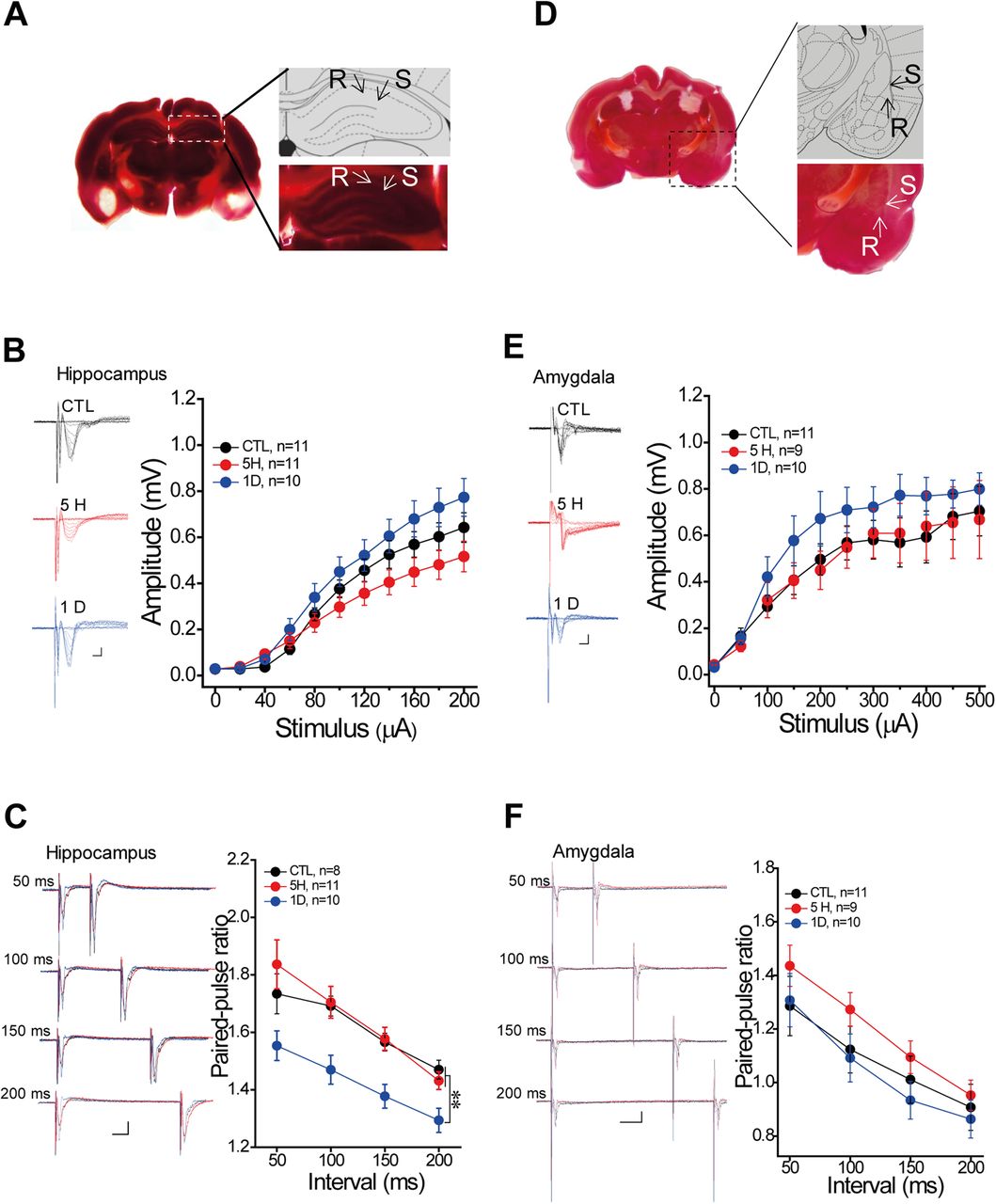
- Figure 1.
Photo-thrombosis induced focal ischemia in freely moving rats. A, Schematic and the representation of focal ischemia injury in unilateral hippocampus or amygdala by TTC staining with different time windows (from 1 h to 3 d), the red area was the normal tissue and the arrow showed the white ischemic area. B, Group data showed the developed ischemia injury after 473-nm laser illumination. C, Schaffer-CA1 pathway recording with focal ischemia injury: S = the stimulus site, R = the recording site. D, Representative traces and the group data of I/O curve, and (E) the traces (left) and group data of PPR from the recording in the Schaffer-CA1 pathway. F, Cortical-amygdala pathway recording with focal ischemia injury. G, The traces and group data of I/O curve, and (H) the traces (Left) and group data of PPR from the recording in cortical-amygdala pathway. Mean ± SEM for each bar. Statistics were performed using one-way ANOVA (B) and repeated ANOVA (D–H). Scale in traces: 2 ms, x-axis = 2 ms, y-axis = 0.2 mV; PPR: x-axis = 25 ms, y-axis = 0.2 mV; *p < 0.05, **p < 0.01, ***p < 0.001, compared with control groups, ##p < 0.01 used for the difference between the 5H and 1D. Each slice was 400 μm. Extended information illustrating the effects of conditions on ischemic induction is available in Extended Data Figure 1-1, the trace of TTC staining from a single rat across the injury site with 1-d ischemia is available in Extended Data Figure 1-2 and the Nissl and anti-GFAP staining at day 7 after focal ischemia in Extended Data Figure 1-3.
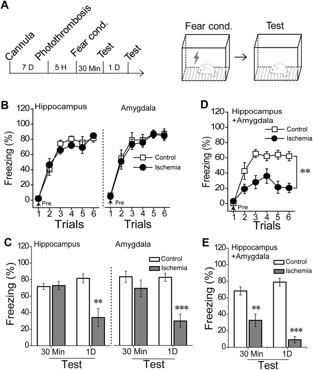
- Figure 2.
Fear memory formation with 5-h ischemia injury in hippocampus or/and amygdala. A, Rats received an ischemia induction 5 h before training, and the schematic of fear conditioning. B, Learning curve during fear conditioning with 5-h ischemic injury in the bilateral hippocampus (left, control, n = 11; ischemia, n = 11) or bilateral amygdala (right, control, n = 8; ischemia, n = 9) and (C) dependent contextual 30-min STM and 1-d LTM. D, The learning curve from the rats with 5-h ischemia in both hippocampus and amygdala before training and (E) the dependent contextual 30-min STM and 1-d LTM (control, n = 8; ischemia, n = 8). Mean ± SEM for each bar, statistics were performed using repeated measure ANOVA (B) and independent t test (C); *p < 0.05, **p < 0.01, ***p < 0.001, compared with control groups. Extended information illustrating the effects of Rose Bengal treatment on contextual fear formation is available in Extended Data Figure 2-1, the trace of TTC staining in a single rat with 1-d ischemia in both hippocampus and amygdala is available in Extended Data Figure 2-2, and the effects of 5-h ischemia in both the hippocampus and amygdala on pain threshold, general activity, and anxiety are available in Extended Data Figure 2-3, and the correlation between the injury size of the adjacent areas and the freezing time during the memory test in Extended Data Figure 2-4.
- Figure 3.
Fear memory acquisition in the rats with 1-d focal ischemia injury in the bilateral hippocampus or bilateral amygdala. A, Rats were conducted fear conditioning following by 1-d ischemia injury. B, Learning curve in the rats with 1-d ischemia injury in the bilateral hippocampus (left: control, n = 9; ischemia, n = 8) or bilateral amygdala (right: control, n = 9; ischemia, n = 9). C, Dependent 30-min STM test. Mean ± SEM for each bar. Statistics were performed using repeated measure ANOVA (B) or independent t test (C); ***p < 0.001, compared with control groups.
- Figure 4.
The basal transmission in the remote site after focal ischemia induction. A, Schaffer-CA1 recording with bilateral amygdala ischemia: S = the stimulus site, R = the recording site. B, Representative traces (left) and group data of I/O curve, and (C) the traces (left) and group data of PPR from hippocampal recording under the ischemia of the bilateral amygdala. D, Cortical-amygdala pathway with bilateral hippocampus ischemia. E, The traces (left) and group data of I/O curve, and (F) the traces (left) and group data of PPR from amygdalar recording under the ischemia of bilateral hippocampus (the left: trace). Control, control groups with surgery and Rose Bengal; 5 H, 5 h; 1 D, 1 d after photo-thrombosis. Mean ± SEM for each bar. Scale in trace: 2 ms, x-axis = 2 ms, y-axis = 0.2 mV; PPR: x-axis = 25 ms, y-axis = 0.2 mV. Statistics were performed using repeated measure ANOVA with post hoc test, **p < 0.01, compared with control groups. Extended information illustrating the interconnection between the hippocampus and amygdala using non-transsynaptic retrograde rabies virus tracing is available in Extended Data Figure 4-1.
- Figure 5.
The effects of focal ischemia on memory retrieval and relearning. A, Schematic: rats received fear conditioning and dependent 18-h memory test and then given photo-thrombosis 5 h before the 1-d test, and the rats received a relearning process 7 d later. B, C, 18-h and 1-d dependent contextual tests, rats were given the photo-thrombosis in the bilateral hippocampus (control, n = 8, ischemia, n = 9) or bilateral amygdala (control, n = 9, ischemia, n = 10) after 18-h test immediately. D, E, Relearning curve 7 d after photo-thrombosis and dependent 1-d contextual test: (left) rats with ischemia in the bilateral hippocampus (control, n = 8, ischemia, n = 8) and (right) rats with ischemia in the bilateral amygdala (control, n = 9, ischemia, n = 9). Repeated measure with post hoc test was used in the (D) and Student’s t test was in (B, C, E) to analyze the difference. Each bar represents mean ± SEM; ***p < 0.001, compared with control groups.


Extended Data
Extended Data Figure 1-1
Focal ischemia induction with different conditions in freely moving rats. A, Three conditions. Left, 30-min irradiation with 593-nm laser. Middle, 30-min irradiation with 565-nm LED. Right, 30-min irradiation with 473-nm laser. B, C, The TTC staining 1 d after irradiation in the hippocampus or amygdala. Each slice was 400 μm. Download Figure 1-1, TIF file.
Extended Data Figure 1-2
Brain sections with TTC staining from one animal showed the whole injury site with 1-d ischemia. A, One-day focal ischemia in the unilateral hippocampus. B, One-day focal ischemia in the unilateral amygdala. Each slice was 400 μm. Download Figure 1-2, TIF file.
Extended Data Figure 1-3
The sustained damage at day 7 after focal ischemia was represented by (A) 7-d Nissl staining and (B) the formation of glial scar indicated by anti-GFAP staining. Each slice was 50 μm. Download Figure 1-3, TIF file.
Extended Data Figure 2-1
The effects of Rose Bengal treatment on contextual fear formation. A, Schematic, rats were injected Rose Bengal (100 mg/kg, i.p.) 1 h before conditioning, and saline (10 ml/kg, i.p.) as control. B, Learning curve during conditioning (repeated measure ANOVA: F(1,8) = 0.08, p = 0.776). C, 30-min and 1-d dependent contextual tests (t test: 30 min, p = 0.156; 1 d, p = 0.725). Control, n = 5, Rose Bengal, n = 5. Mean ± SEM for each bar. Download Figure 2-1, TIF file.
Extended Data Figure 2-2
Brain sections with TTC staining from a single rat one with 1-d ischemia in both hippocampus and amygdala. Each slice was 400 μm. Download Figure 2-2, TIF file.
Extended Data Figure 2-3
The effects of 5-h ischemia in both the hippocampus and amygdala on other behaviors. A, Pain threshold test in fear conditioning box (control, n = 10 rats, ischemia, n = 9 rats; T = 2, p = 0.062, t test). B, Total time moving during EPM test (control, n = 10, ischemia, n = 9; T = 0.679, p = 0.502, t test). C, The percentage of time spend in open arms during EPM test (control, n = 10, ischemia, n = 9; T = 0.846, p = 0.409, t test). Mean ± SEM for each bar. Download Figure 2-3, TIF file.
Extended Data Figure 2-4
The effects of the injury size at adjacent areas on the freezing behaviors. A, The white dash line indicated the damage to the adjacent areas out of the region of interest (ROI). Each slice was 400 μm. There was no correlation between the injury size of adjacent areas and the freezing time during the (B) 30-min (r = 0.003, p = 0.45) and (C) 1-d (r = 0.22, p = 0.57) memory test (n = 4). D, E, One rat with focal ischemia at the out of the ROI (the arrow) showed the same fear acquisition ability to its literature control. Download Figure 2-4, TIF file.
Extended Data Figure 4-1
The retrograde tracing of rabies virus in hippocampus or amygdala. A, Schematic: a non-transsynaptic rabies virus carried the GFP (RV-dg-GFP) as the reporter was injected into the unilateral hippocampus and a rabies virus carried the Dsred (RV-dg-dsRed) as the reporter was injected into the unilateral amygdala. B, C, The dsRed expression in the local injection site of the amygdala. D, E, The dsRed-positive cells expressed in the hippocampus. F, The GFP expression in the local injection site of the hippocampus. G, the GFP-positive cells expressed in the amygdala. Amy, amygdala; Hip, hippocampus. Each slice was 40 μm, confocal microscope scanning. All scale bars were 100 μm. Download Figure 4-1, TIF file.
In this issue


