
Main menu

  • HOME
  • CONTENT
    • Early Release
    • Featured
    • Current Issue
    • Issue Archive
    • Blog
    • Collections
    • Podcast
  • TOPICS
    • Cognition and Behavior
    • Development
    • Disorders of the Nervous System
    • History, Teaching and Public Awareness
    • Integrative Systems
    • Neuronal Excitability
    • Novel Tools and Methods
    • Sensory and Motor Systems
  • ALERTS
  • FOR AUTHORS
  • ABOUT
    • Overview
    • Editorial Board
    • For the Media
    • Privacy Policy
    • Contact Us
    • Feedback
  • SUBMIT

User menu

Search

  • Advanced search
eNeuro
eNeuro

Advanced Search


  • HOME
  • CONTENT
    • Early Release
    • Featured
    • Current Issue
    • Issue Archive
    • Blog
    • Collections
    • Podcast
  • TOPICS
    • Cognition and Behavior
    • Development
    • Disorders of the Nervous System
    • History, Teaching and Public Awareness
    • Integrative Systems
    • Neuronal Excitability
    • Novel Tools and Methods
    • Sensory and Motor Systems
  • ALERTS
  • FOR AUTHORS
  • ABOUT
    • Overview
    • Editorial Board
    • For the Media
    • Privacy Policy
    • Contact Us
    • Feedback
  • SUBMIT
PreviousNext
Research ArticleResearch Article: New Research, Neuronal Excitability

Angiotensin-II Modulates GABAergic Neurotransmission in the Mouse Substantia Nigra

Maibam R. Singh, Jozsef Vigh and Gregory C. Amberg
eNeuro 26 March 2021, 8 (2) ENEURO.0090-21.2021; https://doi.org/10.1523/ENEURO.0090-21.2021
Maibam R. Singh
Department of Biomedical Sciences, Colorado State University, Fort Collins, CO 80523
  • Find this author on Google Scholar
  • Find this author on PubMed
  • Search for this author on this site
Jozsef Vigh
Department of Biomedical Sciences, Colorado State University, Fort Collins, CO 80523
  • Find this author on Google Scholar
  • Find this author on PubMed
  • Search for this author on this site
Gregory C. Amberg
Department of Biomedical Sciences, Colorado State University, Fort Collins, CO 80523
  • Find this author on Google Scholar
  • Find this author on PubMed
  • Search for this author on this site
  • Article
  • Figures & Data
  • Info & Metrics
  • eLetters
  • PDF
Loading

Article Figures & Data

Figures

  • Figure 1.
    • Download figure
    • Open in new tab
    • Download powerpoint
    Figure 1.

    Electrophysiological characteristics of GABAergic and dopaminergic neurons in the mouse substantia nigra. A, SNr GABAergic neurons have a narrower (<1.5 ms) AP width than SNc dopaminergic neurons (>3 ms). B, Representative sustained high-frequency firing of SNr GABAergic neurons recorded in whole-cell (top) and cell-attached configuration (bottom). C, SNr GABAergic neurons, in contrast to SNc dopaminergic neurons, show little to no Ih in response to a series of hyperpolarizing pre pulses (–70 to –140 mV). D, Representative low-frequency, slow, and irregular pacemaker-like firing in SNc dopaminergic neurons.

  • Figure 2.
    • Download figure
    • Open in new tab
    • Download powerpoint
    Figure 2.

    Ang-II decreases SNr GABAergic projection neuron spike firing. A, Evoked APs in response to 150-pA current injection. B, Individual scatter plot of evoked spike frequency in the SNr GABAergic projection neurons in response to 50-, 100-, and 150-pA current injections. Ang-II significantly decreased evoked firing in all stimulus levels combined, 50–150 pA (***p < 0.001, two-way repeated-measures ANOVA). In 10 out of 17 cells, washout partially reversed the effect of Ang-II. C, Ang-II increased the SNr GABAergic interspike interval (ISI; **p = 0.005, mixed effect analysis) and (D) increased the irregularity of firing as quantified by the ISI SD (*p = 0.01, mixed effect analysis). E, F, The AT1-R-specific blocker losartan (1 μm) abolished the suppression of evoked SNr GABAergic spike firing by Ang-II.

  • Figure 3.
    • Download figure
    • Open in new tab
    • Download powerpoint
    Figure 3.

    Ang-II slows the evoked AP kinetics of SNr GABAergic neurons. A, Representative AP waveforms of SNr projection neurons showing that Ang-II reversibly slows AP kinetics. B, Ang-II increased the AP half-width in 11 out of 17 SNr GABAergic neurons (*p = 0.010, mixed-effect analysis). C, Ang-II slowed the rise of APs in SNr GABAergic neurons (**p = 0.005, repeated measures one-way ANOVA). D, Ang-II slowed the decay of APs in SNr GABAergic neurons (**p = 0.004, repeated measure one-way ANOVA). Pooled data are a grouped data of all stimulus levels (50, 100, and 150 pA).
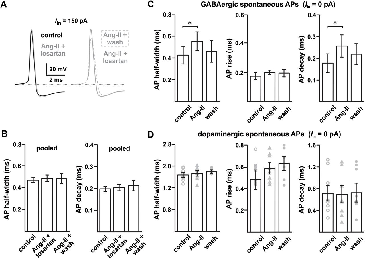
  • Figure 4.
    • Download figure
    • Open in new tab
    • Download powerpoint
    Figure 4.

    Disparate effect of Ang-II on SN GABAergic and dopaminergic neurons. A, Losartan blocks Ang-II-mediated increase in APD in SNr GABAergic neurons; losartan washout with continued Ang-II perfusion prolonged the APD. B, Summary data showing that losartan (1 μm) blocks Ang-II-mediated increase in AP half-width and decay. Data from all stimulus levels (50, 100, and 150 pA) were pooled for each group. C, Ang-II increased the duration of SNr GABAergic neuron spontaneous APs (*p = 0.04, AP half-width; *p = 0.015, AP decay; repeated measures one-way ANOVA). D, Unlike SNr GABAergic neurons, Ang-II had no noticeable effect on the AP kinetics of SNc dopaminergic neurons.
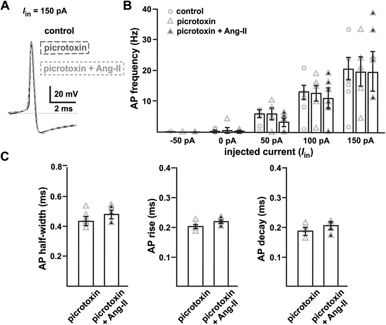
  • Figure 5.
    • Download figure
    • Open in new tab
    • Download powerpoint
    Figure 5.

    GABAAR blockade prevents Ang-II-mediated suppression of evoked spike firing in the SNr GABAergic projection neurons. A, Overlaid spike waveforms for control conditions, in the presence of the GABAAR antagonist picrotoxin (1 μm), and in the presence of picrotoxin plus Ang-II. B, Summary plot showing evoked spike firing of SNr GABAergic neurons under control conditions, in the presence of picrotoxin (1 μm), and in the presence of picrotoxin plus Ang-II. C, GABAAR blockade with picrotoxin attenuates Ang-II-mediated increases in SNr GABAergic neuron APDs.
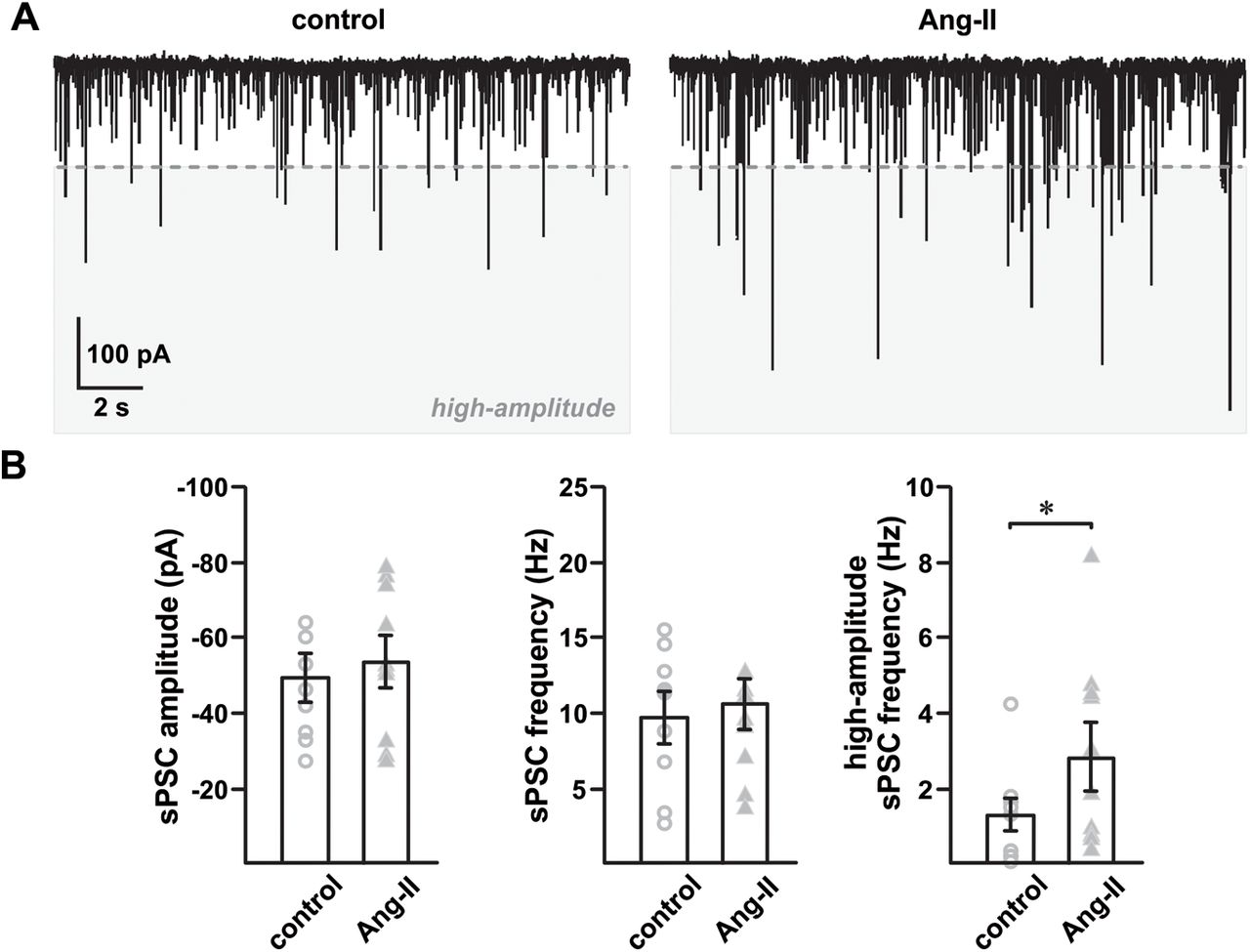
  • Figure 6.
    • Download figure
    • Open in new tab
    • Download powerpoint
    Figure 6.

    Ang-II increases SNr GABAergic neuron spontaneous PSCs. A, At a holding potential of –65 mV (junction potential corrected), spontaneous PSCs were recorded with a high chloride (57.5 nm) internal pipette solution before and after 5 min of Ang-II (500 nm) exposure. B, Ang-II did not markedly change the average PSC amplitude (p = 0.211, paired Student’s t test) and frequency (p = 0.466, paired Student’s t test) of spontaneous postsynaptic Cl– currents. However, Ang-II significantly increased the observed incidence of high-amplitude spontaneous PSCs (right; *p = 0.012; paired Student’s t test). High-amplitude spontaneous PSC, demarcated by the dashed line, were defined a priori as currents with an amplitude more than or equal to three times the mean current amplitude under control conditions.

  • Figure 7.
    • Download figure
    • Open in new tab
    • Download powerpoint
    Figure 7.

    Ang-II increases picrotoxin-sensitive spontaneous MIPSC in SNr GABAergic neurons. Spontaneous mIPSC recorded from SNr GABAergic and SNc dopaminergic neurons in the presence of excitatory synaptic blockers (DNQX, MK-801; 1 μm) and TTX (500 nm). A, Representative traces showing Ang-II-mediated enhancement of high-amplitude postsynaptic GABAAR currents. High-amplitude spontaneous PSCs, demarcated by the dashed line, were defined a priori as currents with an amplitude more than or equal to three times the mean current amplitude recorded in the presence of excitatory synaptic blockers and TTX. B, Summary data showing Ang-II-mediated ∼2.5-fold increase in high-amplitude mIPSC (*p = 0.019, one-way repeated measures ANOVA), but not in average mIPSC amplitude and frequency. C, In contrast to SNr GABAergic neurons, Ang-II had no detectable effect on spontaneous mIPSC in SNc dopaminergic neurons.

  • Figure 8.
    • Download figure
    • Open in new tab
    • Download powerpoint
    Figure 8.

    Ang-II modulates SN GABAergic neurotransmission. A, Light-evoked EPSP (bursts) in SNr GABAergic neurons in response to a 100-ms-long light pulse. B, Ang-II decreased light-evoked intraburst spikes in four out of six SNr GABAergic neurons (*p = 0.024, paired Student’s t test); intraburst spikes shown are enlarged from panel A. C, Summary data showing the effect of Ang-II on intraburst spikes (EPSP) in SNr GABAergic neurons in response to 100-ms-long light pulse. In four out of six SNr GABAergic neurons, Ang-II decreased intraburst spikes by ∼25%. D, IPSP recorded from SNc dopaminergic neurons in response to photostimulation of SNr GABAergic neurons, as shown by downward deflection of membrane potential (blue stars). Ang-II enhanced light-evoked inhibitory input onto SNc dopaminergic neurons as shown by an increase in the amplitude of light-evoked IPSP (E) and IPSC (F, G; *p = 0.049, paired Student’s t test).

Back to top

In this issue

eneuro: 8 (2)
eNeuro
Vol. 8, Issue 2
March/April 2021
  • Table of Contents
  • Index by author
  • Ed Board (PDF)
Email

Thank you for sharing this eNeuro article.

NOTE: We request your email address only to inform the recipient that it was you who recommended this article, and that it is not junk mail. We do not retain these email addresses.

Enter multiple addresses on separate lines or separate them with commas.
Angiotensin-II Modulates GABAergic Neurotransmission in the Mouse Substantia Nigra
(Your Name) has forwarded a page to you from eNeuro
(Your Name) thought you would be interested in this article in eNeuro.
CAPTCHA
This question is for testing whether or not you are a human visitor and to prevent automated spam submissions.
Print
View Full Page PDF
Citation Tools
Angiotensin-II Modulates GABAergic Neurotransmission in the Mouse Substantia Nigra
Maibam R. Singh, Jozsef Vigh, Gregory C. Amberg
eNeuro 26 March 2021, 8 (2) ENEURO.0090-21.2021; DOI: 10.1523/ENEURO.0090-21.2021

Citation Manager Formats

  • BibTeX
  • Bookends
  • EasyBib
  • EndNote (tagged)
  • EndNote 8 (xml)
  • Medlars
  • Mendeley
  • Papers
  • RefWorks Tagged
  • Ref Manager
  • RIS
  • Zotero
Respond to this article
Share
Angiotensin-II Modulates GABAergic Neurotransmission in the Mouse Substantia Nigra
Maibam R. Singh, Jozsef Vigh, Gregory C. Amberg
eNeuro 26 March 2021, 8 (2) ENEURO.0090-21.2021; DOI: 10.1523/ENEURO.0090-21.2021
Twitter logo Facebook logo Mendeley logo
  • Tweet Widget
  • Facebook Like
  • Google Plus One

Jump to section

  • Article
    • Abstract
    • Significance Statement
    • Introduction
    • Materials and Methods
    • Results
    • Discussion
    • Acknowledgments
    • Footnotes
    • References
    • Synthesis
  • Figures & Data
  • Info & Metrics
  • eLetters
  • PDF

Keywords

  • angiotensin II
  • brain RAS
  • dopaminergic
  • GABAergic
  • neurotransmission
  • substantia nigra

Responses to this article

Respond to this article

Jump to comment:

No eLetters have been published for this article.

Related Articles

Cited By...

More in this TOC Section

Research Article: New Research

  • Novel roles for the GPI-anchor cleaving enzyme, GDE2, in hippocampal synaptic morphology and function
  • Upright posture: a singular condition stabilizing sensorimotor coordination
  • EEG Signatures of Auditory Distraction: Neural Responses to Spectral Novelty in Real-World Soundscapes
Show more Research Article: New Research

Neuronal Excitability

  • Novel roles for the GPI-anchor cleaving enzyme, GDE2, in hippocampal synaptic morphology and function
  • Tolerance in Thalamic Paraventricular Nucleus Neurons Following Chronic Treatment of Animals with Morphine
  • Investigating Mechanically Activated Currents from Trigeminal Neurons of Nonhuman Primates
Show more Neuronal Excitability

Subjects

  • Neuronal Excitability
  • Home
  • Alerts
  • Follow SFN on BlueSky
  • Visit Society for Neuroscience on Facebook
  • Follow Society for Neuroscience on Twitter
  • Follow Society for Neuroscience on LinkedIn
  • Visit Society for Neuroscience on Youtube
  • Follow our RSS feeds

Content

  • Early Release
  • Current Issue
  • Latest Articles
  • Issue Archive
  • Blog
  • Browse by Topic

Information

  • For Authors
  • For the Media

About

  • About the Journal
  • Editorial Board
  • Privacy Notice
  • Contact
  • Feedback
(eNeuro logo)
(SfN logo)

Copyright © 2025 by the Society for Neuroscience.
eNeuro eISSN: 2373-2822

The ideas and opinions expressed in eNeuro do not necessarily reflect those of SfN or the eNeuro Editorial Board. Publication of an advertisement or other product mention in eNeuro should not be construed as an endorsement of the manufacturer’s claims. SfN does not assume any responsibility for any injury and/or damage to persons or property arising from or related to any use of any material contained in eNeuro.