

Article Figures & Data
Figures
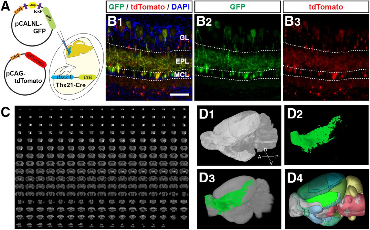
- Figure 1.
Strategy to analyze the axonal projection patterns of OB projection neurons. A, Schematic diagram of in utero electroporation. Plasmid mixture was injected into the lateral ventricle of the mice embryos, and the negative current was applied from posterior to anterior to electroporate the cells in the presumptive OB. B, Medial region of a coronal section of P7 Tbx21-Cre OB electroporated with pCALNL-GFP and pCAG-tdTomato, at E11. OB projection neurons, mitral and tufted cells, express both GFP (green) and tdTomato (red) while tdTomato+ interneurons are negative for GFP. All nuclei were stained with DAPI (blue). Scale bar: 100 μm. C, 270 serial section images acquired in STPT. D, 3D reconstruction from the SPTP imaging (D1), axonal projection signal (D2), registered axonal signals in Allen CCF reference brain (D3), and anatomic labels in the reference brain (D4).
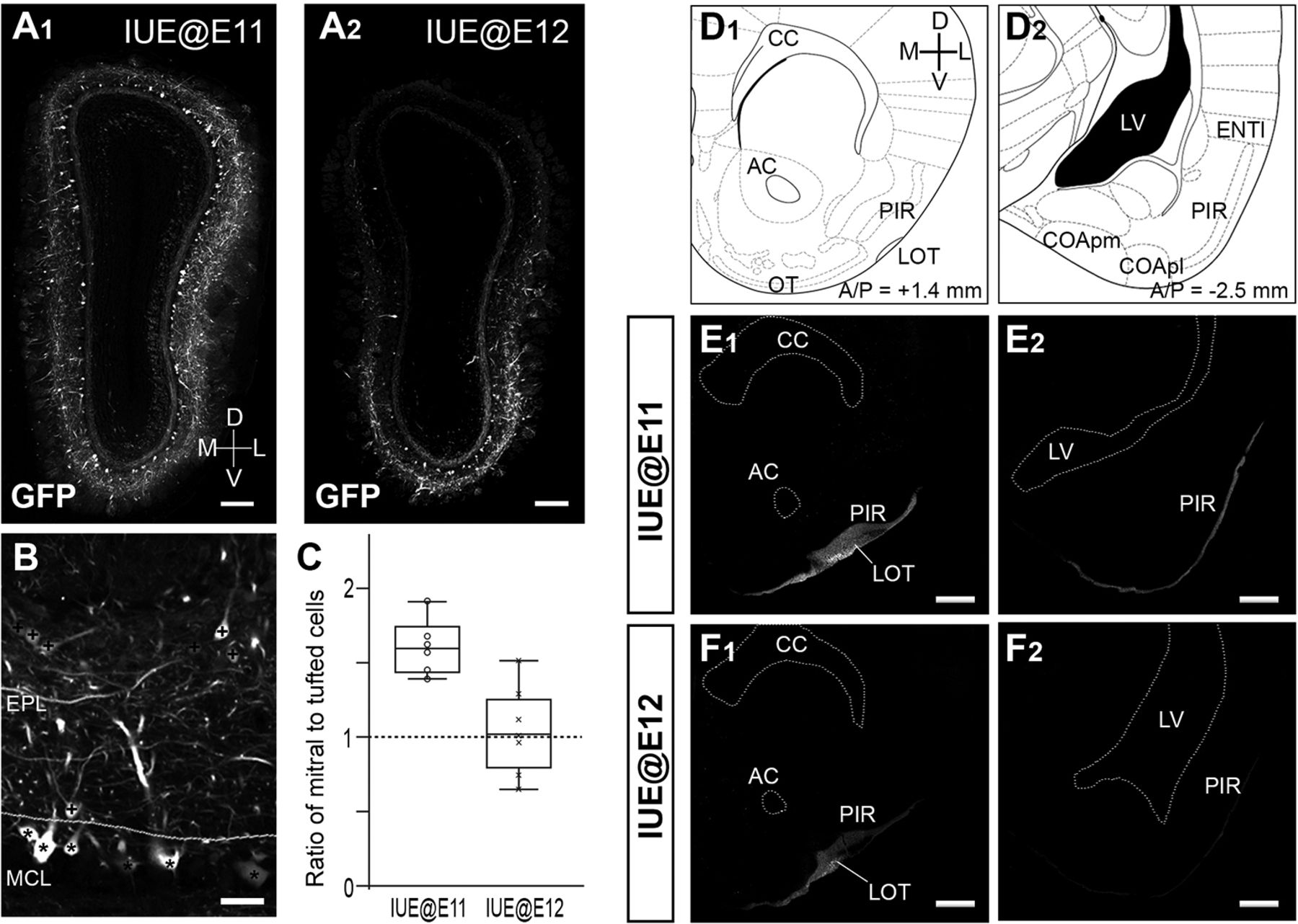
- Figure 2.
Labeling of different subpopulations of OB projection neurons using in utero electroporation. A, Coronal sections of the OBs from adult mice in which electroporations were performed at E11 (A1) or E12 (A2). GFP is expressed only in mitral and tufted cells. IUE@E12 preferentially labeled mitral cells in the ventrolateral part of the OB. B, Quantification of mitral and tufted cells in the OB. Cells that have GFP+ somata in the MCL and EPL were defined as mitral cells (marked with asterisks) and tufted cells (marked with plus signs), respectively. C, Ratios of mitral cells to tufted cells calculated from IUE@E11 (n = 5) and IUE@E12 (n = 7) OBs are shown with box plots. D–F, Projection of GFP+ axons to the anterior (D1, E1, F1) and posterior (D2, E2, F2) part of the olfactory cortex in the IUE@E11 (E) and IUE@E12 brain (F). Reference brain regions observed in E, F are cited from a mouse brain atlas (Paxinos and Franklin, 2001). GFP+ axons are seen in the anterior PIR of both IUE@E11 (E1) and IUE@E12 (F1) brains, whereas only the IUE@E11 brain has a significant GFP signal in the posterior PIR (E2, F2). Scale bars: 200 μm (A), 50 μm (B), and 500 μm (E, F). EPL: external plexiform layer; MCL: mitral cell layer; CC: corpus callosum; AC: anterior commissure; LOT: lateral olfactory tract; PIR: piriform cortex; OT: olfactory tubercle; LV: lateral ventricle; COApl and COApm: posterolateral and posteromedial cortical amygdala; ENTl: lateral entorhinal cortex.
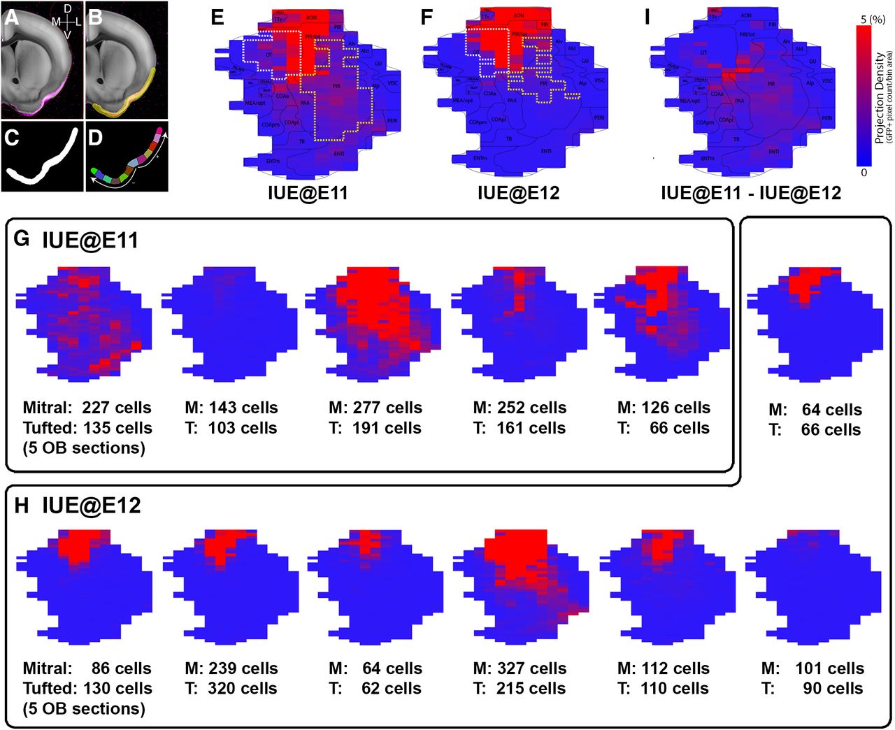
- Figure 3.
Brain-wide axonal projection pattern from OB neurons with different birthdates. A, B, Axonal projection signals from IUE at E11 (A) and IUE at E12 (B) registered on the reference brain. GFP signals were pseudo-colored as red in B to facilitate a comparison between signals from two different birth dates. Bregma anterior/posterior (A/P) coordinates were included. C, 3D rendering of axonal projection from IUE at E11 (C1), E12 (C2), and merged (C3) in the reference brain. Late-generated OB projection neurons labeled with IUE@E12 do not project their axons to the posterior regions of the olfactory cortex.
- Figure 4.
Topographical axonal projection pattern on 2D flatmap. A–D, Creation of 2D flatmap. Axonal projection signal in the reference brain (A) and binary mask to cover areas with projection signal (B), binary mask (C), and evenly spaced bins (D) to create the flatmap (for details, see Materials and Methods). E, F, Averaged axonal projection signal in heatmap from IUE at E11 (E) and E12 (F). Bins that show >5% of GFP+ signals (projection density) in the OT and PIR are encircled with white and yellow dashed lines, respectively. G, H, The 2D flatmaps constructed from five IUE@E11 (G) and seven IUE@E12 (H) individual mouse brains are shown. The numbers of mitral and tufted cells counted from five OB sections are listed under the maps. Dense GFP signals are observed throughout the majority of the olfactory cortex of IUE@E11 brains while only the anterior regions of IUE@E12 brains show dense GFP signals regardless of the numbers of labeled mitral and tufted cells. I, The 2D flatmap in which the averaged IUE@E12 projection (F) was subtracted from the averaged IUE@E11 projection (G) to highlight the difference between two groups.


In this issue


