

Article Figures & Data
Figures
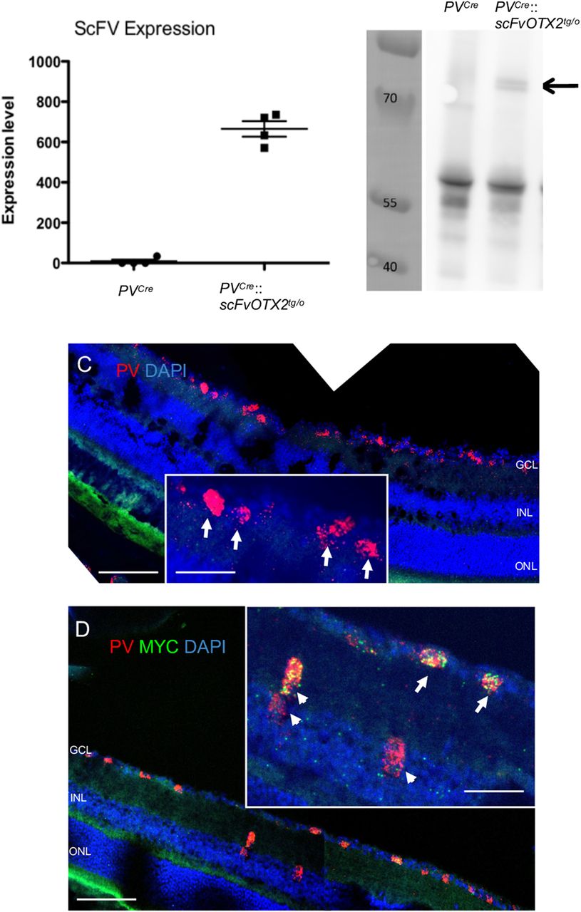
- Figure 1.
Expression of OTX2-scFV in P30 mouse retinal tissue. A, qRT-PCR for OTX2scFV mRNA was conducted on extracts from PVCre and PVCre::scFvOTX2tg/o mice and raw ct values normalized and converted to arbitrary units. B, Retinal lysates from PVCre and PVCre::scFvOTX2tg/o mice were immunoprecipitated for GFP followed by Western blotting with an anti-MYC antibody. Molecular weight markers are in the left lane in kDa. The middle lane contains the lysate from PVCre mice. Right lane contains lysate from PVCre::scFvOTX2tg/o mice. The arrow indicates the presence of the bands at the expected migration position of the full-length OTX2scFv-GFP protein. C, In situ hybridization using RNAscope technology of PVCre retina shows PV expression (red) in the inner retina. DAPI is in blue. Inset arrows show details of PV-expressing cells in the GCL. D, In situ hybridization using RNAscope technology of PVCre::scFvOTX2tg/o retina. mRNA for PV is red, and mRNA for the myc tag of the OTX2scfv is green. Arrows indicate scFv-expressing PV cells in the GCL (i.e., displaced amacrines and/or RGCs); arrowheads indicate scFv-expressing PV cells in the innermost part of the INL in the place of amacrine cells. Scale bars: low-magnification images, 100 μm; insets, 50 μm. Values: mean ± sem.
- Figure 2.
Optomotor test monitoring visual acuity. Thirty-day-old mice were subjected to the optomotor test using an 100% contrast optotype of 0.375c/deg. PVCre and PVCre::scFvPax6tg/o mice made an average of about five head turns during the 2 min test period. PVCre::scFvOTX2tg/o mice made significantly fewer head turns revealing reduced visual acuity. **p < 0.01 or ***p < 0.001, Mann–Whitney U test, two tailed. N = 25 for PVCre, N = 12 for PVCre::scFvPax6tg/o, and N = 13 for PVCre::scFvOTX2tg/o. Values: mean ± sem.
- Figure 3.
Expression of OTX2scFv does not alter retinal organization in the P30 mouse. Top, All retinal layers organization and approximate sizes are similar in retina from mice expressing OTX2scFv and their PVCre littermates. Bottom, The number of cells in the GCL, the INL thickness, and the ONL thickness are similar between the two genotypes. Scale bar, 50 μm. N = 8 for PVCre mice and N = 6 for PVCre::scFvOTX2tg/o mice. Values: mean ± sem.
- Figure 4.
Expression of retinal cell type-specific genes in P30 mice of the two genotypes. Genes specific of RGCs, bipolar cells, amacrine cells, rods, and cones show similar expression levels in the two genotypes. Lim1 mRNA expression in horizontal cells is significantly increased in the OTX2-scFv-expressing mice. *p < 0.05, Mann–Whitney U test, two tailed. N = 4 for each genotype. Values: mean ± sem.
- Figure 5.
ERG of P30 mice expressing OTX2scFV and their PVCre littermates. A, B, Representative traces under scotopic conditions of the right eye at 3.19 cd/m2. C–F, Scotopic ERG parameters plotted against log intensity. Two-way ANOVA for repeated measures showed no significant differences based on genotype or genotype by intensity interaction (see text). N = 7 for PVCre mice and N = 9 for PVCre::scFvOTX2tg/o mice. Values: mean ± sd.
- Figure 6.
The b-wave amplitude in photopic lighting in the two genotypes of 1-month-old mice. N = 7 for PVCre mice and N = 9 for PVCre::scFvOTX2tg/o mice. Values: mean ± sem.
- Figure 7.
Inner retinal function. A, Extracted oscillatory potential traces of a PVCre mouse (left) and PVCre::scFvOTX2tg/o mouse (right). B, The amplitude of an early OP (OP2) is not altered in the PVCre::scFvOTX2tg/o mice, while the amplitude of a late OP (OP4) is significantly reduced by ∼50%. C, Representative traces of 20 Hz flickers of the left eye of mice of the two genotypes. D, The amplitude of the flicker response to 10 Hz stimulation after light adaptation is slightly increased, and at 20 Hz the response is significantly doubled in amplitude. Bottom, There was no difference in the implicit time of the flicker responses between mice expressing OTX2scFv and their PVCre littermates. ***p < 0.005, Mann–Whitney U test, two tailed. N = 7 for PVCre and N = 9 for PVCre::scFvOTX2tg/o mice. Values mean ± sem.


Tables
Data structure Type of test Power Figure 2
PVcre vs PVCre::scFvOTX2tg/o
Not considered normal due to unequal NMann–Whitney U 0.92 Figure 4
PVcre vs PVCre::scFvOTX2tg/o
Data normalizedMann–Whitney U 0.78 Figure 5
PVcre vs PVCre::scFvOTX2tg/o
a-wave latency, F (1,14) = 0.011
a-wave amplitude, F (1,14) = 0.100
b-wave latency, F (1,14) = 3.826
b-wave amplitude, F (1,14) = 0.77
Genotype × stimulus intensity interaction
a-wave latency, F (3,42) = 0.660
a-wave amplitude, F (3,42) = 0.913
b-wave latency, F (3,41) = 1.915
b-wave amplitude, F (3,42) = 0.139,Two-way ANOVA with repeated measures n.s.
n.s.
n.s.
n.s.
n.s.
n.s.
n.s.
n.s.Figure 7 OPs
PVcre vs PVCre::scFvOTX2tg/o
Dissimilar variance
Figure 7D Flickers
PVcre vs PVCre::scFvOTX2tg/o
Dissimilar varianceMann–Whitney U
Mann–Whitney U0.97
0.96
In this issue


