

Article Figures & Data
Figures
- Figure 1.
Illustration of experimental design and the proposed FOV alignment approach. A, In vivo set up for two-photon imaging data collection. B, Experimental procedure. The GCaMP6f was injected into the RFA and CFA of the layer 2/3 motor cortex. Two weeks later, a cranial window surgery was conducted above the RFA and CFA. Behavioral training and two-photon imaging recording began two weeks after the window surgery. The mouse received one session per day and 17 sessions in total. C, Geometric interpretation of the affine decomposition. λ and ψ are the zoom factor and the rotation angle of the camera around the optical axis respectively. ϕ and θ corresponds to the longitude and latitude angles of the optical axis. u0 represents the frontal view of the flat object. D, Generic phases of the ASIFT method. Image1 and Image2 were individually transformed by simulating a large set of affine distortions caused by the change of longitude ϕ and latitude θ. Then, SIFT was used to detect and describe the keypoints on every simulated image. NNDR was used to match the keypoints. RANSAC was used to exclude outliers from initial matches. The remaining inliers were used to estimate the transformation matrix. SIFT was replaced by SURF, AKAZE, BRISK, and ORB to achieve ASURF, AAKAZE, ABRISK, and AORB. E, Outline of the FOV alignment procedure. TurboReg was used to process within-session motion artifacts. The motion-corrected imaging session was averaged and normalized to get the corresponding FOV image. The FOV image of the first session was used as the template, and FOV images of all other sessions were aligned to it. The alignment was achieved by fully affine invariant methods (ASIFT, ASURF, AAKAZE, ABRISK, AORB), the feature-based methods (SIFT, SURF, AKAZE, BRISK, ORB), the conventional methods (LK, ECC, MOCO, TurboReg, NoRMCorre), and the CLAHE-based conventional methods (LK-CLAHE, ECC-CLAHE, MOCO-CLAHE, TurboReg-CLAHE, NoRMCorre-CLAHE).
- Figure 2.
Basic information of the collected FOV images. A, The FOV image of each session from RFA, labeled as A5. B, The ROIs mask of each session from A5. C, The FOV image of each session from CFA, labeled as A6. D, The ROIs mask of each session from A6. The yellow circles represent the detected neurons in each FOV image. The yellow circles filled with black color represent the neurons common to each
- Figure 3.
Comparison of performance between the fully affine invariant group and the feature-based group. The correlation between
- Figure 4.
Comparison of performance between the fully affine invariant group and the conventional group. The correlation between
- Figure 5.
Comparison of performance between the fully affine invariant group and the CLAHE-based conventional group. A, The CLAHE adjusted FOV image of each session from A5 (upper row) and A6 (lower row). CLAHE, contrast limited adaptive histogram equalization. The correlation between
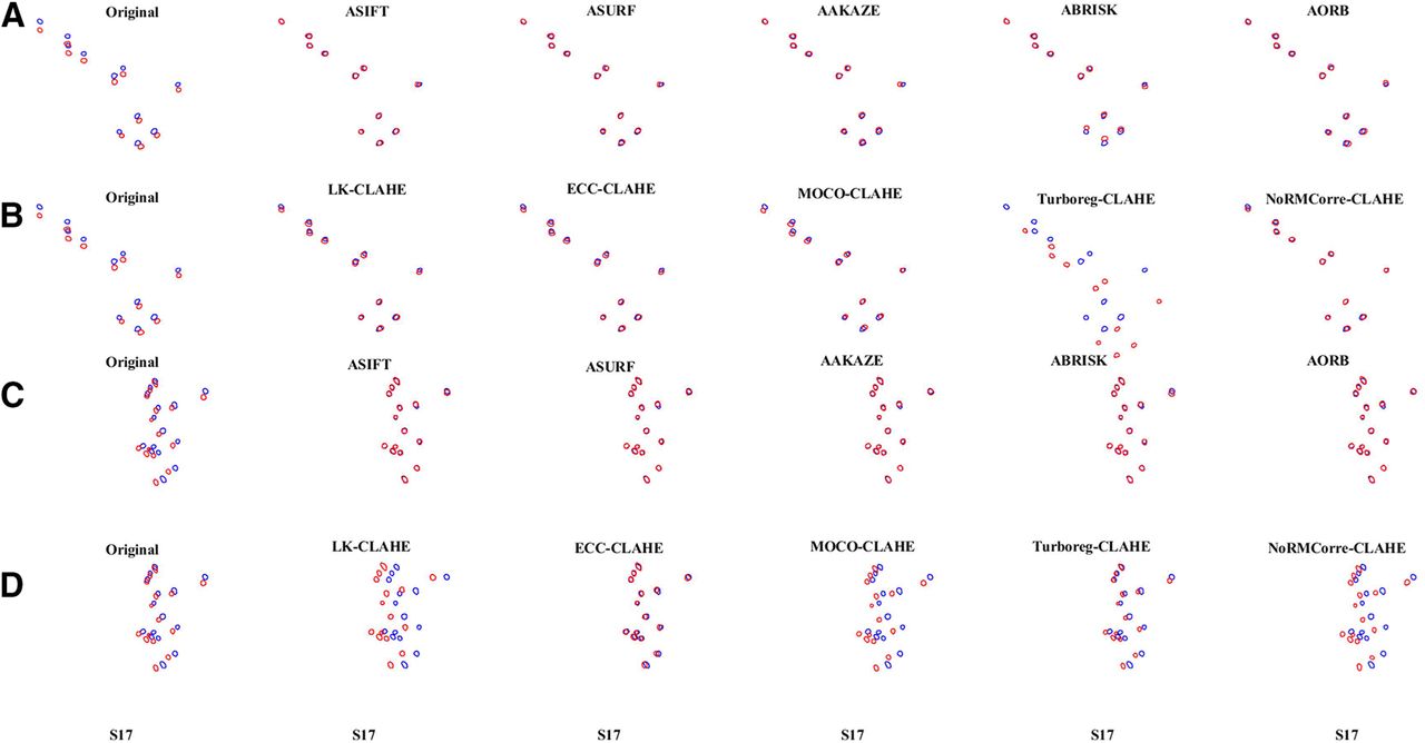
- Figure 6.
Visualization of the overlay of the ROIs mask-pairs on S17 of A5 and A6. The overlay of the
- Figure 7.
The mean ± SEM of the correlations on all registered ROIs mask-pairs


Tables
- Table 1.
Quantitative comparison between the fully affine invariant group and the feature-based group with respect to area A5
Methods Features detectedin image pair Inliers Matchedfeatures Inlierratio Methods Features detectedin image pair Inliers Matchedfeatures Inlierratio Image Template(S06) Image Template(S06) Image pair #1 of A5 (S08, S06) ASIFT 10636 9524 95 241 0.39 SIFT 629 565 10 17 0.59 ASURF 20012 18698 131 427 0.30 SURF 1258 1174 17 42 0.40 AAKAZE 3120 2893 55 89 0.62 AKAZE 236 209 11 18 0.61 ABRISK 8677 7667 32 57 0.56 BRISK 918 894 10 17 0.59 AORB 13927 13427 31 100 0.31 ORB 500 500 9 11 0.82 Image pair #2 of A5 (S09, S06) ASIFT 11357 9524 82 195 0.42 SIFT 690 565 12 22 0.55 ASURF 20398 18698 98 397 0.25 SURF 1346 1174 17 36 0.47 AAKAZE 3267 2893 47 91 0.52 AKAZE 269 209 10 16 0.63 ABRISK 9830 7667 50 80 0.63 BRISK 1169 894 15 26 0.58 AORB 14111 13427 43 96 0.45 ORB 500 500 9 17 0.53 Image pair #3 of A5 (S10, S06) ASIFT 12935 9524 41 174 0.23 SIFT 771 565 10 23 0.43 ASURF 22158 18698 49 408 0.12 SURF 1466 1174 7 45 0.16 AAKAZE 3697 2893 29 67 0.43 AKAZE 270 209 8 15 0.53 ABRISK 10848 7667 21 41 0.51 BRISK 1260 894 10 15 0.67 AORB 14387 13427 18 103 0.17 ORB 500 500 4 8 0.50 Image pair #4 of A5 (S11, S06) ASIFT 13833 9524 61 163 0.37 SIFT 850 565 9 26 0.35 ASURF 22345 18698 83 407 0.20 SURF 1446 1174 8 32 0.25 AAKAZE 3753 2893 30 76 0.39 AKAZE 291 209 7 10 0.70 ABRISK 11149 7667 18 44 0.41 BRISK 1243 894 11 18 0.61 AORB 14514 13427 20 105 0.19 ORB 500 500 4 5 0.80 Image pair #5 of A5 (S12, S06) ASIFT 10106 9524 71 176 0.40 SIFT 588 565 13 20 0.65 ASURF 20415 18698 71 404 0.18 SURF 1293 1174 7 35 0.20 AAKAZE 3239 2893 47 90 0.52 AKAZE 240 209 15 19 0.79 ABRISK 8477 7667 29 50 0.58 BRISK 874 894 6 9 0.67 AORB 14062 13427 56 148 0.38 ORB 500 500 13 25 0.52 Image pair #6 of A5 (S16, S06) ASIFT 9751 9524 17 114 0.15 SIFT 613 565 4 9 0.44 ASURF 20095 18698 28 296 0.095 SURF 1275 1174 6 24 0.25 AAKAZE 3269 2893 12 39 0.31 AKAZE 240 209 5 15 0.33 ABRISK 7416 7667 13 40 0.33 BRISK 811 894 4 8 0.50 AORB 13690 13427 16 97 0.16 ORB 500 500 8 24 0.33 Image pair #7 of A5 (S17, S06) ASIFT 3945 9524 20 69 0.29 SIFT 202 565 4 5 0.80 ASURF 12914 18698 20 242 0.08 SURF 649 1174 6 27 0.22 AAKAZE 1802 2893 13 33 0.39 AKAZE 128 209 5 8 0.63 ABRISK 3092 7667 12 26 0.46 BRISK 201 894 4 4 1.00 AORB 9454 13427 13 85 0.15 ORB 498 500 7 14 0.50 Mean values for all image pairs ASIFT 10366.14 9524 55.29 161.71 0.34 SIFT 620.43 565 8.86 17.43 0.51 ASURF 19762.43 18698 68.57 368.71 0.19 SURF 1247.57 1174 9.71 34.43 0.28 AAKAZE 3163.86 2893 33.28 69.29 0.48 AKAZE 239.14 209 8.71 14.43 0.60 ABRISK 8498.43 7667 25 48.26 0.52 BRISK 925.14 894 8.57 13.86 0.62 AORB 13449.29 13427 28.14 104.86 0.27 ORB 499.71 500 7.71 14.85 0.52 - Table 2.
Quantitative comparison between the fully affine invariant group and the feature-based group with respect to area A6
Methods Features detectedin image pair Inliers Matchedfeatures Inlierratio Methods Features detectedin image pair Inliers Matchedfeatures Inlierratio Image Template(S06) Image Template(S06) Image pair #1 of A6 (S08, S06) ASIFT 6485 6184 1236 1412 0.88 SIFT 431 444 117 143 0.82 ASURF 13192 12398 1705 2351 0.73 SURF 832 843 123 162 0.76 AAKAZE 2864 2586 840 1179 0.71 AKAZE 208 191 95 108 0.88 ABRISK 5221 5164 777 880 0.88 BRISK 417 432 86 93 0.92 AORB 13177 12917 1467 1858 0.79 ORB 500 500 101 127 0.80 Image pair #2 of A6 (S09, S06) ASIFT 6611 6184 191 365 0.52 SIFT 477 444 30 44 0.68 ASURF 13411 12398 322 734 0.44 SURF 886 843 30 59 0.51 AAKAZE 2530 2586 351 490 0.72 AKAZE 219 191 37 56 0.66 ABRISK 4636 5164 203 296 0.69 BRISK 419 432 30 43 0.70 AORB 12477 12917 335 555 0.60 ORB 500 500 32 52 0.62 Image pair #3 of A6 (S10, S06) ASIFT 7681 6184 469 602 0.78 SIFT 521 444 53 66 0.80 ASURF 12154 12398 554 880 0.63 SURF 844 843 62 86 0.72 AAKAZE 2837 2586 422 546 0.77 AKAZE 263 191 48 61 0.79 ABRISK 6476 5164 459 548 0.84 BRISK 643 432 70 74 0.95 AORB 13486 12917 784 1027 0.76 ORB 500 500 61 75 0.81 Image pair #4 of A6 (S11, S06) ASIFT 8556 6184 132 273 0.48 SIFT 581 444 30 40 0.75 ASURF 13982 12398 195 502 0.39 SURF 931 843 35 53 0.66 AAKAZE 3033 2586 200 299 0.67 AKAZE 271 191 31 47 0.66 ABRISK 7262 5164 159 239 0.67 BRISK 720 432 32 42 0.76 AORB 14158 12917 255 396 0.64 ORB 500 500 26 41 0.63 Image pair #5 of A6 (S12, S06) ASIFT 9027 6184 426 624 0.68 SIFT 651 444 58 79 0.73 ASURF 13988 12398 448 772 0.58 SURF 935 843 57 85 0.67 AAKAZE 3031 2586 278 403 0.69 AKAZE 251 191 52 66 0.79 ABRISK 7478 5164 365 452 0.81 BRISK 662 432 50 59 0.85 AORB 13889 12917 553 750 0.74 ORB 500 500 62 77 0.81 Image pair #6 of A6 (S16 S06) ASIFT 6248 6184 392 532 0.74 SIFT 388 444 49 56 0.88 ASURF 12517 12398 399 681 0.59 SURF 782 843 53 75 0.71 AAKAZE 2424 2586 338 359 0.94 AKAZE 197 191 47 56 0.84 ABRISK 4596 5164 240 271 0.89 BRISK 371 432 41 53 0.77 AORB 12004 12917 447 556 0.80 ORB 500 500 49 57 0.86 Image pair #7 of A6 (S17, S06) ASIFT 4343 6184 80 140 0.57 SIFT 279 444 17 22 0.77 ASURF 11170 12398 82 291 0.28 SURF 704 843 13 30 0.43 AAKAZE 1885 2586 62 92 0.67 AKAZE 140 191 16 19 0.84 ABRISK 3449 5164 43 57 0.75 BRISK 254 432 19 20 0.95 AORB 10545 12917 76 143 0.53 ORB 500 500 21 34 0.62 Mean values for all image pairs ASIFT 6993 6184 418 564 0.74 SIFT 475.43 444 50.57 64.29 0.79 ASURF 12916.29 12398 529.29 887.29 0.60 SURF 844.86 843 53.29 78.57 0.68 AAKAZE 2657.71 2586 355.86 481.14 0.74 AKAZE 221.29 191 46.57 59 0.79 ABRISK 5588.29 5164 320.86 391.86 0.82 BRISK 498 432 46.86 54.86 0.85 AORB 12819.42 12917 559.57 755 0.74 ORB 500 500 50.29 66.14 0.76
Extended Data.
A zip file (named “data_code.zip”), including PyPI package (“FAIM_package” folder), example FOV images (within “examples” folder), and codes used to reproduce all results (within “AffineCa2p_reproduce_results” folder) were submitted as Extended Data. Each folder contains a readme file. Download Extended Data, ZIP file.
In this issue


