

Article Figures & Data
Figures
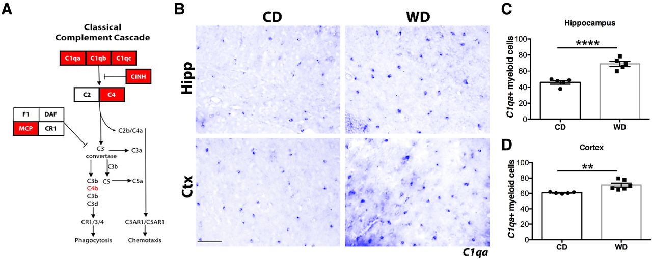
- Figure 1.
Complement components are increased in brains of WD-fed obese mice. A, Expression of complement proteins upregulated in brains of WD-fed mice (red boxes). B–D, Increased number of C1qa+ cells in the hippocampus and cortex of WD-fed mice. Data are presented as mean ± SEM, n ≥ 5, **p < 0.01 and ****p < 0.001 by paired t test. Scale bars: 50 μm.
- Figure 2.
C1QA and C3 deposits increase in white matter tracts of WD-fed mice. A, Increased C1QA immunoreactivity in the corpus callosum of WD-fed compared with CD-fed mice colocalized with IBA1+ cells and MBP+ myelin. B, Increased C3 immunoreactivity in the corpus callosum of WD-fed compared with CD-fed mice colocalized with MBP+ myelin. White dotted lines indicate the boundary between the CA1 region of the hippocampus and the corpus callosum. Scale bars: 40 μm.
- Figure 3.
WD-induced obesity and metabolic profiles were not altered by C1QA deficiency. A–D, Weight and adiposity increased in WD-fed mice independent of genotype and sex. E–H, No differences were detected in total cholesterol (E, G) and triglycerides (F, H) between WD-fed WT and C1qa KO mice. I–L, LDL and HDL plasma levels in male and female WD-fed mice were not changed by C1QA deficiency. M, N, Fatty acids plasma levels were not altered by C1QA deficiency. Data are presented as mean ± SEM, n ≥ 5, *p < 0.05, **p< 0.01, ***p< 0.001, ****p < 0.0001 by one-way ANOVA with Tukey’s post hoc test. n.s., not significant.
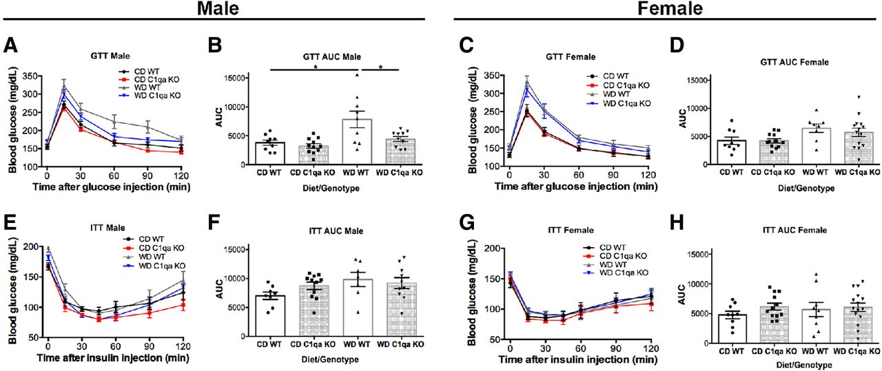
- Figure 4.
C1QA deficiency did not alter glucose or insulin tolerance in obese mice. A–D, No significant changes were detected in glucose tolerance between genotypes and sex. Only WD-fed male mice showed a moderate impaired glucose tolerance that was prevented by C1QA deficiency (B). E–H, No changes were observed in the ITT between groups. Data are presented as mean ± SEM, n ≥ 5, *p < 0.05 by one-way ANOVA with Tukey’s post hoc test.
- Figure 5.
C1QA deficiency prevented WD-induced cognitive deficits in male mice. A, B, WD-fed male mice showed a significant impairment in % correct alternation (A), which was not due to reductions in total activity as measured by total arm entries (B). However, C1qa KO WD-fed male mice showed no significant impairment in % correct alternations when compared with CD-fed male mice. C, D, No differences were observed in % correct alternations between CD-fed and WD-fed female mice. E, G, Activity levels in the open field as measured by cumulative distance traveled revealed no differences between groups in both male and female mice. F, H, Grip strength measurements were not different between groups in both male and female mice. Data are presented as mean ± SEM, n ≥ 8, *p < 0.05 by one-way ANOVA with Tukey’s post hoc test.
- Figure 6.
C1QA deficiency prevented WD-diet induced cerebrovascular damage. A–C, WD-induced decrease in PDGFRβ+ pericytes in WT mice was prevented by C1QA deficiency (A, B). The decrease of laminin+ capillary density caused by WD was prevented by C1QA deficiency (A, C). Representative images are from a region of the parietal cortex (above the CA1 region of the hippocampus; see Materials and Methods). Data are presented as mean ± SEM, n ≥ 5, *p < 0.05 by one-way ANOVA with Tukey’s post hoc test. Scale bars: 30 μm. n.s., not significant.
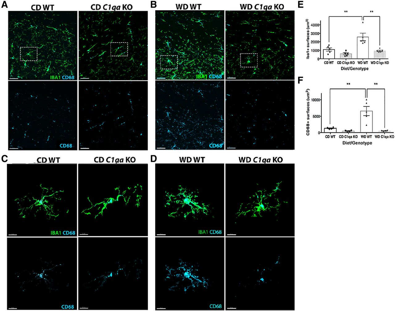
- Figure 7.
WD-induced myeloid cell activation was prevented by C1QA deficiency. A, B, The increase in the number of IBA1+ (A, B, E) and CD68+ (A, B, F) surfaces in WD-fed WT was prevented by C1QA deficiency. C–D, Representative examples of IBA1+CD68+ microglia. Microglia from WD-fed WT mice showed the greatest levels of CD68. Representative images for quantification are shown that contain a portion of the CA1 of the hippocampus, the corpus callosum and the parietal cortex. Data are presented as mean ± SEM, n ≥ 4, **p < 0.01 by one-way ANOVA with Tukey’s post hoc test. Scale bars: 30 μm (A, B) and 10 μm (C, D).
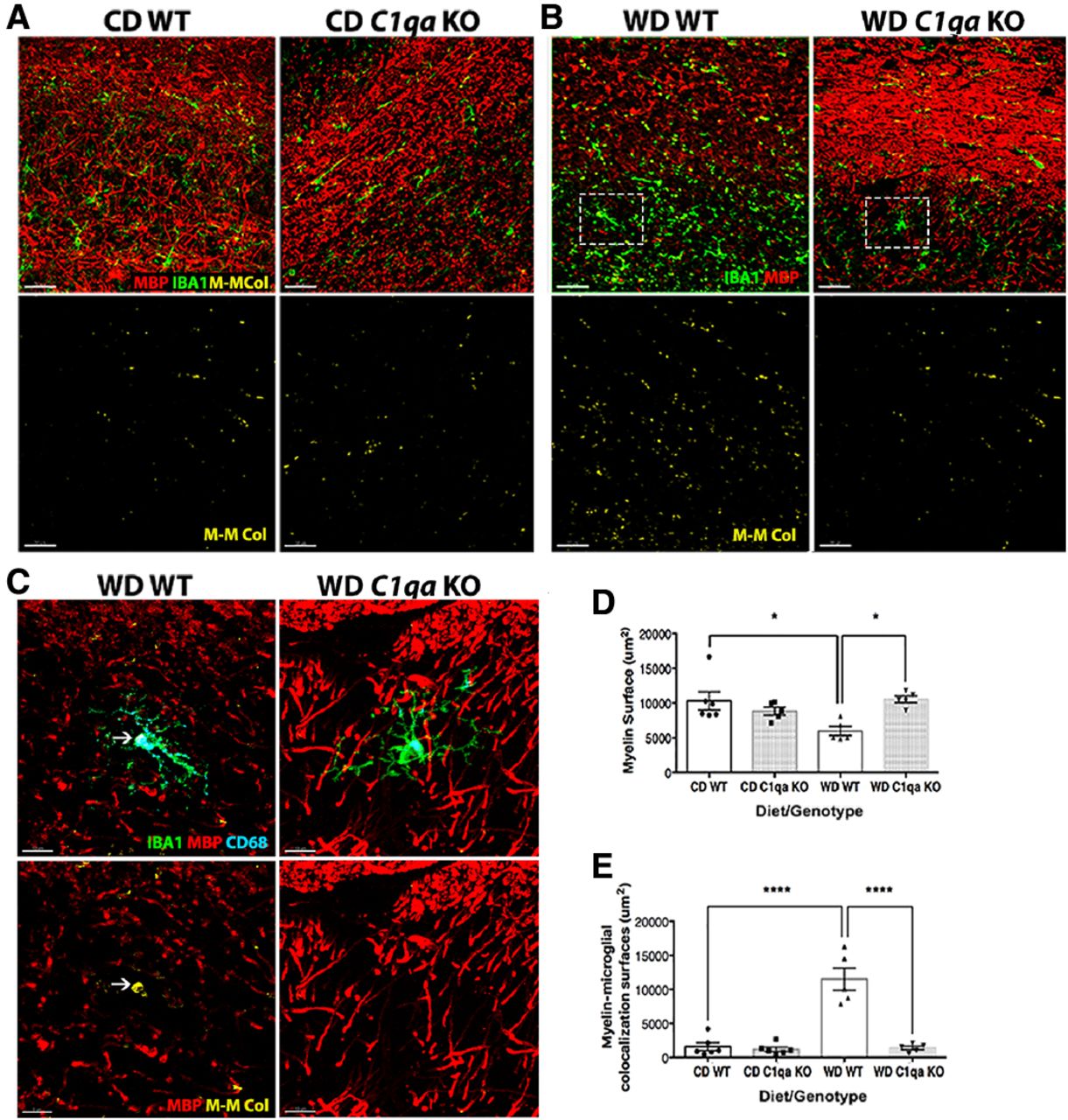
- Figure 8.
WD-induced myelin loss was prevented by C1QA deficiency. A–D, C1QA deficiency in WD-fed mice prevented the decline of MBP+ surfaces. The white arrow (C) indicates an example of CD68+ and MBP+ surfaces surrounded by IBA1+ surfaces, suggesting engulfment. E, C1QA deficiency prevented the increased microglia-myelin interactions observed in WD-fed WT obese mice. Representative images for quantification are shown in A, B that contain a portion of the CA1 of the hippocampus, the corpus callosum and the parietal cortex. The region of the image within the white boxes in B are shown in high resolution in C. Data are presented as mean ± SEM, n ≥ 4, *p < 0.05, ****p < 0.0001 by one-way ANOVA with Tukey post hoc test. M.-M. Col = myelin-microglia colocalization. Scale bars: 30 μm (A, B) and 10 μm (C).


Extended Data
Extended Data Figure 1
C1QA and C3 immunoreactivity in young WT and C1qa or C3 knockout mice. (A) C1QA (upper panel) and C3 (lower panel) immunoreactivity in the young WT corpus callosum is low or absence. (B) No C1QA immunoreactivity is found in the C1qa KO mice. (C) No C3 immunoreactivity is found in the C3 KO mice. Scale bars: 40μm. Download Extended Data Figure 1, DOCX file
Extended Data Figure 2
Creation and validation of the C1qa KO mouse. A conditional C1qa mouse was created as part of the Knockout Mouse Project (KOMP) at The Jackson Labs (https://www.jax.org/research-and-faculty/tools/knockout-mouse-project). (A) Representation of the C1qa alleles that are available including C1qaTm1a (LacZ reporter), C1qaTm1c (floxed allele), C1qaTm1d (null allele). As has been shown for other KOMP alleles, the Tm1a allele is not capable of reporting C1qa expression using the β-galactosidase assay. Mating the C1qaTm1a mice to mice carrying FLP recombinase creates the C1qaTm1c floxed allele. Mating the C1qaTm1c mice to mice carrying CRE recombinase creates the C1qaTm1d null allele. Cell-specific ablation of C1qa can be achieved using a cell-specific Cre line. The locations of genotyping primers are shown as inward facing arrows. (B) Examples of genotyping assays for the different alleles and band sizes. Primers pairs in this example were as follows. C1qa+: F – CCGGAAGAAAAGACATCCTG; R – CTTTCACGCCCTTCAGTCCT. C1qaTm1a: F – GTGGTTTGTCCAAACTCATCAA; R – TCTCTGAGCCTCTGCTTCAA. C1qaTm1c: F – GGACGAGAGGGGAGGAGTTA; R – TTAGGACCCTTTGGCACAAC. C1qaTm1d: F – CCGGAACCGAAGTTCCTATT; R – AGACGGGGATCGTTTATTCC. Note that the C1qa+ primers amplify a larger product in mice carrying the C1qaTm1c allele. Standard PCR conditions were used with an annealing temperature of 59.3oC for C1qa+, C1qaTm1b and C1qaTm1d and 61.0oC for C1qaTm1c. (C) RNA in situ hybridization to visualize C1qa transcripts in brains sections from C1qa+/+ and C1qaTm1d/Tm1d mice. As expected, C1qa expression is absent in C1qaTm1d/Tm1d (C1qa KO) mice. Download Extended Data Figure 2, DOCX file
In this issue


