

Article Figures & Data
Figures
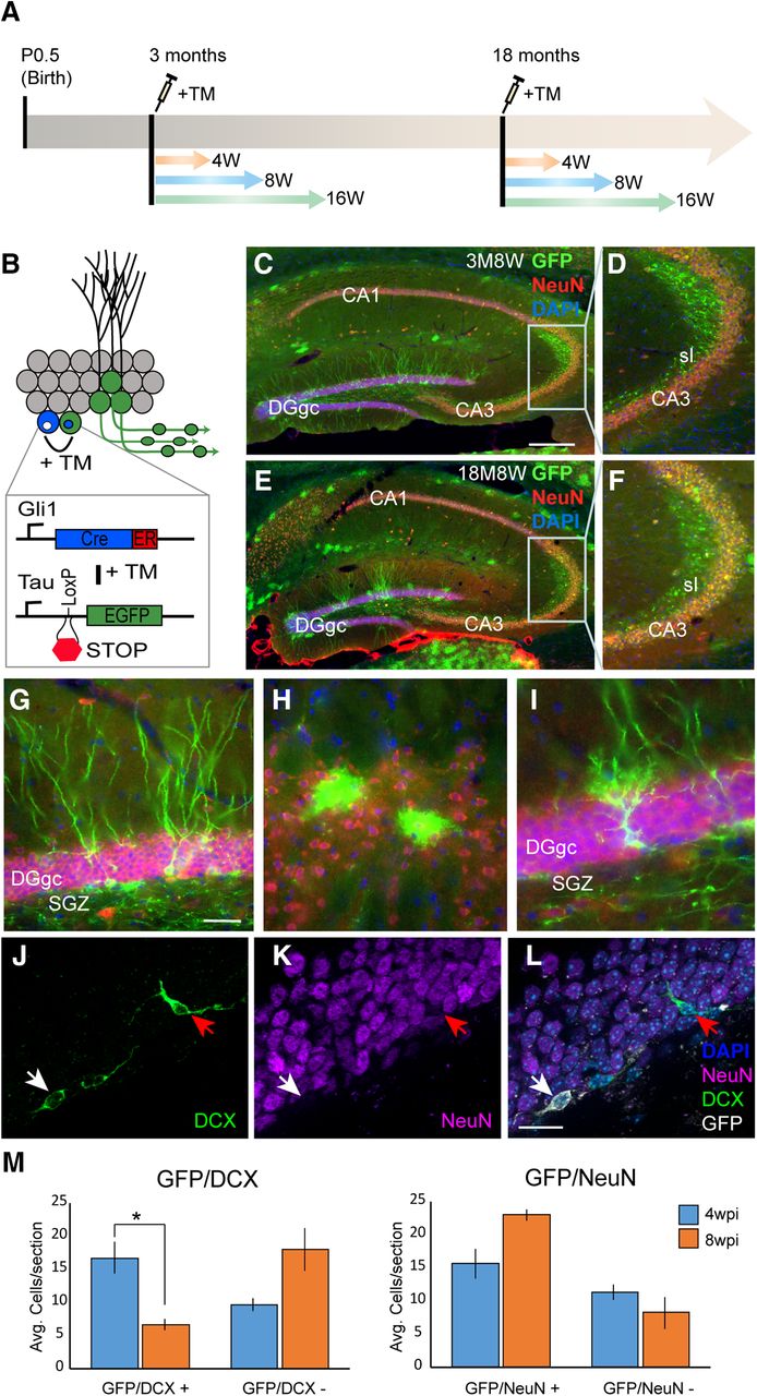
- Figure 1.
GliCreGFP transgenic mice enable conditional birth-dating and morphometric analysis of newborn hippocampal GCs in adult and aged animals. A, Strategy for newborn GC birth dating. Administration of TM at different ages turns on GFP expression in progenitors and enables tracking of subsequent daughter GCs at various time points (e.g., 4, 8, or 16 weeks) postinjection. B, Schematic illustration of conditional GFP expression strategy in Gli1 responsive progenitors post-TM administration. C, Epifluorescent images of immunofluorescent labeling for GFP and NeuN in a 3 M adult hippocampus 8W post-TM administration. D, Higher magnification image taken from boxed region in C. E, Epifluorescent images of immunofluorescent labeling for GFP and NeuN in an 18 M-aged hippocampus 8W post-TM administration. F, Higher magnification image taken from boxed region in E. G, Morphologically mature looking dentate gyrus GCs immunolabeled for GFP 8W post-TM. H, Astrocyte glia in subiculum immunolabeled for GFP 8 wpi. I, Radial glial-like progenitors immunolabeled for GFP are observed in the dentate gyrus bordering the subgranular zone 8W post-TM. J–L, Cell-type marker analysis of GFP-labeled cells by immunolabeling. At 4W post-TM, some GFP cells expressed the immature neuronal marker DCX but not mature neuronal marker NeuN (white arrow). Not all DCX cells were GFP-positive (red arrow). M, Cell counts of DCX/NeuN/GFP cells shows a progressive decrease in number of DCX/GFP cells and increase in NeuN/GFP cells from 4W to 8W post-TM (GFP/DCX+: 4 wpi: n = 9, 8 wpi: n = 9, p < 0.05, Student’s t test). DGgc, dentate gyrus GC; CA1-3, cornu ammonis region1-3; SGZ, subgranular zone; sl, stratum lucidum; sr, stratum radiatum. Scale bars = 1 mm (C–F), 100 μm (G–I), 50 μm (J–L); *p < 0.05.
- Figure 2.
Spatiotemporal maturation of adult-born GC mossy fiber axon terminals in adult and aged mice. A, Examples of large complex MFBs labeled by immunohistochemistry with an antibody against GFP. B, Example of a lollipop bouton. Note short thin collateral and small bulbous terminal. C, Example of an immunolabeled en passant swelling in newborn mossy fiber axon. Note the small diameter of the swelling and that it is in line with the axon. D, Low-power image of immunolabeled adult-born mossy fibers and boutons in region CA3. E, Neurolucida reconstructions of adult-born mossy fibers and boutons taken from image in D. F, Spatial density of labeled MFBs in stratum lucidum at 8W post-TM administration in adult (3 M; n = 400), midlife (6 M; n = 252), and aged (12 M and 15 M; n = 249 and n = 261, respectively) animals. G, Axonal density measures of MFBs at various times post-TM administration in adult and aged animals (3 M: n = 135, 12 M: n = 146, 18 M: n = 128). H, Interbouton spacing measures of MFBs at various times post-TM administration in adult and aged animals (3 M: n = 135, 12 M: n = 146, 18 M: n = 128). I, Axonal density measures of en passant swellings at various times post-TM administration in adult and aged animals (3 M: n = 110, 12 M: n = 116, 18 M: n = 90). J, Axonal density measures of mossy fiber lollipop boutons at various times post-TM administration in adult and aged animals (3 M: n = 25, 12 M: n = 30, 18 M: n = 38). CA3, cornu ammons region 3; DGgc, dentate gyrus GCL; sr, stratum radiatum; sl, stratum lucidum; sp, stratum pyramidale; so, stratum oriens. Scale bars = 5 μm (A), 10 μm (B, C), 500 μm (E); *p < 0.05, **p < 0.01, ***p < 0.005, two-way ANOVA followed by Student’s t test. All values are mean ± SEM.
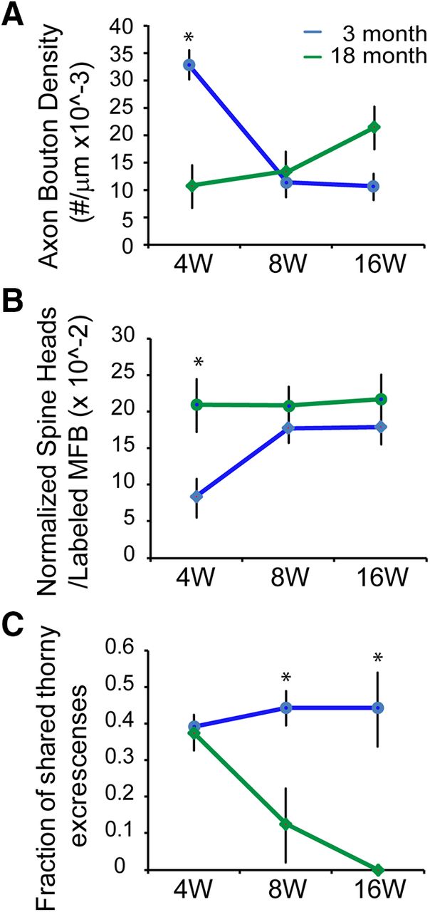
- Figure 3.
Opposing developmental trajectories in MFB size during adult and aged hippocampal neurogenesis. A, Newborn MFB size distributions in 3 M adult hippocampus, 4W, 8W, and 16W post-TM administration (4 wpi; n = 47, 8 wpi: n = 37, 16 wpi: n = 51). B, Newborn MFB size distribution in 18 M-aged hippocampus, 4W, 8W, and 16W post-TM administration (4 wpi: n = 38, 8 wpi: n = 28, 16 wpi: n = 62). C, Comparison of the relative newborn MFB size distribution 4W post-TM administration in 3-M-, 12-M-, and 18-M-old animals. Note the significantly smaller size distribution in 3 M animals relative to 12 M and 18 M animals (p < 0.005 Kolmogorov–Smirnov test). D, Developmental changes in MFB size occurred in opposite directions between 3 M and 18 M brain. Changes in average size plotted relative to 4W post-TM show increases in MFB size in 3 M but decreases in 18 M animals. Average MFB size remained relatively unchanged in 12 M animals. E, Averaged MFB sizes. The size is determined by measuring the diameter of each MFB. Values in D, E are mean ± SEM.
- Figure 4.
Quantitative analysis of serial EM reconstructed adult-born MFB onto postsynaptic TEs. A, Axonal bouton density (n = 50–100 boutons per time point; 3M4 wpi: n = 63, 18M4 wpi, n = 78, p < 0.05). B, Normalized average number of TE spine heads in the GFP-labeled newborn MFB (n = 3–4 reconstructed MFBs per time point; 3M4 wpi: n = 4, 18M4 wpi: n = 3). C, Percentage of TEs shared by GFP-labeled and non-labeled MFBs were analyzed in adult (3 M) and aged (18 M) hippocampus, 4W, 8W, and 16W post-TM administration (n = 8–15 reconstructed TEs per time point). *p < 0.05, Student’s t test. Values are mean ± SEM (A, B) or ±SD (C).
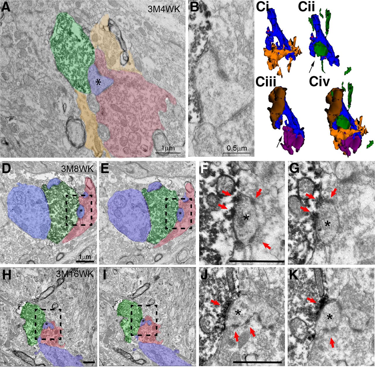
- Figure 5.
Electron micrographs and 3D reconstructions demonstrating sharing of postsynaptic TEs by newborn and mature MFBs in 3 M adult mice at different times post-TM injection. A–C, Representative 3D reconstruction of a 3M4W newborn MFB. Shown in A is an electron micrograph of GFP-labeled newborn MFB (green) at 3M4W and non-GFP-labeled mature MFB (red) both forming asymmetrical synaptic contacts with the same spine head (blue, asterisk). Surrounding glia processes are also marked (yellow). Note the glia processes are not seen in the proximity to the postsynaptic density and synaptic cleft. The synaptic contact region and the shared spine head are magnified and shown in B. Different combinations of 3D reconstruction of this 3M4W newborn MFB with postsynaptic dendrite, neighboring mature MFBs and associated glial cell are shown in Ci–Civ. These include: (Ci) postsynaptic dendrite (blue) giving rise to two TEs and associated glial processes (yellow), (Cii) GFP-labeled newborn mossy fiber and its bouton (green) forming synaptic contact with the spine head of the smaller TE (arrow), (Ciii) two non-GFP-labeled mature MFBs (brown and purple) forming contacts with the two TEs, respectively, and (Civ) a combination of all components. Note the small TE (arrow) is shared by the newborn MFB (green) and mature MFB (purple) as seen in the electron micrograph in A. D–G, Representative electron micrographs of a 3M8W newborn MFB. Shown in D, E are two serial EM sections of a GFP-labeled MFB (green) at 3M8W shares a postsynaptic spine head (blue, asterisk) with a non-GFP-labeled mature MFB (red). Higher magnification boxed regions in D, E are shown in F, G, respectively. The asymmetric synaptic contacts are marked with red arrowheads. H–K, Representative electron micrographs of a 3M16W newborn MFB. Shown in H, I are two serial EM sections of a GFP-labeled MFB (green) at 3M16W shares a postsynaptic spine head (blue, asterisk) with a non-GFP-labeled mature MFB (red). Higher magnification boxed regions in H, I are shown in J, K, respectively. The asymmetric synaptic contacts are marked with red arrowheads. Scale bars = 1 μm.
- Figure 6.
Smaller newborn MFB at 3M4W can share a postsynaptic spine head with mature MFB. A–D, 3D reconstruction of a newborn mossy fiber at 3M4W forming contacts with postsynaptic dendrite of a CA3 pyramidal cell. The 3M4W GFP-labeled mossy fiber (green) forms two small MFB contact sites, labeled as 1 and 2, with TEs on the dendrite of a CA3 pyramidal cell (blue). The two TE also form contacts with mature MFBs at site 1 (purple) and site 2 (red). E, Electron micrograph shows that MFB at site 1 (green) does not form asymmetric synaptic contact with TE (blue), which in turn forms multiple asymmetric synaptic contacts with the non-GFP-labeled mature MFB. F, Electron micrograph shows that MFB at site 2 (green) forms an asymmetrical synaptic contact with a spine head of TE (blue) that is also contacted by another non-GFP-labeled mature MFB. Inset shows a high magnification of the shared spine head and the asymmetrical synaptic contacts. Scale bar = 1 μm.
- Figure 7.
Electron micrographs and 3D reconstructions demonstrating sharing of postsynaptic TEs by newborn and mature MFBs in 18 M adult mice at 8W post-TM but not at 16W post-TM. A–C, Representative electron micrograph shows an 18M8W GFP-labeled MFB (green) shares a small postsynaptic spine head (blue, asterisk) with a non-GFP-labeled mature MFB (red). Serial micrographs of the boxed area in A are magnified and shown in B, C. Note the labeled newborn MFB and non-labeled mature MFB are very close to each other, but the axoplasm membranes clearly separate two boutons, the serial section analysis also show that the two postsynaptic densities are separated. Red arrowheads mark the asymmetric synaptic contacts by the newborn MFB. D–F, Representative electron micrographs of a 18M16W newborn MFB. A GFP-labeled 18M16W newborn MFB (green) and a non-GFP-labeled mature MFB (red) form asymmetric synaptic contacts with spine heads derived from different TEs (blue). They do not share contacts with spine heads (asterisk). Serial micrographs of the boxed area in D are magnified and shown in E, F. Red arrowheads mark the asymmetric synaptic contacts by the newborn MFB. G–I, 3D reconstruction of a 18M16W newborn MFB that does not show sharing. The two reconstructed postsynaptic dendrites (blue and yellow) have a total of three TEs (G). The GFP-labeled MFB (green) forms synaptic contacts with one TE from the top dendrite (blue; H). This MFB does not form synaptic contact with spine heads from other TEs. Combined image (I) show that the other two TEs are contacted by two non-GFP-labeled MFBs (brown and purple). Again, these two mature MFBs confine their synaptic contacts to only the TE they cover; no sharing is observed among them. Scale bars = 1 μm.


In this issue


