

Article Figures & Data
Figures
- Figure 1.
At birth, CB1 is prominently expressed in axon tracts in hindbrain. A–A”, Immunolocalization of CB1 in sagittal hindbrain sections spanning cerebellar vermis and paravermis at P0 shows robust expression in developing axons, including long-range axon tracts in the brainstem and in the fibers cruising through the inner regions of the developing cerebellar cortex. Immediately relevant for the developing pontocerebellar system, CB1 immunolocalization is seen in mcp and scp [mcp (A”) and scp (A’), highlighted by green arrowheads], and in axons filling pWM (A–A”, green arrowheads). CB1 expression in icp (A”, green dotted outline) is very weak or absent. B–B”, Sagittal sections showing localization of CB1 mRNA in the hindbrain at E18 from the Allen Brain Atlas Developmental Mouse database (2008 Allen Institute for Brain Science. Allen Developing Mouse Brain Atlas, available from http://developingmouse.brain-map.org/experiment/show/100024806). Notably for pontocerebellar development, CB1 mRNA expression is prominent both in the Pn (B, maroon arrowhead) and the IO (B, maroon arrowhead), among many other brainstem nuclei which project axons to the cerebellum (marked by maroon triangles and anatomic abbreviations in summary diagrams in panels C–C”). In the cerebellum, CB1 mRNA is most prominent in deeper layers (B, maroon arrowhead). CB1 mRNA positive region spans developing IGL and pWM, marked in summary diagram in C. CB1 mRNA expression is also prominent in vestibulocerebellar nucleus (VeCb; B”, maroon arrowhead). In addition, restricted regions of the iEGL (B–B”, maroon arrow) show thin layer of CB1-positive cells. C–C”, Traced outlines of sections shown in A, summarizing perinatal distribution of CB1 expression relative to anatomic landmarks. Green shapes mark CB1 immunolocalization in axon tracts as shown in A, red triangles mark brain regions with robust CB1 mRNA expression as shown in B. Lines in coronal insets represent mediolateral plane of sectioning. Anatomical locations are adapted from Franklin and Paxinos (2008) and from Paxinos et al. (2007). bsc, brachium of superior colliculus; CAT, nucleus of the central acoustic tract; CB, cerebellum; CnF, cuneiform nucleus; cp, cerebral peduncle; csc, commissure of superior colliculus; Cu, cuneate nucleus; DC, dorsal cochlear nucleus; DTg, dorsal tegmental nucleus; fr, fasciculus retroflexus; Gi, gigantocellular reticular nucleus; GiV, gigantocellular reticular nucleus, ventral part; g, gracile fasciculus; Gr, gracile nucleus; icp, inferior cerebellar peduncle; IC, inferior colliculus; IO, inferior olivary nucleus; IP, interpeduncular nucleus; IRt, intermediate reticular nucleus; ll, lateral lemniscus; LRt, lateral reticular nucleus; mcp, middle cerebellar peduncle; mcp, middle cerebellar peduncle; MiTg, microcellular tegmental nucleus; mlf, medial longitudinal fasciculus; MPB, medial parabrachial nucleus; MVe, medial vestibular nucleus, magnocellular part; PDTg, posterodorsal tegmental nucleus; PMn, paramedian reticular nucleus; Pn, pontine nucleus; PnC, pontine reticular nucleus, caudal part; PrCnF, precuneiform area; py, pyramidal tract; RLi, rostral linear nucleus (midbrain); RR, retrobulbar nucleus; Rt, reticular nucleus; RtTg, reticulotegmental nucleus of the pons; SC, superior colliculus; scp, superior cerebellar peduncle; Sol, solitary nucleus; sp5, spinal trigeminal tract; Tz, nucleus of the trapezoid body; tz, trapezoid body; VeCb, vestibulocerebellar nucleus; vsc, ventral spinocerebellar tract; VTg, ventral tegmental nucleus; X, nucleus X, dorsal nucleus of the vagus nerve.
- Figure 2.
Zoomed-in view of CB1 distribution is the brainstem and the cerebellum at P0. Top row: diagram showing distribution of CB1 staining relative to the layers of the developing cerebellum (superimposed on top of the tracing of medial cerebellar section). A, A’, Thick CB1-positive fibers are prominent in the scp and pWM (darker green in the diagram). Thinner CB1-positive structures (lighter and more diffuse staining) span iEGL through PCL and IGL (turquoise in the diagram). CB1 staining is very week or absent from the oEGL, and DN. B, CB1-positive fibers are radially oriented in Pn. CB1 is prominently expressed within multiple axon tracts visible in this section through the brainstem (ns, cp, py, tz). C, CB1-positive fibers can be seen within the territory of the IO, and running through the cuneate tract (cu). Anatomical locations are adapted from Franklin and Paxinos (2008) and from Paxinos et al. (2007). CB, cerebellum; cp, cerebral peduncle; cu, cuneate fasciculus; DN, deep cerebellar nuclei; ECu, external cuneate nucleus; iEGL, inner EGL; IGL, inner GC layer; IO, inferior olivary nucleus; ns, nigrostriatal tract; oEGL, outer EGL; PCL, PC layer; Pn, pontine nucleus; pWM, presumptive white matter; py, pyramidal tract; scp, superior cerebellar peduncle; tz, trapezoid body; vsc, ventral spinocerebellar tract.
- Figure 3.
At E17.5, CB1 is highly expressed in cerebellar peduncles and is required for orderly arrangement of axons within the peduncles. Coronal sections at E17.5 through anterior cerebellar zone, scp and mcp are clearly seen in this plane of sectioning. Top row, left: outlines of a representative coronal section showing anatomic landmarks. Dotted square indicates the regions shown in panels A–D’’’. Top row, right: black line through sagittal diagram shows plane of sectioning. Anatomical locations are adapted from Paxinos et al. (2007). A–A’’’, In WTs, CB1 co-localizes with GAP43, a marker of elongating axons, and with axonal neurofilaments (C–C”). Large fascicles of CB1-positive axons run through scp and mcp (highlighted by arrowheads). B–B’’’, D–D’’’, CB1 staining is absent in KO littermates. Furthermore, density of GAP43 staining within the peduncles is increased in CB1 KOs (A”, A’’’ vs B”, B’’’), and distribution of axons as judged by neurofilament staining appears abnormal (C”, C’’’ vs D”, D’’’). Panels in the bottom row (A’’’, B’’’, C’’’, D’’’) show zoomed-in views from regions indicated by maroon dotted outlines in (A”, B”, C”, D”).
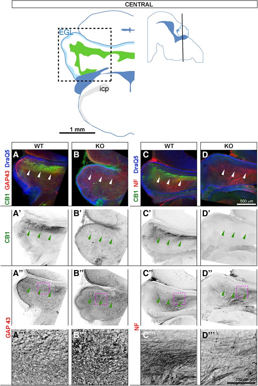
- Figure 4.
At E17.5, CB1 is expressed in the developing white matter tracts (pWM), which show increased GAP43 accumulation and altered axon distribution in CB1 KOs. Coronal sections at E17.5 through central cerebellar zone. Top row, left: outlines of a representative coronal section showing anatomic landmarks. Regions imaged in (A–D’’’) are indicated by dotted square. Top row, right: black line through sagittal diagram shows plane of sectioning. Anatomical locations are adapted from Paxinos et al. (2007). In WTs, CB1 co-localizes with axonal markers GAP43 (A–A’’’) and NF (C–C’’’). In KO littermates (B–B’’’) and (D–D’’’) CB1 staining is absent, density of GAP43 staining within pWM is increased (B”, B’’’), and distribution of NF-positive axons (D”, D’’’) is abnormal. Panels in the bottom row (A’’’–D’’’) show zoomed-in views from regions indicated by maroon dotted outlines in A”–D”.
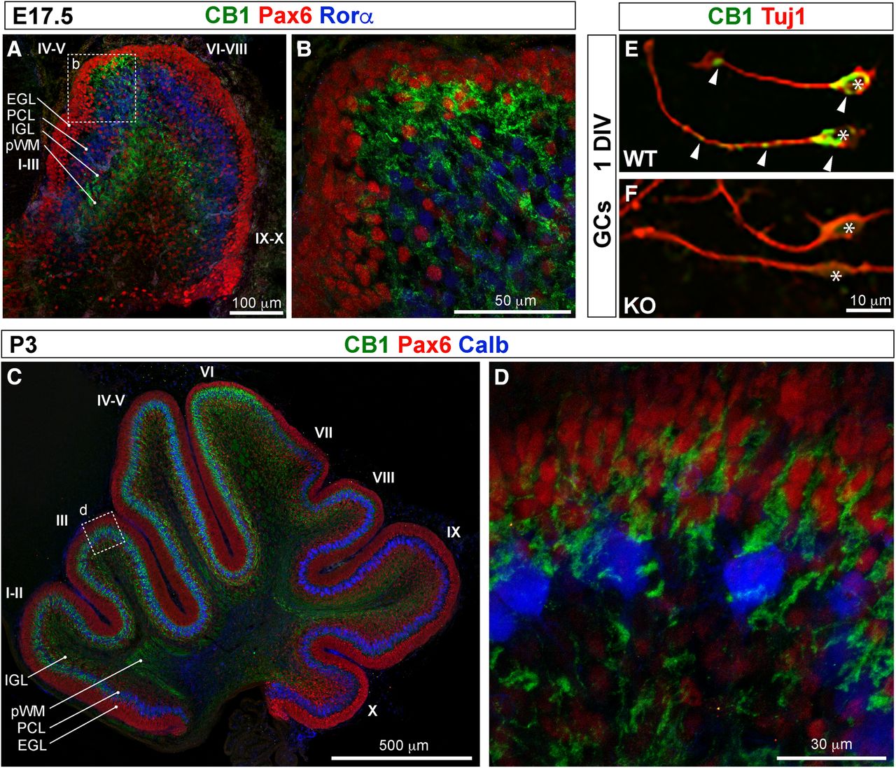
- Figure 5.
CB1 is highly expressed in a subset of differentiating GCs during the first postnatal week. Midsagittal sections of cerebellar vermis at E17.5 (A, B) and at P3 (C, D). B, D, Zoomed-in views from regions highlighted by white dotted outlines in A, C. During the first postnatal week, CB1 expression (green) becomes pronounced in differentiating GCs in the anterior and central cerebellar vermis. Postmitotic GCs in the EGL and GCs radially migrating through the PCL are marked by expression of transcription factor Pax6 (red in A–D). PCs (PCL) are identified by expression of Rorα at E17.5 (blue in A, B) and calbindin (Calb) expression at P3 (blue in C, D). A, At E17.5, CB1 expression is most prominent in a patch of radially migrating GCs clustered at the tip of developing lobes IV and V. C, At P3 the territory of radially migrating GCs that express high levels of CB1 expands to occupy the extent of lobes I–VI, but not the posterior and nodular zones (lobes VII–X). Higher magnification images (B, D) highlight CB1 expression in neurites surrounding Pax6-positive nuclei at the inner boundary of EGL and throughout the PCL and the developing IGL. E, CB1 is expressed in neurites and somata of isolated and purified 24-h-cultured (1 DIV) GCs. GC neurites and somata are identified by expression of neuronal tubulin (Tuj1, red). Somata of GCs are indicated by asterisks. Arrowheads highlight CB1 expression in somata, axons, and growth cone of 1 DIV GCs. F, Confirming specificity of CB1 expression in differentiating GCs, the staining is absent in sister GC cultures isolated from CB1 KOs.
- Figure 6.
CB1 expression in developing cerebellum is dynamic and region specific, while DAGLα is expressed primarily in the PCL throughout development. CB1 expression in long-range axons within pWM becomes weak/undetectable after P5 (A’ compared to B’, C’). At P3 CB1 staining can be detected in PCL-IGL, corresponding to expression in migrating and differentiating GCs (A, A’); however, at P5 (B, B’) and P12 (C, C’, D), CB1 expression becomes restricted to ML in the anterior and central vermis. DAGLα (maroon) is expressed in PCL throughout cerebellar cortex (grayscale DAGLα channel in A”, B”, C”). D, In coronal sections, at P12, CB1 expression in ML is prominent in the vermis and paravermis (lobes II, III, IV–V, and Sim), but weak in the hemispheres (Crus 2) and in the nodular zone (PFL and FL). Stripes of high-DAGLα-expressing PCs are apparent in lobes III and IV–V (D, asterisks).
- Figure 7.
CB1 is expressed in developing PFs during the second postnatal week. High-magnification views of CB1 localization relative to cerebellar layers. Images were taken at the tips of lobe III. A–A”, C–C”, Coronal sections, in-plane with trajectories of PFs and orthogonal to the plane of branching of PC dendrites. B–B”, D–D”, Sagittal sections, orthogonal to the trajectories of PFs, and in-plane with branching of PC dendritic trees. During the second postnatal week (P8, top two rows; P10, bottom two rows) strong CB1 expression (green) is most prominent in ML, in thin fibers stacked above PCL (marked by Calb, maroon) running parallel to the coronal plane of sectioning, i.e., in the PFs, axons of GCs. No co-localization of CB1 is seen with PC dendrites, somata, or proximal axons. CB1 expression was not detected in EGL. Thin CB1-positive fibers and week somatic staining in the IGL (more prominent at P10; C’, D’) most likely correspond to weak expression in GC somata and in vertical portions of GC axons.
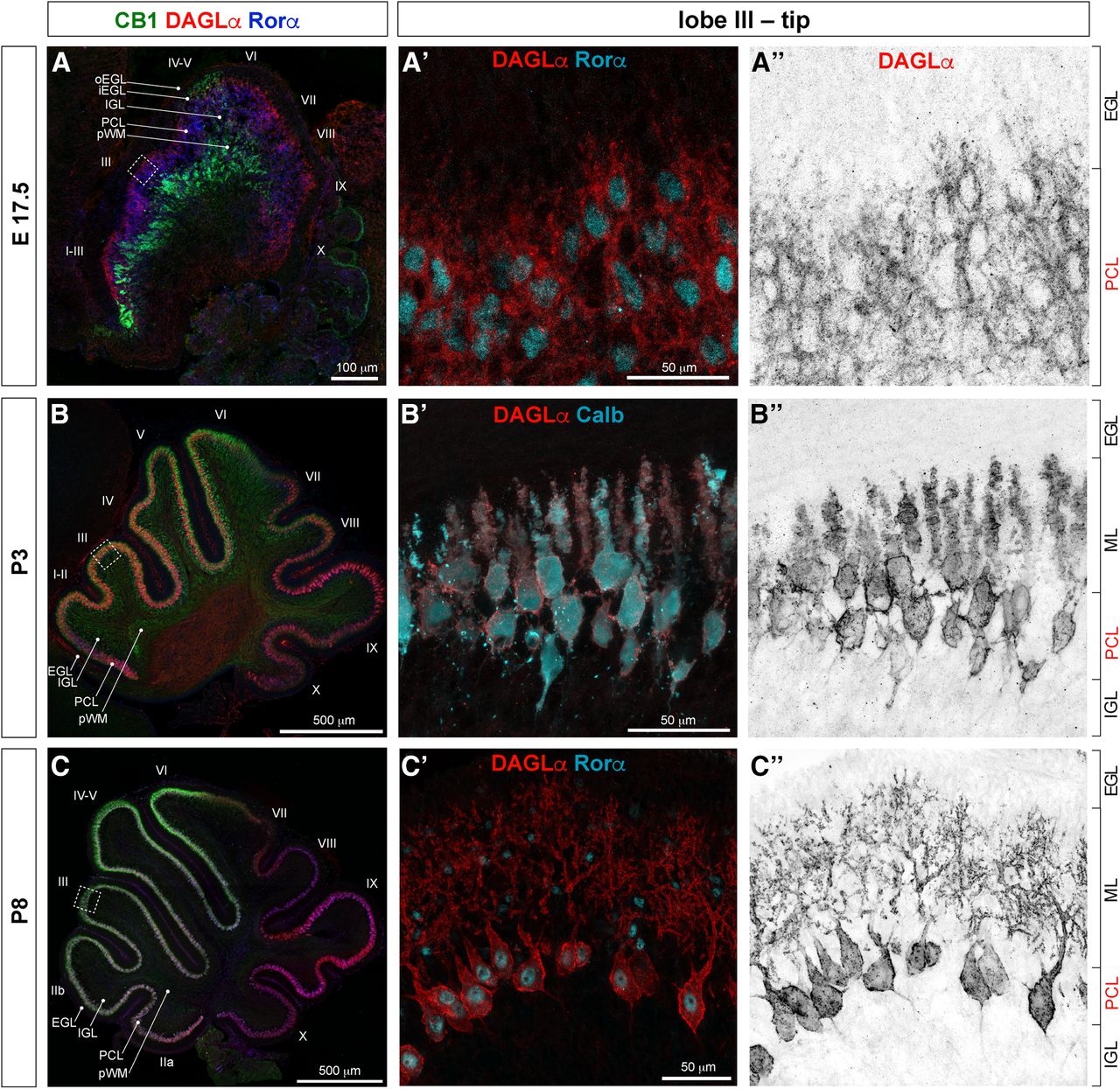
- Figure 8.
DAGLα is expressed in PCs during cerebellar development. Midsagittal sections of cerebellar vermis at (A–A”) E17.5, (B–B”) P3, and (C–C”) P8. Middle and right columns, Zoomed-in views at the tip of lobe III in the anterior zone. At all developmental stages investigated, immunoreactivity for DAGLα (red) within cerebellar cortex was restricted to somata, dendrites, and initial axon segments of PCs (identified by morphology and by expression of Rorα and Calb, blue/cyan).
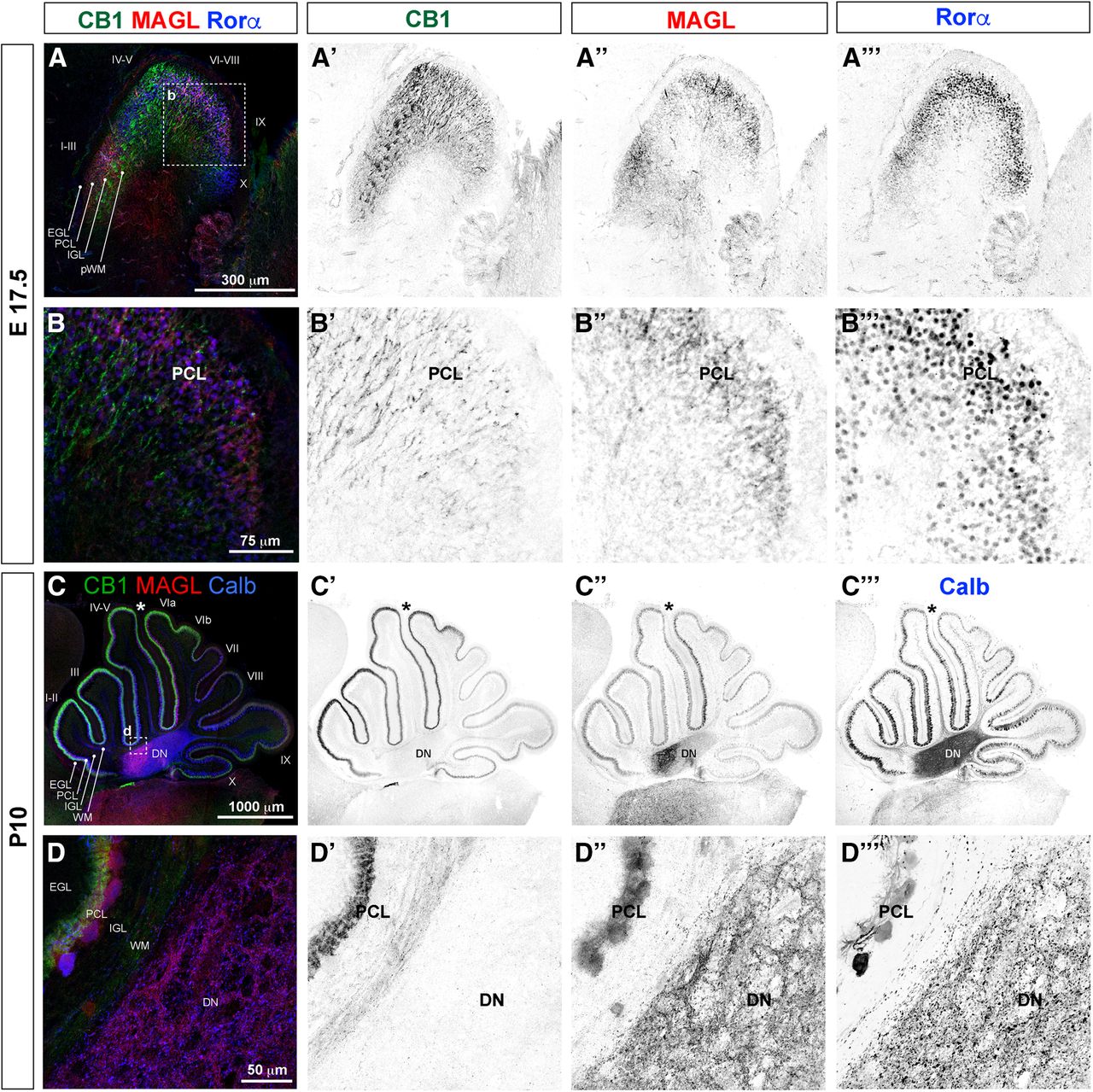
- Figure 9.
MAGL is expressed in PCs. Midsagittal vermal sections at (A–A’’’, B–B’’’) E17.5 and (C–C’’’, D–D’’’) P10. B–B’’’, D–D’’’, are zoomed-in views from regions indicated by white dotted lines in A, C. PCs are identified by Rorα (A, B, blue) and Calb (C, D, blue). MAGL (red) expression is seen primarily in the PCL (A, A”, C, C”). Higher magnification views show that MAGL is expressed in the somata and dendrites of PCs (B, B”, D, D”, PCL) and in axon terminals in DN (C”, D”). MAGL expression at P10 is uneven across the midvermal region, with the highest levels expressed within PCs lining the primary fissure, which separates lobes V and VI (C–C’’’, asterisks).
- Figure 10.
Midvermal area is reduced in CB1 KOs. A, Representative Nissl-stained midvermal cerebellar sections from WTs, showing regions used for area quantification and illustrating postnatal developmental stages analyzed. Territories of anterior lobes I–III, and nodular lobes IX–X are outlined. Scale bars: 200 μm. B–D, The mean difference for (B) four comparisons (P3, P5, P12, two months) and (C, D) five comparisons (lobes I–III P3, lobes I–III P5, lobes I–III P12, lobes I–III two months, lobes IX–X two months) are shown in Cumming estimation plots. The raw data are plotted on the upper axes; each mean difference is plotted on the lower axes as a bootstrap sampling distribution. Mean differences are depicted as dots; 95% confidence intervals are indicated by the ends of the vertical error bars. A total of 5000 bootstrap samples were taken; the confidence interval is bias-corrected and accelerated. Details of statistical analysis are shown in Table 3. B, Stunted increase in total midvermal area in CB1 KOs becomes more pronounced with age. C, D, Reduction in midvermal area in CB1 KOs is primarily contributed by reduced size of anterior zone, quantified for lobes I–III.
- Figure 11.
Selective impairments in motor behaviors in CB1 KOs at two-month-old. A, Time course of rotarod performance (latency to fall from rotating rod accelerating from 4 to 40 rpm) was plotted per trial as average performance of all WT animals (24, sexes combined) and all KO animals (30, sexes combined). Three trials were performed per day over the course of 4 d (12 trials total). Both groups performed at comparable levels in the beginning of the time course (day 1, trials 1–3), in the subsequent trials (days 2 and 3, trials 4–9), and achieved similar performance in the fourth (last) day of the time course (trials 10–12). Statistical hypothesis of equivalent latency to fall between genotypes was shown to be true by Tukey multiple comparison, and by comparing areas under the curve for all trials (statistical table in Table 4). B, Improvement in rotarod performance was evaluated by fitting linear regression curves over the first six trials; differences in slopes between genotypes were compared with evaluate rate of learning, and found to be similar between CB1 KOs and WTs. C, No difference in grip strength was detected between genotypes, and performance in rotarod does not show grip-strength-dependent bias. D, Mean differences in latency to open sunflower seeds between WTs (25 animals, sexes combined) and KOs (26 animals, sexes combined) are shown in Gardner–Altman estimation plot. Both groups are plotted on the left axes; the mean difference is plotted on floating axes on the right as a bootstrap sampling distribution. The mean difference is depicted as a dot; the 95% confidence interval is indicated by the ends of the vertical error bar. A total of 5000 bootstrap samples were taken; the confidence interval is bias-corrected and accelerated. Average time to open sunflower seeds is significantly longer in CB1 KOs. Details of statistical analysis are shown in Table 4.


Tables
- Table 1
Primary antibodies against CB1, DAGLα, and MAGL: target epitopes, prior publications, RRIDs, and working dilutions
eCB signaling machinery Antibody Immunogen Source RRID Working
dilutionReference Rabbit anti-CB1 Synthetic peptide,
aa 443–473ImmunoGenes,
polyclonalAB_2813823 1:300 Dudok et al. (2015) Rabbit anti-CB1 L15 CB1-GST fusion protein, aa 460–473 of rat CB1 Custom made,
polyclonalAB_2315250 1:300 Bodor et al. (2005) Guinea pig anti-CB1 L15 Last 15 aa on L-term of human CB1 Custom made,
polyclonalAB_2813824 1:600 Berghuis et al. (2007) Guinea pig anti-DAGLα aa 790–908 of rat DGLα Custom made,
polyclonalAB_2813825 1:600 Katona et al. (2006) Rabbit anti-MAGL GST fusion protein, aa 172–206 of mouse MAGL Custom made,
polyclonalAB_2813826 1:600 Straiker et al. (2009) - Table 2
Commercially available primary antibodies used to identify cerebellar cell types: immunogens, sources, RRIDs, and working dilutions
Cerebellar cell type markers Antibody Immunogen Source RRID Working dilution Mouse anti-SMI312 (pan-axonal neurofilament cocktail) SMI 312 is directed against highly phosphorylated axonal epitopes of neurofilaments BioLegend, #837904, monoclonal AB_2566782 1:1000 Rabbit anti-Pax-6 Peptide QVPGSEPDMSQYWPRLQ from the C terminus of mouse Pax-6 protein BioLegend, clone poly-19013, #901301, polyclonal AB_2565003 1:600 After antigen retrieval Goat anti-RORα Peptide mapping at the C terminus of RORα1 of human origin Santa Cruz, sc-6062, polyclonal AB_655755 1:100 at E17.5–P0 1:300 at P3–P20 Mouse anti- TuJ-1 (neuron-specific beta-III tubulin) Monoclonal mouse IgG2A clone #TuJ-1; detects mammalian and chicken neuron-specific beta–III tubulin; raised against rat brain-derived microtubules R&D Systems, #MAB1195, monoclonal AB_357520 1:1000 at 1 DIV Mouse anti-Calb Monoclonal anti-calbindin D-28k is a mouse IgG1 produced by hybridization of mouse myeloma cells with spleen cells from mice immunized with calbindin D-28k purified from chicken gut Swant, #300, monoclonal AB_10000347 1:500 at P0–P5 1:1000 at P6–P15 1:3000 at P16–adult Rabbit anti-Calb Against recombinant rat calbindin D-28k Swant, #CB38, polyclonal AB_2721225 Differences in area means between WT and KO Age WT (control); n = animals; N = litters KO (test); n = animals; N = litters Difference 95% CI of difference p value Mann– Whitney *<0.05
**<0.01
***<0.005
****<0.0001Total Midvermal Area P3 n = 6
N = 2n = 8
N = 10.22046525 0.1005809
to
0.338003710.02386844 * Total Midvermal Area P5 n = 23
N = 10n = 19
N = 9–0.5345564 –1.1207783
to
–0.00944940.08802591 ns Total Midvermal Area P12 n = 8
N = 2n = 8
N = 2–0.8547641 –1.3264183
to
–0.36904210.01008169 * Total Midvermal Area 2 months n = 23
N = 6n = 11
N = 5–1.544694 –2.3539983
to
–0.9299240.00040931 *** lobes I–III P3 n = 6
N = 2n = 8
N = 10.00060315 –0.0444497
to
0.041752210.94853252 ns lobes I–III P5 n = 23
N = 10n = 19
N = 9–0.3107878 –0.4960554
to
–0.12198110.00145207 *** lobes I–III P12 n = 8
N = 2n = 8
N = 2–0.5471343 –0.7196807
to
0.41790970.00093911 *** lobes I–III 2 months n = 23
N = 6n = 11
N = 5–0.9491027 –1.1671892
to
–0.75930151.1904E-06 **** lobes IX–X 2 months n = 21
N = 6n = 11
N = 5–0.384772 –0.5777334
to
–0.22500310.00064456 *** Differences in ratio of subregions over total midvermal areas between WT and KO I–III/total P3 n = 6
N = 2n = 8
N = 1–0.0465324 –0.0774791
to
–0.01817340.02386844 * I–III/total P5 n = 23
N = 10n = 19
N = 9–0.0725127 –0.0951794
to
–0.04568562.4409E-05 **** I–III/total P12 n = 8
N = 2n = 8
N = 2–0.0728148 –0.0866715
to
–0.05844680.00093911 *** I–III/total 2 months n = 21
N = 6n = 11
N = 5–0.0391941 –0.0483767
to
–0.02892591.275E-05 **** IX–X/total 2 months n = 21
N = 6n = 11
N = 5–0.0046574 –0.012602
to
0.00425590.40473443 ns Difference in latency to open sunflower seeds between WT and KO Condition n; N Number of trials per animal Mean Difference betweenmeans SE ofdifference 95% CI ofdifference p value,Mann–Whitney *<0.05,**<0.01,***<0.005, ****<0.0001 WTn = animals,N = litters n = 25;N = 10 2–5 61 27.9297959 30.1743641 12.0735231to42.2478872 0.00050806 *** KOn = animals,N = litters n = 26;N = 9 2–5 88 Difference in latency to fall from rotarod between WT and KO Condition n; N Number of trials(3 per day) Mean Differencebetweenmeans SE ofdifference 95% CI ofdifference p value (mixedeffects analysis) *<0.05, **<0.01,***<0.005,****<0.0001 Area under thecurve (all trials) WT/KO ratio of areas underthe curve WTn = animals,N = litters n = 24;N = 10 12 170.7 –1.24 13.97 –29.28 to26.80 0.9296 ns 1775 0.986 KOn = animals,N = litters n = 30;N = 10 12 172.0 1799.5
In this issue


