

Article Figures & Data
Figures
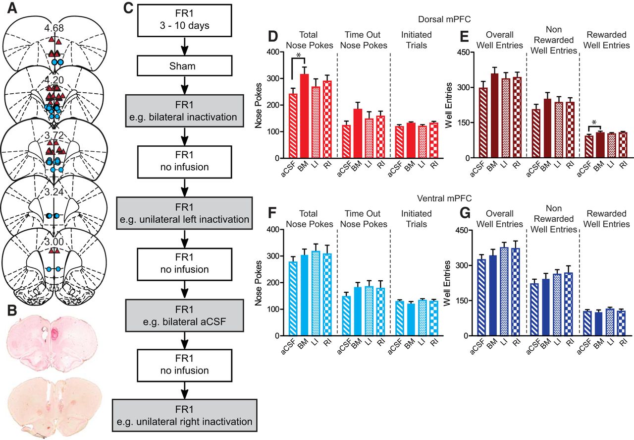
- Figure 1.
Cannula placements, test design, and FR1 data. A, Cannula placements for FR1 cohort. Dorsal mPFC cannula placements (triangles) and ventral mPFC cannula placements (circles). Numbers are A/P distance from bregma. B, Histology of coronal slices stained with neutral red showing cannula tracks for dorsal (top) and ventral (bottom) mPFC. C, Timeline for FR1 testing. Rats were retrained for 3–10 d after surgery. They then received sham infusions followed by 8 d of FR1 tests. Rats received one of four infusions every other day of testing: bilateral inactivation, bilateral aCSF, unilateral left, or right inactivation, counterbalanced across rats. All rats received all four conditions. aCSF (stripes) = control infusion, BI (solid) = bilateral inactivation, LI (dots) = inactivation of left hemisphere, RI (checkers) = inactivation of right hemisphere. D, F, Total number of nose pokes, time-out nose pokes, and initiated trials. E, G, Total number of well entries, non-rewarded well entries, and rewarded well entries. D, E, There was a significant increase in total number of nose pokes and total number of rewarded well entries when the dorsal mPFC was bilaterally inactivated (*). F, G, Ventral mPFC inactivation did not affect nose poking or well entries; *p < 0.05, Dunnett’s test for planned multiple comparison.
- Figure 2.
Cannula placements, test design, and extinction data for extinction cohort. A, Dorsal mPFC cannula placements (triangles) and ventral mPFC cannula placements (circles). B, Timeline for extinction task. Extinction rats were trained on FR1 but only received bilateral infusions during early and late extinction. C, G, There was a significant decrease in number of nose pokes between last day of FR1 and aCSF treatment during extinction (#). C, D, G, H, Bilateral inactivation of dorsal or ventral mPFC did not significantly affect nose pokes or well entries during early extinction. E, F, Inactivation of dorsal mPFC during late extinction decreased nose pokes and well entries (*). I, There was no effect of ventral mPFC inactivation for number of nose pokes during late extinction. J, However, there was a decrease in number of well entries during ventral mPFC inactivation during late extinction (*). # and *p < 0.05, paired t test.
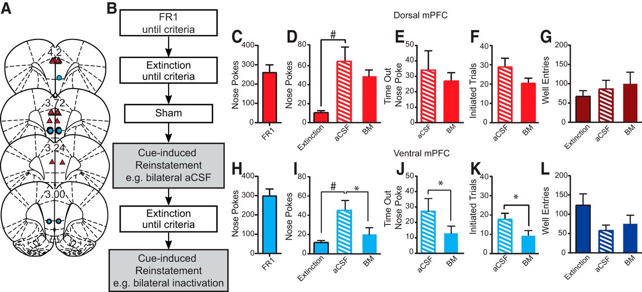
- Figure 3.
Cannula placements, test design, and reinstatement data for reinstatement cohort. A, Dorsal mPFC cannula placements (triangles) and ventral mPFC cannula placements (circles). B, Timeline for reinstatement task. Reinstatement rats were trained on FR1 and extinction but only received bilateral infusion during reinstatement. C, H, Number of nose pokes during FR1 session the day before extinction training. D, I, There was a significant increase in nose pokes on aCSF reinstatement infusion day compared to last day of extinction (#). D–G, Bilateral inactivation of dorsal mPFC did not significantly affect nose pokes, time-out nose pokes, initiated trials, or well entries. I–L, Bilateral ventral mPFC inactivation significantly decreased total number of nose pokes, time out nose pokes, and initiated trials (*), but not rewarded well entries; # and *p < 0.05, paired t test.
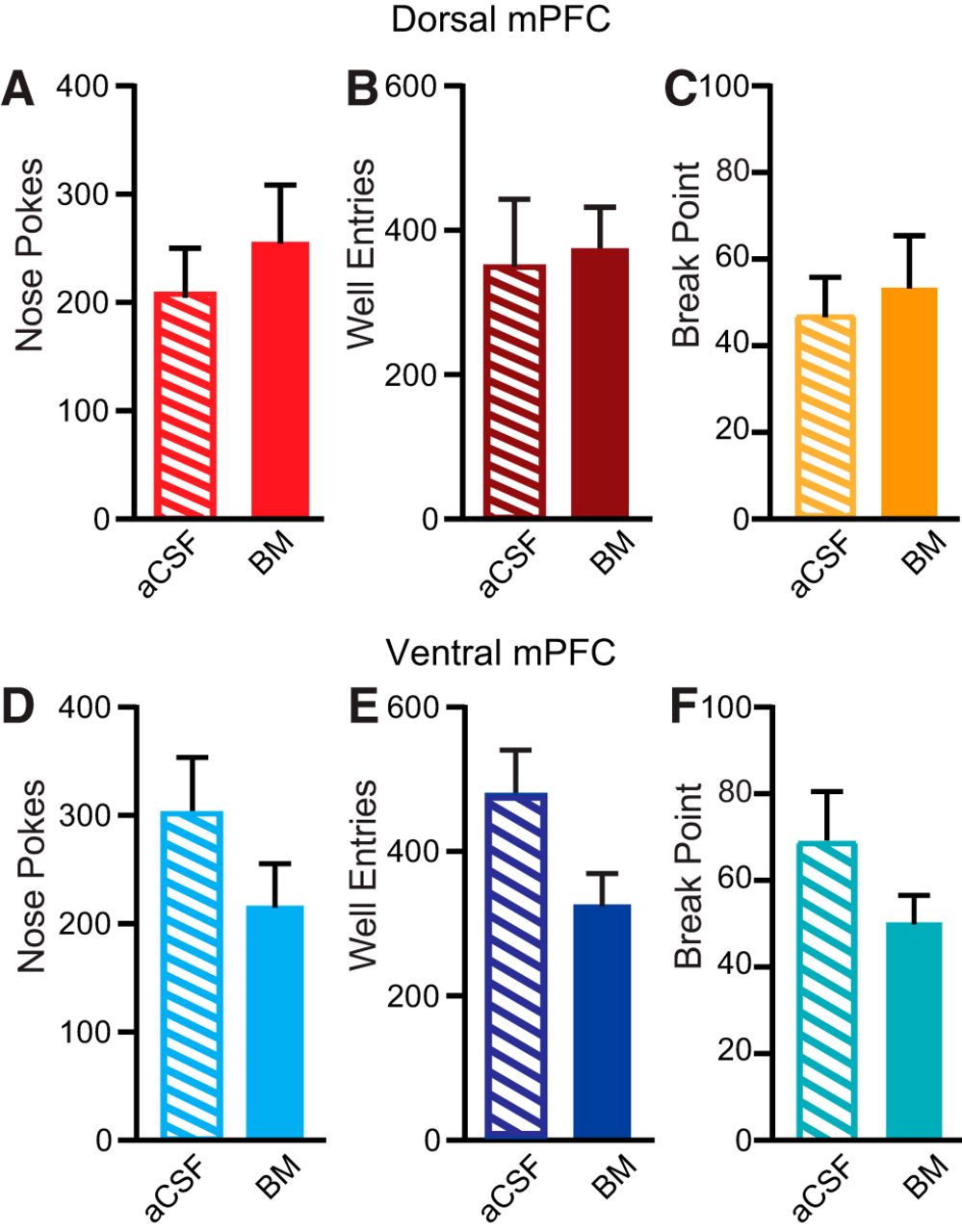
- Figure 4.
PR data. No significant effects of dorsal mPFC (A–C) or ventral mPFC (D–F) inactivation on nose pokes, well entries, or break point.
- Figure 5.
Average number of nose pokes per quartile for FR1 (A, F), early extinction (B, G), late extinction (C, H), cue-induced reinstatement (D, I), and PR (E, J) for during inactivation of dosal mPFC (A–E) or ventral mPFC (F–J). Dorsal mPFC inactivation increased FR1 nose pokes, notably in the first half of the session. Dorsal mPFC inactivation decreased late extinction nose pokes, primarily early in the session. Ventral mPFC inactivation decreased cue-induced reinstatement nose pokes, but the effect was distributed across the session; *p < 0.05, **p < 0.01, two-factor ANOVA (treatment × quartile); #p < 0.05, Sidak’s MCT.
- Figure 6.
Behavioral and physiological verification of BM efficacy. BM infusion in NAc disrupted spontaneous locomotion, and in vitro BM infusion decreased sPSCs in mPFC neurons. A, Cannula placements for locomotion study. B, aCSF-infused rats decreased locomotion over time, but this effect was not observed for rats receiving BM infusions; *p < 0.05, RM ANOVA. C, sPSCs of one representative neuron. D, Mean sPSC frequency before BM, after BM, and after washout; *p < 0.05, Tukey’s multiple comparison test. E, Example recorded rat mPFC neuron stained with Alexa Fluor 488.


In this issue


