
Main menu

  • HOME
  • CONTENT
    • Early Release
    • Featured
    • Current Issue
    • Issue Archive
    • Blog
    • Collections
    • Podcast
  • TOPICS
    • Cognition and Behavior
    • Development
    • Disorders of the Nervous System
    • History, Teaching and Public Awareness
    • Integrative Systems
    • Neuronal Excitability
    • Novel Tools and Methods
    • Sensory and Motor Systems
  • ALERTS
  • FOR AUTHORS
  • ABOUT
    • Overview
    • Editorial Board
    • For the Media
    • Privacy Policy
    • Contact Us
    • Feedback
  • SUBMIT

User menu

Search

  • Advanced search
eNeuro
eNeuro

Advanced Search


  • HOME
  • CONTENT
    • Early Release
    • Featured
    • Current Issue
    • Issue Archive
    • Blog
    • Collections
    • Podcast
  • TOPICS
    • Cognition and Behavior
    • Development
    • Disorders of the Nervous System
    • History, Teaching and Public Awareness
    • Integrative Systems
    • Neuronal Excitability
    • Novel Tools and Methods
    • Sensory and Motor Systems
  • ALERTS
  • FOR AUTHORS
  • ABOUT
    • Overview
    • Editorial Board
    • For the Media
    • Privacy Policy
    • Contact Us
    • Feedback
  • SUBMIT
PreviousNext
Research ArticleNew Research, Cognition and Behavior

The Basolateral Nucleus of the Amygdala Executes the Parallel Processes of Avoidance and Palatability in the Retrieval of Conditioned Taste Aversion in Male Rats

Tadashi Inui, Tomoaki Sugishita, Chizuko Inui-Yamamoto, Yasunobu Yasoshima and Tsuyoshi Shimura
eNeuro 24 June 2019, 6 (4) ENEURO.0004-19.2019; https://doi.org/10.1523/ENEURO.0004-19.2019
Tadashi Inui
1Division of Behavioral Physiology, Department of Behavioral Sciences, Graduate School of Human Sciences, Osaka University, Osaka 565-0871, Japan
  • Find this author on Google Scholar
  • Find this author on PubMed
  • Search for this author on this site
Tomoaki Sugishita
1Division of Behavioral Physiology, Department of Behavioral Sciences, Graduate School of Human Sciences, Osaka University, Osaka 565-0871, Japan
  • Find this author on Google Scholar
  • Find this author on PubMed
  • Search for this author on this site
Chizuko Inui-Yamamoto
2Department of Oral Anatomy and Developmental Biology, Osaka University Graduate School of Dentistry, Osaka 565-0871, Japan
  • Find this author on Google Scholar
  • Find this author on PubMed
  • Search for this author on this site
Yasunobu Yasoshima
1Division of Behavioral Physiology, Department of Behavioral Sciences, Graduate School of Human Sciences, Osaka University, Osaka 565-0871, Japan
  • Find this author on Google Scholar
  • Find this author on PubMed
  • Search for this author on this site
Tsuyoshi Shimura
1Division of Behavioral Physiology, Department of Behavioral Sciences, Graduate School of Human Sciences, Osaka University, Osaka 565-0871, Japan
  • Find this author on Google Scholar
  • Find this author on PubMed
  • Search for this author on this site
  • Article
  • Figures & Data
  • Info & Metrics
  • eLetters
  • PDF
Loading

Article Figures & Data

Figures

  • Tables
  • Figure 1.
    • Download figure
    • Open in new tab
    • Download powerpoint
    Figure 1.

    The experimental chamber. a, The box was separated into a start box and an arena. Animals were put in the start box at the start of the trials. The animals were allowed to enter the arena through the door opened and closed by an experimenter. There was a bay window on the opposite side of the door in the arena. b, The photograph shows a rat approach the bay window after the opening of the door at the start of a trial. c–e, A diagram and photograph of the bay window. The rats put their heads into the bay window as shown in d to lick a spout in front of the bay window. Infrared LEDs below and sensors above the bay window detected the entry of the heads. The metal spout was connected with a touch sensor via a metal clip and wire.
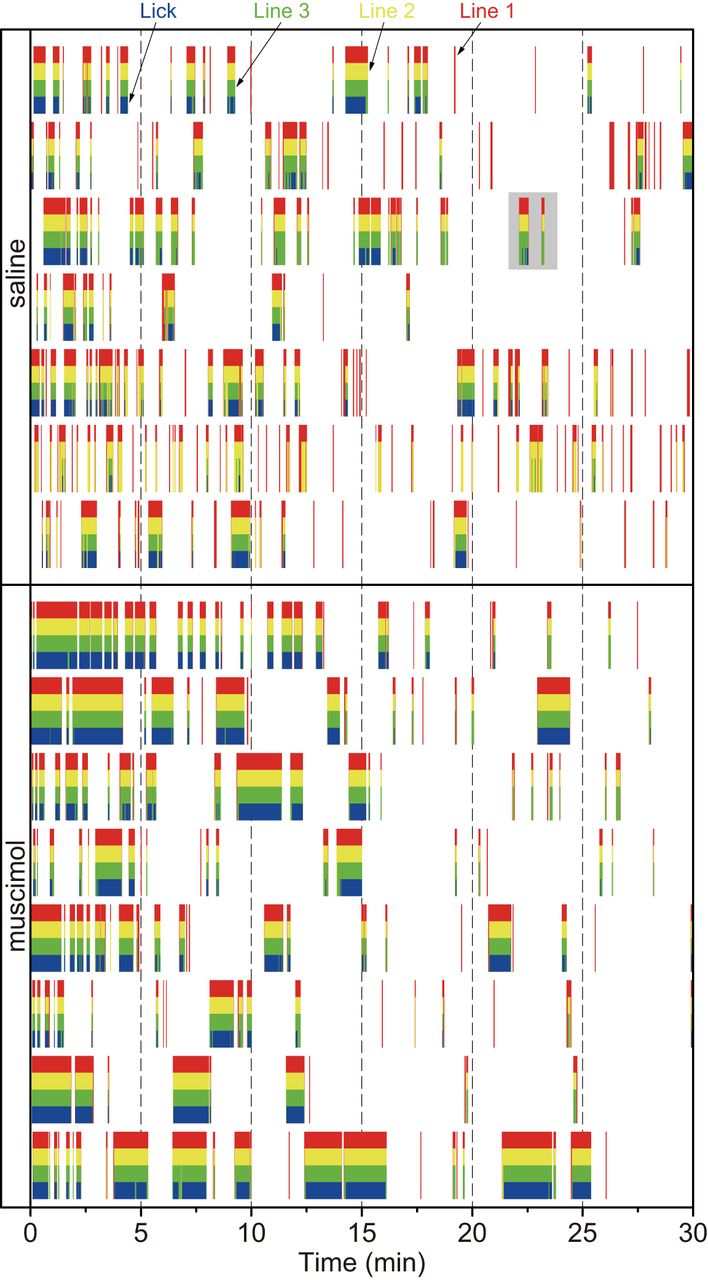
  • Figure 2.
    • Download figure
    • Open in new tab
    • Download powerpoint
    Figure 2.

    Raster plots of the responses of the passing and touch sensors in each animal on Test 2. The red, yellow, and green vertical lines indicate Line 1 (far from the spout), Line 2 (middle of the bay window), and Line 3 (close to the spout), respectively. The red color bars indicate that the rat’s head (tip of nose) stopped at the entrance of the bay window. Both the red and yellow color bars indicate the stop at the middle of the bay window, and the sets of the red, yellow, and green bars indicate the stop just before the spout. The plots with a gray background in the saline group are enlarged in Figure 3 for explaining behavioral analyses.

  • Figure 3.
    • Download figure
    • Open in new tab
    • Download powerpoint
    Figure 3.

    Scheme for behavioral analyses. a, The analysis of the entrance of the rat’s heads into the bay window (Entry). Top, The blocks containing all the color bars indicate Entry-Lick, while the ones containing all the colors without blue indicate Entry-Stop. Bottom, Illustrations showing the animal’s behavior on Entry-Lick and Entry-Stop. b, Schematic diagram of the licking pattern. The vertical lines indicate licks.

  • Figure 4.
    • Download figure
    • Open in new tab
    • Download powerpoint
    Figure 4.

    Histologic findings. The locations of the injection cannulae tips were reconstructed based on the Paxinos and Watson brain atlas (Paxinos and Watson, 2007). Orange circles, muscimol-injected rats; black squares, saline-injected rats.

  • Figure 5.
    • Download figure
    • Open in new tab
    • Download powerpoint
    Figure 5.

    The intra-BLA muscimol altered Entry. a, The mean duration of all Entry in the saline-injected (SAL) and muscimol-injected (MUS) groups on Conditioning, Test 1, and Test 2. After the CS-US pairings, the mean duration of Entry decreased in both groups. The microinjections of muscimol into the BLA on Test 2 impaired the decline in the mean duration of Entry. b, The proportion of the frequency of Entry-Lick and Entry-Stop. The conditioning decreased Entry-Lick and increased Entry-Stop. The MUS group showed smaller proportion of Entry-Stop and larger Entry-Lick than the SAL group on Test 2; ***p < 0.001 (vs SAL), *p < 0.05, †p < 0.05 (vs Conditioning), $$p < 0.01 (vs Test 1).
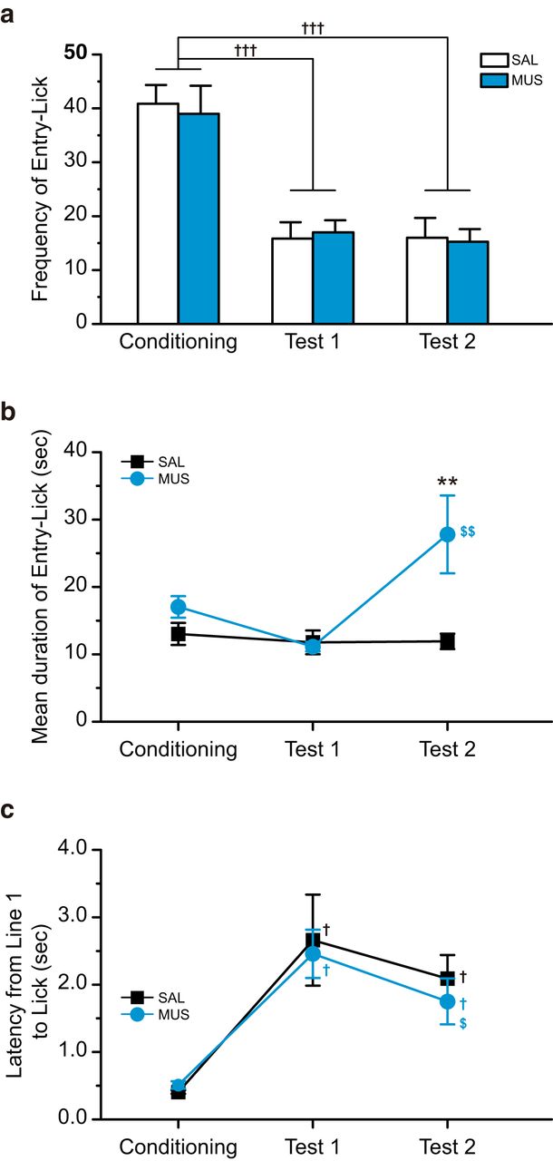
  • Figure 6.
    • Download figure
    • Open in new tab
    • Download powerpoint
    Figure 6.

    The intra-BLA muscimol decreased the mean duration of Entry-Lick. a, The frequency of Entry-Lick in the saline-injected (SAL) and muscimol-injected (MUS) groups on Conditioning, Test 1, and Test 2. The CS-US pairings considerably decreased the frequency of Entry-Lick. Both the SAL and MUS groups showed suppressed frequency of Entry-Lick on Test 2. There was no group difference. b, The mean duration of Entry-Lick. The rats showed a slightly shorter mean duration of Entry-Lick on Test 1 than Conditioning. On Test 2, the SAL group showed the same level of mean duration with Test 1, while the MUS group demonstrated significantly longer mean duration than Test 1 and the SAL group on Test 2. c, The latency from the cut of Line 1 to Lick. The CS-US pairings significantly increased latency. The microinjections of muscimol into the BLA did not alter the latency in Test 2; **p < 0.01 (vs SAL), †††p < 0.001, †p < 0.05 (vs Conditioning), $$p < 0.01, $p < 0.05 (vs Test 1).
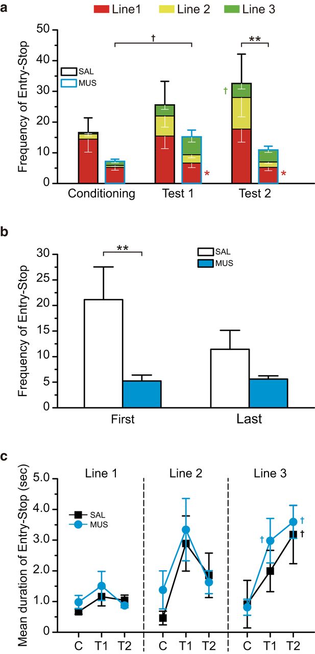
  • Figure 7.
    • Download figure
    • Open in new tab
    • Download powerpoint
    Figure 7.

    The intra-BLA muscimol decreased the frequency of Entry-Stop. a, The frequency of Entry-Lick in the saline-injected (SAL) or muscimol-injected (MUS) group in Conditioning, Test 1, and Test 2. The CS-US pairings increased entry-stop. On test 2, the MUS group showed significantly smaller stops than the SAL group. b, The frequency of Entry-Stop during the first and last half of Test 2. The muscimol-injected group showed smaller frequency of the stop on both the time windows. There was a significant group difference for the first half. c, Mean duration of Entry-Stop at each line. The CS-US pairings significantly increased mean duration at lines 2 and 3. The mean duration at Line 2 decreased in both groups for Test 2 and increased at Line 3 for Test 2. However, there were no differences between the groups. C, Conditioning; T1, Test 1; T2, Test 2; **p < 0.01, *p < 0.05 (vs SAL), †p < 0.01 (vs Conditioning).
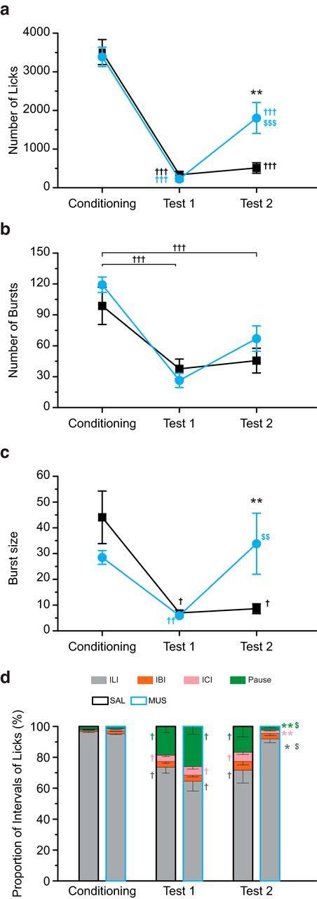
  • Figure 8.
    • Download figure
    • Open in new tab
    • Download powerpoint
    Figure 8.

    The intra-BLA muscimol attenuated aversion to the CS. a, The total number of licks in the saline-injected (SAL) and muscimol-injected (MUS) groups on Conditioning, Test 1, and Test 2. The CS-US pairings significantly decreased the total number of licks. The MUS group showed larger CS licking than the SAL group on Test 2. b, The number of bursts. The CS-US pairings decreased the burst number. There was no group difference for Test 2. c, The size of the burst. The CS-US pairings decreased the burst size. The MUS group demonstrated a larger burst size than the SAL group for Test 2. d, The proportion of ILI. The CS-US pairings increased the proportions of IBI, ICI, and pause, which were smaller in the MUS group than in the SAL group; **p < 0.01, *p < 0.05 (vs SAL), †††p < 0.001, ††p < 0.01, †p < 0.05 (vs Conditioning), $$$p < 0.001, $$p < 0.01, $p < 0.05 (vs Test 1).

  • Figure 9.
    • Download figure
    • Open in new tab
    • Download powerpoint
    Figure 9.

    The intra-BLA muscimol increased larger burst size. Plots for all burst sizes in the saline-injected (SAL) and muscimol-injected (MUS) groups for Test 2. a, The size of almost all the bursts was <100 licks in the SAL group. b, The MUS group showed a larger number of lick bursts containing over 100 licks. c, There was a significant group difference in the average number of >100 lick burst size; *p < 0.05 (vs SAL).

Tables

  • Figures
    • View popup
    Table 1.

    Latency, frequency, and total duration of entry

    ConditioningTest 1Test 2
    SALMUSSALMUSSALMUS
    Latency of 1st entry4.66 ± 0.845.91 ± 0.638.73 ± 0.934.18 ± 0.9413.34 ± 4.274.46 ± 0.87*
    Frequency of entry57.43 ± 5.8946.13 ± 4.8641.43 ± 7.3032.13 ± 4.0267.86 ± 21.426.13 ± 2.91††**
    Total duration of entry534.6 ± 64.14625.93 ± 35.99243.19 ± 41.56239.63 ± 41.57255.67 ± 32.15393.18 ± 62.49
    • ** p < 0.01.

    • ↵* p < 0.05 (vs SAL).

    • †† p < 0.01 (vs Conditioning).

Back to top

In this issue

eneuro: 6 (4)
eNeuro
Vol. 6, Issue 4
July/August 2019
  • Table of Contents
  • Index by author
  • Ed Board (PDF)
Email

Thank you for sharing this eNeuro article.

NOTE: We request your email address only to inform the recipient that it was you who recommended this article, and that it is not junk mail. We do not retain these email addresses.

Enter multiple addresses on separate lines or separate them with commas.
The Basolateral Nucleus of the Amygdala Executes the Parallel Processes of Avoidance and Palatability in the Retrieval of Conditioned Taste Aversion in Male Rats
(Your Name) has forwarded a page to you from eNeuro
(Your Name) thought you would be interested in this article in eNeuro.
CAPTCHA
This question is for testing whether or not you are a human visitor and to prevent automated spam submissions.
Print
View Full Page PDF
Citation Tools
The Basolateral Nucleus of the Amygdala Executes the Parallel Processes of Avoidance and Palatability in the Retrieval of Conditioned Taste Aversion in Male Rats
Tadashi Inui, Tomoaki Sugishita, Chizuko Inui-Yamamoto, Yasunobu Yasoshima, Tsuyoshi Shimura
eNeuro 24 June 2019, 6 (4) ENEURO.0004-19.2019; DOI: 10.1523/ENEURO.0004-19.2019

Citation Manager Formats

  • BibTeX
  • Bookends
  • EasyBib
  • EndNote (tagged)
  • EndNote 8 (xml)
  • Medlars
  • Mendeley
  • Papers
  • RefWorks Tagged
  • Ref Manager
  • RIS
  • Zotero
Respond to this article
Share
The Basolateral Nucleus of the Amygdala Executes the Parallel Processes of Avoidance and Palatability in the Retrieval of Conditioned Taste Aversion in Male Rats
Tadashi Inui, Tomoaki Sugishita, Chizuko Inui-Yamamoto, Yasunobu Yasoshima, Tsuyoshi Shimura
eNeuro 24 June 2019, 6 (4) ENEURO.0004-19.2019; DOI: 10.1523/ENEURO.0004-19.2019
Twitter logo Facebook logo Mendeley logo
  • Tweet Widget
  • Facebook Like
  • Google Plus One

Jump to section

  • Article
    • Abstract
    • Significance Statement
    • Introduction
    • Materials and Methods
    • Results
    • Discussion
    • Conclusion
    • Footnotes
    • References
    • Synthesis
  • Figures & Data
  • Info & Metrics
  • eLetters
  • PDF

Keywords

  • avoidance
  • conditioned taste aversion
  • new method of behavioral experiment
  • palatability
  • retrieval
  • the basolateral nucleus of the amygdala

Responses to this article

Respond to this article

Jump to comment:

No eLetters have been published for this article.

Related Articles

Cited By...

More in this TOC Section

New Research

  • A Very Fast Time Scale of Human Motor Adaptation: Within Movement Adjustments of Internal Representations during Reaching
  • Optogenetic Activation of β-Endorphin Terminals in the Medial Preoptic Nucleus Regulates Female Sexual Receptivity
  • Hsc70 Ameliorates the Vesicle Recycling Defects Caused by Excess α-Synuclein at Synapses
Show more New Research

Cognition and Behavior

  • EEG Signatures of Auditory Distraction: Neural Responses to Spectral Novelty in Real-World Soundscapes
  • Excess neonatal testosterone causes male-specific social and fear memory deficits in wild-type mice
  • The effects of mindfulness meditation on mechanisms of attentional control in young and older adults: a preregistered eye tracking study
Show more Cognition and Behavior

Subjects

  • Cognition and Behavior
  • Home
  • Alerts
  • Follow SFN on BlueSky
  • Visit Society for Neuroscience on Facebook
  • Follow Society for Neuroscience on Twitter
  • Follow Society for Neuroscience on LinkedIn
  • Visit Society for Neuroscience on Youtube
  • Follow our RSS feeds

Content

  • Early Release
  • Current Issue
  • Latest Articles
  • Issue Archive
  • Blog
  • Browse by Topic

Information

  • For Authors
  • For the Media

About

  • About the Journal
  • Editorial Board
  • Privacy Notice
  • Contact
  • Feedback
(eNeuro logo)
(SfN logo)

Copyright © 2025 by the Society for Neuroscience.
eNeuro eISSN: 2373-2822

The ideas and opinions expressed in eNeuro do not necessarily reflect those of SfN or the eNeuro Editorial Board. Publication of an advertisement or other product mention in eNeuro should not be construed as an endorsement of the manufacturer’s claims. SfN does not assume any responsibility for any injury and/or damage to persons or property arising from or related to any use of any material contained in eNeuro.