

Article Figures & Data
- Figure 1.
Schematic illustration of the experimental setup. The ∼1-mm-thick aluminum panels of the electrically-grounded Faraday shielding provides an electromagnetically “quiet” environment. Three orthogonal sets of square coils ∼2 m on edge, following the design of Merritt et al. (1983), allow the ambient geomagnetic field to be altered around the participant’s head with high spatial uniformity; double-wrapping provides an active-sham for blinding of experimental conditions (Kirschvink, 1992b). Acoustic panels on the wall help reduce external noise from the building air ventilation system as well as internal noise due to echoing. A non-magnetic chair is supported on an elevated wooden base isolated from direct contact with the magnetic coils. The battery-powered EEG is located on a stool behind the participant and communicates with the recording computer via an optical fiber cable to a control room ∼20 m away. Additional details are available in Figure 2. This diagram and the center figure for the visual abstract was modified from the figure “Center of attraction,” by C. Bickel (Hand, 2016), with permission.
- Figure 2.
Additional images of critical aspects of the human magnetic exposure at Caltech. A, Partially complete assembly of the Faraday cage (summer of 2014) showing the nested set of orthogonal, Merritt square four-coils (Merritt et al., 1983) with all but two aluminum walls of the Faraday cage complete. B, Image of a participant in the facility seated in a comfortable, non-magnetic wooden chair and wearing the 64-lead BioSim EEG head cap. The EEG sensor leads are carefully braided together to minimize electrical artifacts. The chair is on a raised wooden platform that is isolated mechanically from the magnet coils and covered with a layer of synthetic carpeting; the height is such that the participant’s head is in the central area of highest magnetic field uniformity. C, Schematic of the double-wrapped control circuits that allow active-sham experiments (Kirschvink, 1992b). In each axis of the coils, the four square frames are wrapped in series with two discrete strands of insulated copper magnet wire and with the number of turns and coil spacing chosen to produce a high-volume, uniform applied magnetic field (Merritt et al., 1983). Reversing the current flow in one of the wire strands via a DPDT switch results in cancellation of the external field with virtually all other parameters being the same. This scheme is implemented on all three independently controlled coil axes (Up/Down, East/West, and North/South). D, Fluxgate magnetometer (Applied Physics Systems 520A) three-axis magnetic field sensor attached to a collapsing carbon-fiber camera stand mount. At the start of each session, the fluxgate is lowered to the center of the chamber for an initial current/control calibration of the ambient geomagnetic field. It is then raised to a position ∼30 cm above the participant’s head during the following experimental trials, and the three-axis magnetic field readings are recorded continuously in the same fashion as the EEG voltage signals. E, Air duct. A 15 cm in diameter aluminum air duct ∼2-m-long connects a variable-speed (100 W) electric fan to the upper SE corner of the experimental chamber; this is also the conduit used for the major electrical cables (power for the magnetic coils, sensor leads for the fluxgate, etc.). F, G, An intercom/video monitoring system was devised by mounting a computer-controlled loudspeaker (F) outside the Faraday shield on the ceiling North of the chamber coupled with (G) a USB-linked IR video camera/microphone system mounted just inside the shield. Note the conductive aluminum tape shielding around the camera to reduce Rf interference. During all experimental trials a small DPDT relay located in the control room disconnects the speaker from computer and directly shorts the speaker connections. A second microphone in the control room can be switched on to communicate with the participant in the experimental chamber, as needed. An experimenter monitors the audio and video of participants at all times, as per Caltech IRB safety requirements. H, LED lights, 12 VDC array, arranged to illuminate from the top surface of the magnetic coils near the ceiling of the chamber. These are powered by rechargeable 11.1-V lithium battery packs (visible in E) and controlled by an external switch. I, Ferrite chokes. Whenever possible, these are mounted in a multiple-turn figure-eight fashion (Counselman, 2013) on all conductive wires and cables entering the shielded area and supplemented with grounded aluminum wool when needed. J, Image of the remote-control area including (from left to right): the PC for controlling the coils, the DPDT switches for changing between active and sham modes, the fluxgate control unit, the three power amplifiers that control the current in the remote coil room, and the separate PC that records the EEG data. Participants seated in the experimental chamber do not report being able to hear sounds from the control room and vice versa. Additional guidance for the design of biomagnetic experiments is given by Kirschvink et al. (2010) and Schwarze et al. (2016).
- Figure 3.
Magnetic field rotations used in these experiments. In the first ∼100 ms of each experimental trial, the magnetic field vector was either: (1) rotated from the first preset orientation to the second (SWEEP), (2) rotated from the second preset orientation to the first (also SWEEP), or (3) left unchanged (FIXED). In all experimental trials, the field intensity was held constant at the ambient lab value (∼35 μT). For declination rotations, the horizontal rotation angle was +90° or –90°. For inclination rotations, the vertical rotation angle was either +120°/–120°, or +150°/–150°, depending on the particular inclination rotation experiment. A, Inclination rotations between ±60° and ±75°. The magnetic field vector rotates from downwards to upwards (Inc.UP.N, red) and vice versa (Inc.DN.N, blue), with declination steady at North (0°). B, Declination rotations used in main assay (solid arrows) and vector opposite rotations used to test the quantum compass hypothesis (dashed arrows). In the main assay, the magnetic field rotated between NE (45°) and NW (315°) with inclination held downwards (+60° or +75°) as in the Northern Hemisphere (DecDn.CW.N and DecDn.CCW.N); vector opposites with upwards inclination (−60° or −75°) and declination rotations between SE (135°) and SW (225°) are shown with dashed arrows (DecUp.CW.S and DecUp.CCW.S). C, Identical declination rotations, with static but opposite vertical components, used to test the electrical induction hypothesis. The magnetic field was shifted in the Northerly direction between NE (45°) and NW (315°) with inclination held downwards (+75°, DecDn.CW.N and DecDn.CCW.N) or upwards (−75°, DecUp.CW.S and DecUp.CCW.S). The two dotted vertical lines indicate that the rotations started at the same declination values. In both B, C, counterclockwise rotations (viewed from above) are shown in red, clockwise in blue.
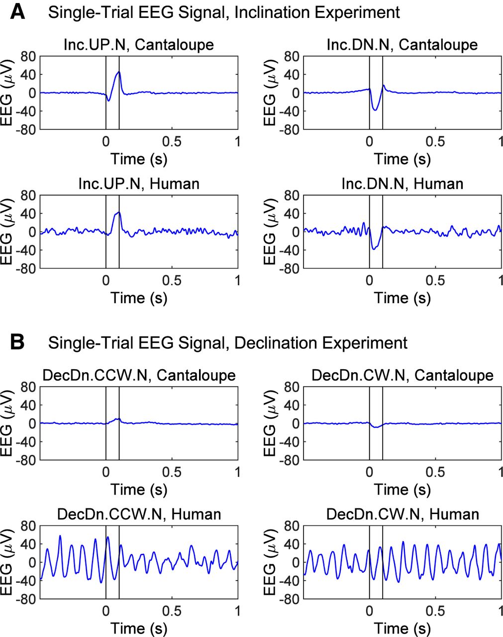
- Figure 4.
Examples of single-trial, time-domain, bandpass-filtered (1–50 Hz) EEG traces at electrode Fz from phantom (cantaloupe) and human participants (one with low and one with high baseline alpha-power) that illustrate the type of data gathered in this study. A, Effect of a 0.1 s inclination sweep of a Northward-pointing, 35-µT magnetic field rotating between a dip of 75° down to 75° up (Inc.UP.N, left panels) and the reverse (Inc.DN.N, right panels). This is the largest stimulus used in our experiments (150° arc, effective frequency 4.2 Hz, with the full vector of 35-µT undergoing rotation). The cantaloupe records an ∼40-µV artifact during the sweep interval but is otherwise flat. A similar artifact can be seen on humans with low alpha-power but is invisible in humans with high alpha-power without trial-averaging. B, Effect of a 0.1-s declination sweep of the horizonal magnetic component (inclination = +75°, total field = 35 µT, so horizontal component = 9.1 µT) rotating from NE to NW in the presence of a static, downward directed vertical magnetic field (33.8 µT; DecDn.CCW.N) and the reverse (DecDn.CW.N). This is a weaker electrical stimulus than used in A (only a 90° arc, a lower effective frequency of 2.5 Hz, and a quarter the field intensity). The cantaloupe shows only a weak artifact of <10 µV during the rotation. In most humans with high or low alpha-power, this artifact is hard to detect without extensive averaging. Artifacts of this sort are phase-locked to the stimulus and are easily removed using standard techniques for analyzing non-phase-locked power as noted in the EEG Methods section. Note that this human example shows an obvious drop in the alpha-power following the CCW rotation but not the CW rotation.
- Figure 5.
Alpha-ERD as a neural response to magnetic field rotation. Post-stimulus power changes (dB) from a pre-stimulus baseline (−500 to −250 ms) plotted according to the ±3-dB color bar at bottom. A, Scalp topography of the alpha-ERD response in an inclination experiment, showing alpha-power at select time points before and after field rotation at 0 s. Alpha-ERD (deep blue) was observed in SWEEP (top row), but not FIXED (bottom row), trials. B, Scalp topography of the alpha-ERD response for two runs of the declination experiment, tested six months apart in a different strongly-responding participant. DecDn.CCW.N condition is shown. In both runs, the response peaked around +500 ms post-stimulus and was widespread over frontal/central electrodes, demonstrating a stable and reproducible response pattern. C, Time-frequency maps at electrode Fz for the same runs shown in B. Black vertical lines indicate the 0- to 100-ms field rotation interval. Pink/white outlines indicate significant alpha-ERD at the p < 0.05 and p < 0.01 statistical thresholds, respectively. Separate runs shown side by side. Significant alpha-ERD was observed following downwards-directed counterclockwise rotations (outlines in top row) with no other power changes reaching significance. Significant power changes appear with similar timing and bandwidth, while activity outside the alpha-ERD response and activity in other conditions is inconsistent across runs.
- Figure 6.
Group results from repeated-measures ANOVA for the effects of geomagnetic stimulation on post-stimulus alpha-power. A, Average alpha-ERD (dB) at electrode Fz in the SWEEP and FIXED conditions of inclination experiments run in active or sham mode. Two-way ANOVA showed an interaction (p < 0.05, N = 29) of inclination rotation (SWEEP vs FIXED) and magnetic stimulation (active vs sham). According to post hoc testing, only inclination sweeps in active mode produced alpha-ERD above background fluctuations in FIXED trials (p < 0.01) or sham mode (p < 0.05). B, Average alpha-ERD (dB) at electrode Fz in the declination experiment with inclination held downwards (DecDn). One-way ANOVA showed a significant main effect of declination rotation (p < 0.001, N = 26). The downwards-directed counterclockwise rotation (DecDn.CCW.N) produced significantly different effects from both the corresponding clockwise rotation (DecDn.CW.N, p < 0.001) and the FIXED control condition (DecDn.FIXED.N, p < 0.001). C, Comparison of the declination rotations with inclination held downwards (DecDn) or upwards (DecUp) in a subset (N = 16 of 26) of participants run in both experiments. Two-way ANOVA showed a significant interaction (p < 0.01) of declination rotation (CCW vs CW vs FIXED) and inclination direction (Dn vs Up). Post hoc testing showed significant differences (p < 0.01) between the DecDn.CCW.N condition and every other condition, none of which were distinct from any other. This is a direct test and rejection of the quantum compass hypothesis. D, Grand average of time-frequency power changes across the 26 participants in the DecDn experiment from B. Black vertical lines indicate the 0- to 100-ms field rotation interval. A post-stimulus drop in alpha-power was observed only following the downwards-directed counterclockwise rotation (left panel). Wider spread of desynchronization reflects interindividual variation. Convolution involved in time/frequency analyses causes the early responses of a few participants to appear spread into the pre-stimulus interval. E, Grand average of time-frequency power changes across the 18 participants with sham data in the declination experiments; no significant power changes were observed.
- Figure 7.
Histogram of alpha-ERD responses over all participants. The panels show the histogram of individual responses for each condition. Frequency is given in number of participants. Because we looked for a drop in alpha-power following magnetic stimulation, the histograms are shifted toward negative values in all conditions. A, Standard DecDn experiment (N = 26). The CCW condition shows the most negative average in a continuous distribution of participant responses with the most participants having a >2-dB response. B, DecUp experiment (N = 16). No significant magnetosensory response was observed in any condition, and no clear difference is apparent between the three distributions. C, Sham declination experiment (N = 18). No significant magnetosensory response was observed in any condition, and no clear difference is apparent between the three distributions.
- Figure 8.
Repeated results from two strongly-responding participants. In both A, B, participants were tested weeks or months apart under the same conditions (run 1 and run 2). Time/frequency maps show similar timing and bandwidth of significant alpha-power changes (blue clusters in outlines) after counterclockwise rotation, while activity outside the alpha-ERD response, and activity in other conditions is inconsistent across runs. Pink/white outlines indicate significance at the p < 0.05 and p < 0.01 thresholds. The participant in A had an alpha-peak frequency at >11 Hz and a lower-frequency alpha-ERD response. The participant in B had an alpha-peak frequency <9 Hz and a higher-frequency alpha-ERD response. Minor power fluctuations in the other conditions or in different frequency bands were not repeated across runs, indicating that only the alpha-ERD was a repeatable signature of magnetosensory processing.


- Table 1.
Group results from two-way, repeated-measures ANOVA for the effects of inclination rotation × magnetic stimulation on post-stimulus alpha-power
ANOVA 1. Effects of inclination rotation and magnetic stimulation on post-stimulus alpha-power Two-way repeated measures ANOVA (N = 29) Inclination rotation × magnetic stimulation F p ηp 2 Main effect of inclination rotation (SWEEP vs FIXED) 3.26 0.08 0.19 Main effect of magnetic stimulation (active vs sham) 2.47 0.13 0.09 Inclination rotation × magnetic stimulation (interaction) 5.67 0.02* 0.17 ANOVA 1 shows a significant interaction of inclination rotation (SWEEP vs FIXED) and magnetic stimulation (active vs sham) in the inclination experiments. Based on post hoc testing, alpha-ERD was significantly greater in SWEEP trials in active mode, compared with all other conditions (p < 0.05). In this table, F is the F-ratio statistic, p the probability value, and ηp 2 the partial η2 value from the ANOVA.
- Table 2.
Group results from one-way, repeated-measures ANOVA for the effects of declination rotation at downwards inclination on post-stimulus alpha-power
ANOVA 2. Effects of declination rotation at downwards inclination on post-stimulus alpha-power One-way repeated measures ANOVA (N = 26) F P ηp 2 Main effect of declination rotation (CCW vs CW vs FIXED) 13.09 0.00003*** 0.34 ANOVA 2 shows a significant main effect of declination rotation when the inclination is static and downwards as in the Northern Hemisphere. Based on post hoc testing, alpha-ERD was significantly greater in CCW trials than in CW or FIXED trials (p < 0.001). F is the F-ratio statistic, p the probability value, and ηp 2 the partial η2 value from the ANOVA.
- Table 3.
Group results from two-way, repeated-measures ANOVA for the effects of declination rotation × inclination direction on post-stimulus alpha-power
ANOVA 3. Effects of declination rotation and inclination direction on post-stimulus alpha-power Two-way repeated measures ANOVA (N = 16) Declination rotation × inclination direction F p ηp 2 Main effect of declination rotation (CCW vs CW vs FIXED) 3.77 0.03* 0.24 Main effect of inclination direction (Dn vs Up) 0.89 0.36 0.06 Declination rotation × inclination direction (interaction) 6.49 0.004*** 0.30 ANOVA 3 shows a significant interaction of declination rotation and inclination direction in declination experiments designed to test the “quantum compass” mechanism of magnetoreception. A significant alpha-ERD difference (p < 0.05) between counterclockwise down (DecDn.CCW.N) and counterclockwise up (DecUp.CCW.S) argues against this hypothesis in humans. F is the F-ratio statistic, p the probability value, and ηp 2 the partial η2 value from the ANOVA.
- Movie 1.
Test of the electrical induction mechanism of magnetoreception using data from a participant with a strong, repeatable alpha-ERD magnetosensory response. Bottom row shows the DecDn.CCW.N, DecDn.CW.N and DecDn.FIXED.N conditions (64 trials per condition) of the DecDn.N experiment; top row shows the corresponding conditions for the DecUp.N experiment. Scalp topography changes from –0.25 s pre-stimulus to +1 s post-stimulus. The CCW rotation of a downwards-directed field (DecDn.CCW.N) caused a strong, repeatable alpha-ERD (lower left panel, p < 0.01 at Fz); weak alpha-power fluctuations observed in other conditions (DecDn.CW.N, DecDn.FIXED.N, DecUp.CW.N, DecUp.CCW.N, and DecUp.FIXED.N) were not consistent across multiple runs of the same experiment. If the magnetoreception mechanism is based on electrical induction, the same response should occur in conditions with identical ∂B/∂t (DecDn.CCW.N and DecUp.CCW.N), but the response was observed only in one of these conditions: a result that contradicts the predictions of the electrical induction hypothesis.
- Movie 2.
Test of the quantum compass mechanism of magnetoreception using data from another strongly-responding participant. Bottom versus top rows compare the DecDn.N and DecUp.S experiments in the CCW, CW, and FIXED conditions (DecDn.CCW.N, DecDn.CW.N, DecDn.FIXED.N, DecUp.CW.S, DecUp.CCW.S, and DecUp.FIXED.S with 100 trials per condition). The quantum compass is not sensitive to magnetic field polarity, so magnetosensory responses should be identical for the DecDn.CCW.N and DecUp.CCW.S rotations sharing the same axis. Our results contradict this prediction. A significant, repeatable alpha-ERD is only observed in the DecDn.CCW.N condition (lower left panel, p < 0.01 at Fz), with no strong, consistent effects in the DecUp.CCW.S condition (top left panel) or any other condition.
In this issue


