

Article Figures & Data
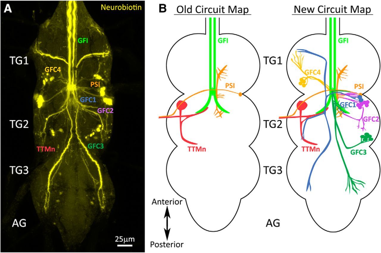
- Figure 1.
Giant fiber interneuron dye injection reveals coupled neurons. A, The GFI iontophoretically injected with neurobiotin (yellow) shows extensive dye coupling to neurons in the TG. The established GFI-coupled neurons are (1) the PSI (orange) and (2) the TTMn (red). The newly identified GFCs project into all three TG segments (TG1–3), but do not extend into the AGs. B, Left, The old GF circuit map showing both of the previously characterized GFI (green) dye-coupled neurons: PSI (orange) and TTMn (red). Right, The new GF circuit map with the addition of all the newly identified GFC neurons from this study: GFC1 (blue), GFC2 (purple), GFC3 (dark green), and GFC4 (yellow).
- Figure 2.
Transgenic Gal4 drivers for the newly identified GFC neurons. Gal4-driven expression of UAS-mCD8::GFP (green, column 1) overlapping with the GFI injection of neurobiotin dye (yellow, column 2) showing the identification of GFC drivers (merge, column 3). Arrows indicate processes with overlapping GFP and NB labeling, and arrowheads show the GFC cell bodies. The GFC neurons are drawn both in color (Fig. 1 color scheme) and perforated outlines to show their bilateral pattern (column 4). TG segments are selected to best show GFC projection architecture. All injections were performed on females. A, 78A06-Gal4 labels GFC1. The driver strength is relatively weak, with a somewhat stochastic labeling of the GFC1 neurons. B, 73C07-Gal4 labels GFC2. This driver is moderately strong, but also labels other neurons. C, 24H07-Gal4 labels GFC3. This driver strength is moderate, with labeling of other neurons. D, 42A06-GAL4 labels both GFC3 and GFC4 neurons. The driver is relatively weak, with stochastic labeling of GFC4 neurons.
- Figure 3.
The GFI interacts with the GFC neurons at the inframedial bridge. Gal4 lines driving UAS-mCD8::GFP (column 1) intersect with the GFI axon revealed by injection of TRITC (column 2), at the GFI IB and the GFI axonal bend (merge, column 3). The first two columns use depth color coding to represent the Z-position within the TG, with more dorsal regions displaying cool colors and ventral regions displaying warm colors (see color scale bar in A, column 2). Arrows indicate overlapping membrane contact between GFCs and GFI at the IB. Arrowheads indicate GFC contact at the GFI axon bend. All injected flies are female. A, GFC1 (78A06-Gal4) interacts with the GFI exclusively at the IB. B, GFC2 (73C07-Gal4) interacts with the GFI at the IB and the GFI axonal bend. C, GFC3 (24H07-Gal4) interacts with the GFI extensively at the IB and the GFI axonal bend. The GFI also produces small side projections that contact GFC3 (inset, arrowheads). D, GFC4 (42A06-Gal4) interacts with the GFI at the IB.
- Figure 4.
GFCs form electrical synapses with the GFI at the inframedial bridge. Electrical synapses between GFI and GFC neurons are shown in Gal4-driven UAS-mCD8::GFP animals (green, column 1) with TRITC dye injection into the GFI (magenta, column 2), while colabeling with the Shaking-B antibody (cyan, column 3). Images were taken using the AiryScan mode of the microscope. The three merged channels (column 4) show the regions of shared ShakB contact between GFI-GFCs. Arrows indicate sites of the GFI–GFC ShakB synaptic contacts (magnified in insets). All injected flies are female. A, GFC1 (78A06-Gal4) makes ShakB electrical synapse contacts with the GFI at the IB. B, GFC2 (73C07-Gal4) forms several ShakB electrical synapse contacts with the GFI. C, GFC2 (73C07-Gal4) contacts the GFI along the axonal bend. D, GFC3 (24H07-Gal4) contacts the GFI with multiple ShakB electrical synapses. E, GFC3 (24H07-Gal4) minimally contacts the GFI along the axonal bend (arrow).
- Figure 5.
Presynaptic and postsynaptic polarity of the newly identified GFC neurons. GFC neuronal polarity is shown using the dendrite/soma label DenMark (magenta) and the presynaptic label synaptotagmin::GFP (Syt::GFP, green). Substacks of the regions of interest for each GFC are shown for DenMark (column 1) and Syt::GFP (column 2), with above and below paired comparisons (image column 3). Arrows indicate the position of the IB. GFC schematic representations are shown (center column), with regions of interest outlined in black boxes. A, GFC1 (78A06-Gal4) processes are labeled by presynaptic Syt::GFP in both TG1 (top) and TG2 (bottom) segments, while the IB is labeled by postsynaptic DenMark. B, GFC2 (73C07-Gal4) processes in TG2 (column 1) are colabeled by both DenMark (column 1) and the Syt::GFP marker (column 2). The IB is labeled by presynaptic Syt::GFP, but also has the DenMark signal (column 4). C, GFC3 (24H07-Gal4) has punctate Syt::GFP within the finger-like processes in TG3 (column 2). The IB is labeled by DenMark, with no Syt::GFP marker (column 4). GFC3 processes along the GFI axonal bend also express the DenMark label (arrowheads).
- Figure 6.
GFC neurons support GF circuit architectural development. A, The GFI labeled by iontophoretically injected TRITC (magenta) reveals the soma (arrow) and dendritic branches (arrowheads) in the brain (top), and descending axon in thoracic ganglion (bottom). Split Gal4 (spGal4) 10B11-AD ∩ 14A06-DBD drives UAS-mCD8::GFP (green) in GFC1 (bottom, arrow) and PSI (bottom, arrowhead). B, Iontophoretic NB injection into the GFI (yellow) in the UAS-hid/+ control reveals the GFI (arrows) interconnected by the GCI (arrowheads) in the brain (top) and normal dye coupling in the thoracic ganglion (bottom). C, Driving UAS-hid with spGal4 10B11-AD ∩ 14A06-DBD results in the loss of GFC1 with occasional PSI survival (arrowhead). When GFC1 is ablated, the GCI labeling is often lost (top), one of the GFI axons is typically absent, and the remaining GFI axon always extends a compensatory contralateral axon projection (arrow). All NB injections were performed on males. D, Schematic representations of GF circuit outcomes with UAS-hid/+ controls and spGal4 10B11-AD ∩ 14A06-DBD-driven UAS-hid cell ablation. Not pictured are instances where neither GFC1 nor PSI are ablated, and instances where both GFIs are absent. E, Frequency of each GF circuit outcome with the targeted spGal4 10B11-AD ∩ 14A06-DBD-driven UAS-hid cell ablation. The pie chart color is coded to dots at the bottom of schematics in D. The sample size for UAS-hid/+ genetic controls is 21 animals, and for the spGal4 cell ablation it is 20 animals.


GFI GCI TTMn PSI GFC1 GFC2 GFC3 R14A01 R32C04 R25D08 R26E04 R93E07 R13C08 R44D02 VT004455 R74E09 R88F07 R75E05 R87D02 R77C12 R58E04 VT042336 VT002209 VT038335 VT030598 VT059438 VT043662 R75D03 New Gal4 drivers (distinct from those used in this study) that express selectively within the GF circuit, as compiled from the Janelia FlyLight and Vienna Tiles library collections. Selective lines for GFC4 have not been uncovered and thus are not reported here.
- Movie 1.
3D animation of GFC1 and GFI interaction. Animated 3D reconstruction of mCD8::GFP-labeled GFC1 (green) and TRITC-injected GFI (magenta) in thoracic ganglion segments 1 and 2 (TG1/2). GFC1 intersects with the GFI in a narrow projection that crosses the IB. This projection then splits to create claw-like synaptic terminals in TG1–3 (TG3 not pictured). Scale bar, 20 μm.
- Movie 2.
3D animation of GFC2 and GFI interaction. Animated 3D reconstruction of mCD8::GFP-labeled GFC2 (green) and TRITC-injected GFI (magenta) in TG1/2. GFC2 extends a large TG2 loop with dorsal projections. GFC2 intersects with the GFI extensively at the IB and to a lesser extent at the tip of the TG2 axonal bend. Scale bar, 20 μm.
- Movie 3.
3D animation of GFC3 and GFI interaction. Animated 3D reconstruction of mCD8::GFP-labeled GFC3 (green) and TRITC-injected GFI (magenta) in TG2/3. GFC3 cell bodies project processes to the IB and contact the GFI, with extensive branching, including along the GFI axonal bends. GFC3 then projects into TG3 to terminate. Scale bar, 20 μm.
- Movie 4.
3D animation of GFC4 and GFI interaction. Animated 3D reconstruction of mCD8::GFP-labeled GFC4 (green) and TRITC-injected GFI (magenta) in TG1/2. GFC4 cell bodies project processes from TG1 to the IB, then reverse course and return to TG1 where they terminate. Scale bar, 20 μm.
- Movie 5.
3D animation of ShakB electrical synapses between GFC1 and GFI at IB. Animated 3D reconstruction of mCD8::GFP-labeled GFC1 (green), TRITC-injected GFI (magenta), and anti-ShakB electrical synapse labeling (cyan). The simple passing dendrite of GFC1 interacts with the GFI at multiple locations within the IB. Multiple sites of ShakB electrical synapses indicate direct GFC1–GFI coupling. Scale bar, 5 μm.
- Movie 6.
3D animation of ShakB synapses between GFC3 and GFI at the axonal bend. Animated 3D reconstruction of mCD8::GFP-labeled GFC3 (green), TRITC-injected GFI (magenta), and anti-ShakB electrical synapse labeling (cyan). GFC3 extensively contacts the GFI along the GFI axonal bends in TG2. Despite this extensive contact, there are minimal ShakB punctae (cyan) shared between the neurons. Scale bar, 5 μm.
- Movie 7.
3D animation of ShakB synapses between GFC2 and GFI at IB. Animated 3D reconstruction of mCD8::GFP-labeled GFC2 (green), TRITC-injected GFI (magenta), and anti-ShakB electrical synapse labeling (cyan). The GFC2 field interacts in multiple locations with the GFI, including several side projections from the IB. Several sites of ShakB electrical synapses indicate GFC2–GFI coupling. Scale bar, 5 μm.
- Movie 8.
3D animation of ShakB synapses between GFC2 and GFI at axonal bend. Animated 3D reconstruction of mCD8::GFP-labeled GFC2 (green), TRITC-injected GFI (magenta), and anti-ShakB electrical synapse labeling (cyan). GFC2 contacts the GFI along the TG2 axonal bends, mostly at the tips. Along these contact sites, there are few to no ShakB contacts (cyan) shared between the neurons. Scale bar, 5 μm.
- Movie 9.
3D animation of ShakB synapses between GFC3 and GFI at IB. Animated 3D reconstruction of mCD8::GFP-labeled GFC3 (green), TRITC-injected GFI (magenta), and anti-ShakB electrical synapse labeling (cyan). GFC3 extends the largest dendritic field at the IB, with extensive GFC3–GFI contact. Several of these contact points are positive for ShakB electrical synapses. Scale bar, 5 μm.
In this issue


