

Article Figures & Data
Figures
- Figure 1.
Survey of cryopreservation media for the storage of primary cortical neurons. A, Experimental design and timeline for validation of cryostored neuron performance. Neurons were concurrently plated from a fresh dissection or cryostored aliquot and evaluated on indicated DIV. B, Percentage of recovered and lost cells after cryostorage in test media CS10, CS5, SAF, or 50% FBS-40% DMEM-10% DMSO (50:40:10). Percentage of “recovered” cells was calculated with ((# cells alive + # cells dead) ÷ (# cells in initial aliquot)) × 100. Percentage of “lost” cells was calculated with 100% recovered. Mean ± SD, N = 3 source dissections. C, Post-thaw viability of cells cryostored in test media compared to the viability of freshly dissected cells as evaluated by Trypan blue exclusion. Mean ± SD, N = 3 source dissections. D, Table displaying summarized data from A, B (white columns) and calculated data (gray columns). Viable cell yield for each experiment was calculated by (percentage recovered × percentage viable) ÷ 100. Efficiency was determined by normalizing viable cell yield to the average viability of a fresh dissection. E, Immunofluorescence labeling of neuron-specific β-III-tubulin (orange) and fluorescently-conjugated phalloidin labeling of the actin cytoskeleton (blue) of DIV7 cells cryostored in test media. Scale bar = 100 μm.
- Figure 2.
Long-term survival of neurons cryostored in CS10 only slightly lower than a fresh dissection. A, Cell survival after cryostorage and recovery in test media compared to freshly dissected cells at DIV3. Live cells were labeled by calcein (green), and dead cells are indicated by ethidium homodimer labeling (magenta). Scale bar = 100 μm. B, Quantification of live/dead assay at DIV3. Table displays average change in percentage survival of cryostored cells versus fresh cells. Mean ± SD, N = 3 source dissections, n > 2300 cells per condition. C, D, Immunofluorescence labeling of fresh or cryostored cells fixed at DIV4. Cells were labeled with antibodies against the neuron-specific marker β-III-tubulin (C, D; orange), astrocyte marker S100 (C; blue), or microglia marker Iba1 (D; magenta). Scale bar = 100 μm. E, Percentage of cell population at DIV4 labeled with β-III-tubulin or S100. Mean ± SD, N = 3 source dissections, n > 1250 cells per condition. F, Percentage of cell population at DIV4 labeled with β-III-tubulin or Iba1. Mean ± SD, N = 3 source dissections, n > 250 cells per condition. All statistical comparisons made by unpaired parametric t test (B, E) or Mann–Whitney U test (F). n.s. = not significant, *p < 0.05, **p < 0.01, ***p < 0.001.
- Figure 3.
Expression of key neurodevelopmental genes is unchanged following recovery from cryostorage. RT-qPCR of RNA samples from fresh or CS10-cryostored cortical cultures collected at DIV12. CTs were normalized to housekeeping genes and relative copy numbers were generated using 2-ΔCT × 106. Floating bar graph spans the minimum and maximum data points, vertical line denotes mean. N = 3 source dissections. All statistical comparisons made by unpaired parametric t test with Holm–Sidak correction for multiple comparisons. Multiplicity adjusted p value for each comparison listed in table (right).
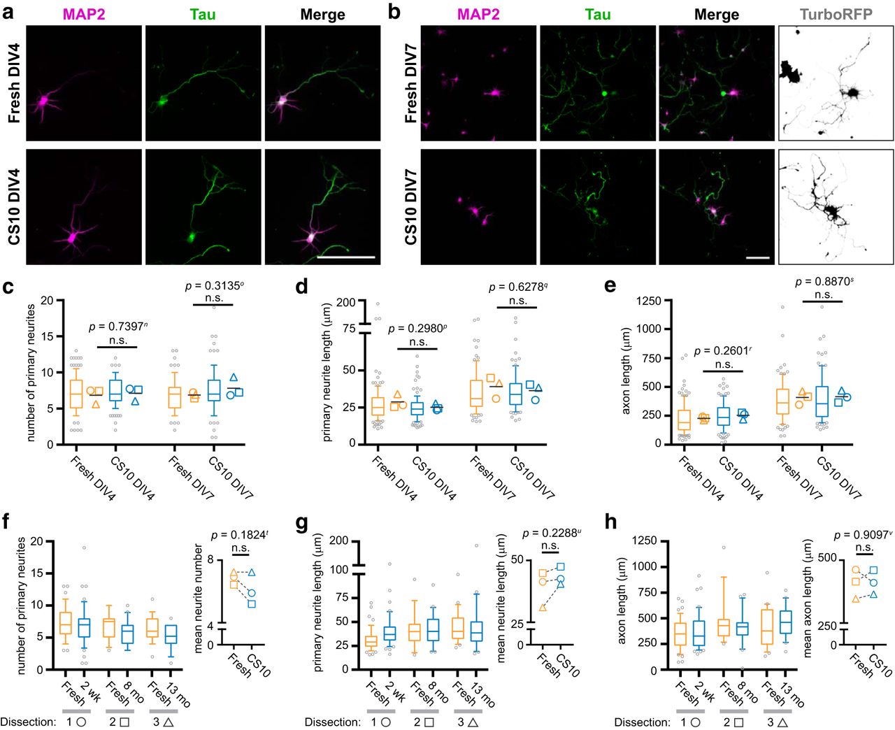
- Figure 4.
Cryostorage does not alter neuron morphogenesis. A, B, Immunofluorescence labeling of freshly dissected or CS10-cryostored cells, using antibodies against the dendrite marker MAP2 (magenta) and axon marker Tau (green). Cells were fixed on DIV4 (A) or DIV7 (B). Cultures grown to DIV7 were also infected with a sub-saturating titer of cytosolic TurboRFP-encoding lentivirus to aid in tracing (inverted white). Scale bar = 100 μm. C–E, Quantification of number of primary neurites (C), primary neurite length (D), and axon length (E) of traced cortical neurons at indicated timepoints and conditions. Box-and-whisker plots show data pooled from three independent experiments; whiskers indicate 10th to 90th percentile, box indicates 1st to 3rd quartile, center line is median, gray points are data outside 10th to 90th percentile. Symbols (○◻△) denote the mean value calculated for each biological replicate, horizontal line is the grand mean. N = 3 source dissections, n > 90 neurons per condition and time point. Statistical comparisons between biological replicate means made by unpaired parametric t test. F–H, Quantification of number of primary neurites (F), primary neurite length (G), and axon length (H) of freshly dissected cortical neurons compared to aliquots from the same source dissection cryostored for indicated durations, and traced at DIV7. Box-and-whisker plots show data from each experiment. n > 14 neurons per condition and time point. Inset graph plots mean for each dataset, symbols (○◻△) denote each source dissection, dashed line connects fresh-CS10 matched pairs. Statistical comparisons between matched means made by paired parametric t test. n.s. = not significant. Fresh DIV7 morphometric data for each dissection were extracted from C–E.
- Figure 5.
Neuron arborization and branch complexity are unaffected by cryostorage. A, Diagram of Sholl analysis with indicated metrics: enclosing radius, maximum number of intersections (Nm), critical radius (rc), and Sholl regression coefficient (k). B, C, Representative masks of neuron tracings illustrate number of intersections (spectrum scale bar) detected by Sholl analysis of DIV4 (B) and DIV7 (C) cortical neurons from indicated conditions. Scale bar = 100 μm. D, E, Sholl profiles of freshly dissected (orange) or CS10-cryostored (blue) neurons at DIV4 (D) and DIV7 (E). Data are represented as mean (solid line, left y-axis) ± SD (shading); number of neurons (n; dashed line, right y-axis). F–I, Quantification of enclosing radius (F), Nm (G), rc (H), and k (I) of Sholl-analyzed tracings at indicated timepoints and conditions. Box-and-whisker plots show data pooled from three independent experiments; whiskers indicate 10th to 90th percentile, box indicates 1st to 3rd quartile, center line is median, gray points are data outside 10th to 90th percentile. Symbols (○◻△) denote the mean value calculated for each biological replicate, horizontal line is the grand mean. N = 3 source dissections, n > 90 neurons per condition and time point. Statistical comparison between mean Sholl profiles at each radius made by unpaired parametric t test with Holm–Sidak correction for multiple comparisons (D, E), and statistical comparison between biological replicate means for Sholl metrics made by unpaired parametric t test (F–I). n.s. = not significant.
- Figure 6.
Synaptogenesis occurs normally in neurons following cryostorage and recovery. A–C, Neurons expressing Lifeact-mRuby2 (inverted white) from indicated conditions were fixed at DIV12 (A), DIV14 (B), and DIV16 (C), and immunolabeled using antibodies against excitatory postsynaptic marker PSD95 (NeuroMab; green), inhibitory postsynaptic marker gephyrin (GPHN; cyan), and presynaptic marker synaptophysin (SYP; magenta). Bottom panel is magnified segment of boxed dendrite region. Scale bar = 100 μm (top) and 5 μm (bottom panel). D, Density of all actin-rich protrusions from secondary dendrites at indicated timepoints and conditions. E, Ratio of dendritic spines to dendritic filopodia as determined by presence or absence of PSD95 on actin-rich protrusions at indicated timepoints and conditions. Box-and-whisker plots (D, E) show data pooled from three independent experiments; whiskers indicate 10th to 90th percentile, box indicates 1st to 3rd quartile, center line is median, gray points are data outside 10th to 90th percentile. Symbols (○◻△) denote the mean value calculated for each biological replicate, horizontal line is the grand mean. F, Percentage of PSD95-positive excitatory synapses and GPHN-positive inhibitory synapses at DIV16 from indicated conditions. Mean ± SD, N = 3 source dissections, n = 6–7 neurons per condition. All statistical comparisons between biological replicate means made by unpaired parametric t test. n.s. = not significant.
- Figure 7.
Cryopreserved cortical neurons have normal spontaneous and stimulated activity. A, Resting membrane potential of fresh and CS10-cryostored neurons recorded by whole-cell patch clamp. Scatterplot of data points pooled from one to two biological replicates. Mean ± SD, N = 1–2 source dissections, n = 10–14 neurons per condition. B, Examples of whole-cell patch clamp recordings of neurons exhibiting spontaneous activity from fresh or CS10-cryostored neurons at DIV12–DIV14. C, Representative tracings of evoked APs after 100-pA current injection for 50 ms (red). D–F, Quantification of evoked AP parameters including amplitude (D), rise time (E), and half-width (F). Scatterplot of data points pooled from one to two biological replicates. Mean ± SD, N = 1–2 source dissections, n = 7–11 neurons per condition. G, Fluorescent images of calcium imaging experiments showing the representative response of CS10-cryostored neurons to the indicated trigger at DIV14. Baseline (left column) Fura-2 fluorescence F340/F380 ratio, and peak (right column) Fura-2 fluorescence F340/F380 ratio, in response to 15-s stimulation with the indicated trigger. Scale bar = 100 μm. H, Traces of mean change in Fura-2 fluorescence F340/F380 ratio over time for the indicated triggers and conditions. I, Summary bar graphs showing the peak fluorescence response (as variation from baseline, adjusted for background) for the indicated triggers. Mean ± SEM, N = 1 source dissection, n > 75 neurons per condition. J, Glutamate release by DIV14 neurons from indicated conditions in non-depolarizing solution (basal), after 15 min of stimulation with KCl (stimulated), and after return to non-depolarizing solution (post-stim). Glutamate release at each time point is presented as a percentage of the total glutamate released. K, Fold change of total glutamate released by CS10-cryostored neurons relative to fresh. Mean ± SEM, N = 3 source dissections. Statistical comparisons made by unpaired parametric t test (A, I–K) or Mann–Whitney U test (D–F). n.s. = not significant.
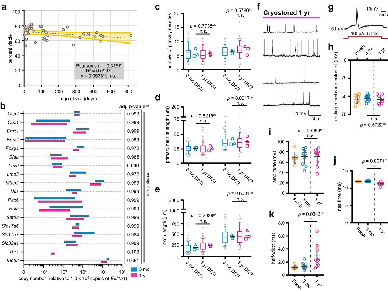
- Figure 8.
Duration of cryostorage does not impact several essential neuronal properties but may affect electrophysiological performance. A, Viability of cells cryostored in CS10 media and recovered at various times after freezing. Dashed gray line = linear regression; yellow shaded area = 95% confidence band. N = 12 source dissections, n = 38 cryostored aliquots. Statistical analysis performed by Pearson’s correlation test. n.s. = not significant. B, RT-qPCR of RNA samples from DIV12 cortical cultures cryostored for three months or one year. CTs were normalized to housekeeping genes and relative copy numbers were generated using 2-ΔCT × 106. Floating bar graph spans the minimum and maximum data points, vertical line denotes mean. N = 2 source dissections, n = 3 thawed aliquots. Statistical comparisons made by unpaired parametric t test with Holm–Sidak correction for multiple comparisons. Multiplicity adjusted p value for each comparison listed in table (right). C–E, Quantification of number of primary neurites (C), primary neurite length (D), and axon length (E) of traced cortical neurons at indicated timepoints. Box-and-whisker plots show data pooled from three independent experiments; whiskers indicate 10th to 90th percentile, box indicates 1st to 3rd quartile, center line is median, gray points are data outside 10th to 90th percentile. Symbols (○◻△) denote the mean value calculated for each biological replicate, horizontal line is the grand mean. N = 2 source dissections, n > 65 neurons per condition and time point. Statistical comparisons between biological replicate means made by unpaired parametric t test (C–E). F, Examples of whole-cell patch clamp recordings of spontaneous activity from neurons recovered after one-year cryostorage in CS10 at DIV12–DIV14. G, Representative tracing of evoked APs after 100-pA current injection for 50 ms (red). H, Resting membrane potential of neurons recovered after one-year cryostorage in CS10 recorded by whole-cell patch clamp, alongside freshly dissected and three month-cryostored neurons reproduced from Figure 7 for comparison (separated by dashed line). Scatterplot of data points pooled from one to two biological replicates. Mean ± SD, N = 1–2 source dissections, n = 8–14 neurons per condition. I–K, Quantification of evoked AP parameters including amplitude (I), rise time (J), and half-width (K) of neurons recovered after one-year cryostorage in CS10, alongside freshly dissected and three month-cryostored neurons reproduced from Figure 7 for comparison (separated by dashed line). Scatterplot of data points pooled from one to two biological replicates. Mean ± SD, N = 1–2 source dissections, n = 7–11 neurons per condition. Statistical comparisons made by one-way ANOVA with Tukey’s test for multiple comparisons (H) or Kruskal–Wallis test with Dunn’s test for multiple comparisons (I–K). n.s. = not significant, *p < 0.05, **p < 0.01.
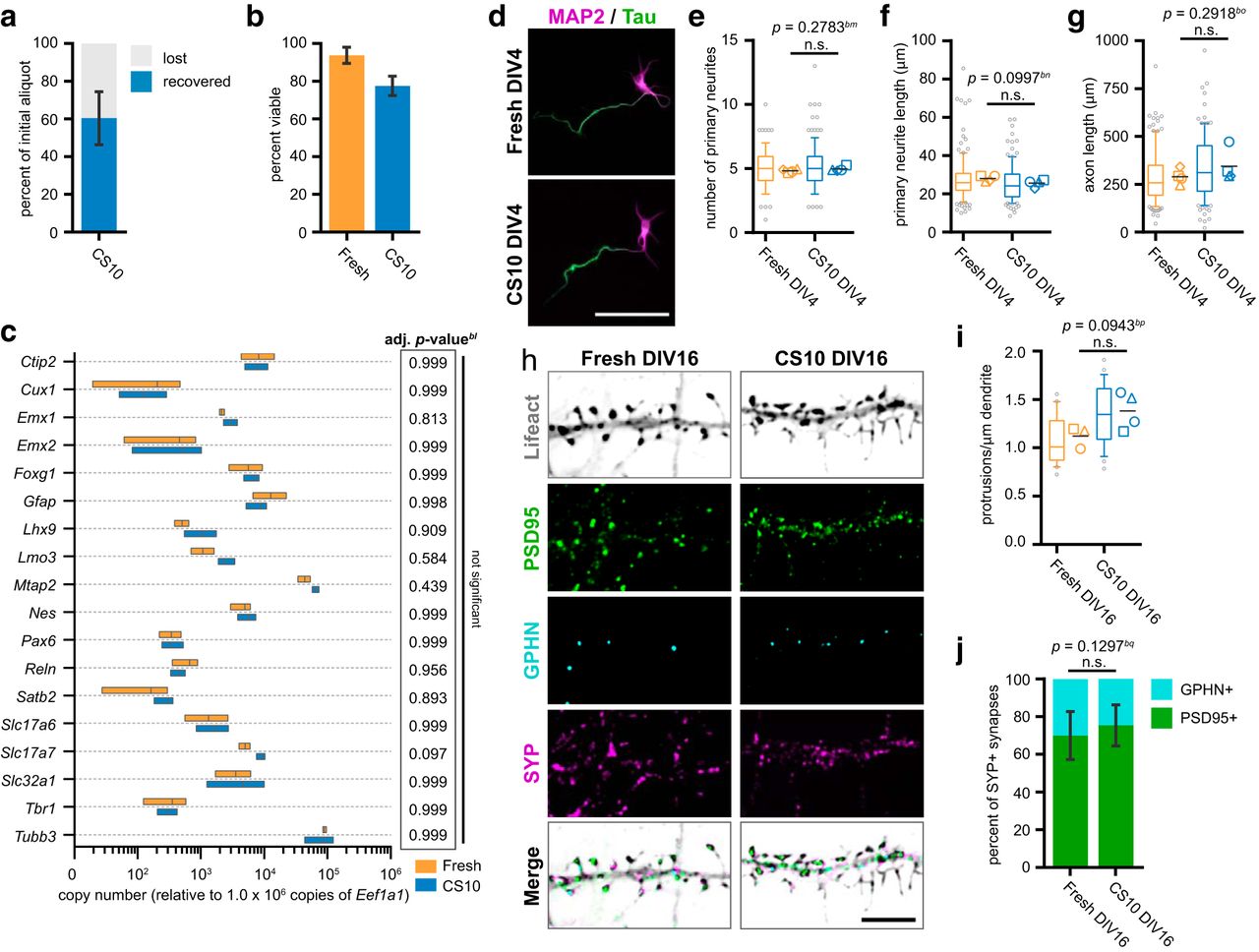
- Figure 9.
CS10 is effective for the cryostorage of primary hippocampal neurons. A, Percentage of recovered and lost hippocampal cells after cryostorage in CS10. Percentage of “recovered” cells was calculated with ((# cells alive + # cells dead) ÷ (# cells in initial aliquot)) × 100. Percentage of “lost” cells was calculated with 100% recovered. Mean ± SD, N = 7 source dissections. B, Post-thaw viability of CS10-cryostored hippocampal cells compared to the viability of freshly dissected cells as evaluated by Trypan blue exclusion. Mean ± SD, N = 7 source dissections. C, RT-qPCR of RNA samples from fresh or CS10-cryostored hippocampal cultures collected at DIV12. CTs were normalized to housekeeping genes and relative copy numbers were generated using 2-ΔCT × 106. Floating bar graph spans the minimum and maximum data points, vertical line denotes mean. N = 3 source dissections. Statistical comparisons made by unpaired parametric t test with Holm–Sidak correction for multiple comparisons. Multiplicity adjusted p value for each comparison listed in table (right). D, Immunofluorescence labeling of freshly dissected or CS10-cryostored hippocampal neurons at DIV4, using antibodies against the dendrite marker MAP2 (magenta) and axon marker Tau (green). Scale bar = 100 μm. E–G, Quantification of number of primary neurites (E), primary neurite length (F), and axon length (G) of freshly dissected or CS10-cryostored hippocampal neurons at DIV4. Box-and-whisker plots show data pooled from four independent experiments; whiskers indicate 10th to 90th percentile, box indicates 1st to 3rd quartile, center line is median, gray points are data outside 10th to 90th percentile. Symbols (○◻△◇) denote the mean value calculated for each biological replicate, horizontal line is the grand mean. N = 4 source dissections, n > 115 neurons per condition. H, Immunofluorescence labeling of freshly dissected or CS10-cryostored hippocampal neurons expressing Lifeact-mRuby2 (inverted white) at DIV16, using antibodies against excitatory postsynaptic marker PSD95 (Novus; green), inhibitory postsynaptic marker gephyrin (GPHN; cyan), and presynaptic marker synaptophysin (SYP; magenta). Scale bar = 5 μm. I, Density of actin-rich protrusions from secondary dendrites ay DIV16 from indicated conditions. J, Percentage of PSD95-positive excitatory synapses and GPHN-positive inhibitory synapses at DIV16 from indicated conditions. Mean ± SD, N = 4 source dissections, n > 16 neurons per condition. All statistical comparisons between biological replicate means made by unpaired parametric t test (E–G, I, J). n.s. = not significant.


Tables
Target name Source RRID Dilution Iba1 Rb polyclonal, Wako (019-19741) RRID:AB_839504 1:500 S100 Rb polyclonal, Dako (Z0311) RRID:AB_10013383 1:400 Olig2 Ms monoclonal, MilliporeSigma (MABN50, 211F1.1) RRID:AB_10807410 1:500 β-III-tubulin Rb monoclonal, Cell Signaling (5568, D71G9) RRID:AB_10694505 1:200 β-III-tubulin Ms monoclonal, MilliporeSigma (MAB1637, TU-20) RRID:AB_2210524 1:200 Tau Ms monoclonal, MilliporeSigma (MAB3420, PC1C6) RRID:AB_94855 1:750 MAP2 Rb monoclonal, Cell Signaling (8707, D5G1) RRID:AB_2722660 1:200 PSD951 Ms monoclonal, UC Davis/NIH NeuroMab Facility (75-028 K28/43) RRID:AB_2292909 1:100 PSD952 Ms monoclonal, Novus Biologicals (NB300-556, 6G6-1C9) RRID: AB_2092366 1:500 Synaptophysin Ck polyclonal, Synaptic Systems (101 006) RRID:AB_2722661 1:500 Gephyrin Rb polyclonal, Alomone (AIP-005) RRID:AB_2722662 1:500 Target name Sequence Direction Source Eef1a1 CAACATCGTCGTAATCGGACA Fwd PrimerBank, Harvard GTCTAAGACCCAGGCGTACTT Rev Rpl29 CAAGTCCAAGAACCACACCAC Fwd PrimerBank, Harvard GCAAAGCGCATGTTCCTCAG Rev Pax6 TACCAGTGTCTACCAGCCAAT Fwd PrimerBank, Harvard TGCACGAGTATGAGGAGGTCT Rev Emx1 GAAGAATCACTACGTGGTGGG Fwd Terrigno et al. (2008) CCGTTTGTATTTTGTCCTCCGA Rev Emx2 GGCTAGAGCACGCTTTTGAG Fwd Terrigno et al. (2008) CACCGGTTAATGTGGTGTGT Rev Mtap2 CTCCTCGCAGGGGTGTATCA Fwd PrimerBank, Harvard GTCCGTCGTGCTGAAGAGA Rev Tbr1 CGCCCTCCTCCATCAAATCCATCG Fwd Terrigno et al. (2008) GCAGTTCTTCTCGCAGTCCCGC Rev Reln TTACTCGCACCTTGCTGAAAT Fwd PrimerBank, Harvard CAGTTGCTGGTAGGAGTCAAAG Rev Sat2b GCCGTGGGAGGTTTGATGATT Fwd PrimerBank, Harvard ACCAAGACGAACTCAGCGTG Rev Cux1 TGACCTGAGCGGTCCTTACA Fwd PrimerBank, Harvard TGGGGCCATGCCATTTACATC Rev Lhx9 TCCAAAACGCACGAGCCAA Fwd Terrigno et al. (2008) CAGGTCTGTTAAAGTGGTCGC Rev Lmo3 ACACGAAGGCTAACCTTATCCT Fwd Terrigno et al. (2008) AGTTTCCCGTTACACCAAACAG Rev Gap43 TGGTGTCAAGCCGGAAGATAA Fwd PrimerBank, Harvard GCTGGTGCATCACCCTTCT Rev Slc17a6 CTGAGAAGAAGGCTCCGCTAT Fwd PrimerBank, Harvard ATGCCGAAGGATATGCAGAAG Rev Slc32a1 ACCTCCGTGTCCAACAAGTC Fwd PrimerBank, Harvard TCAAAGTCGAGATCGTCGCAG Rev Tubb3 GCCAAGTTCTGGGAGGTCAT Fwd PrimerBank, Harvard GGGCACATACTTGTGAGAGGA Rev Gfap ACCAGCTTACGGCCAACAG Fwd PrimerBank, Harvard CCAGCGATTCAACCTTTCTCT Rev Bcl11b CCCGACCCTGATCTACTCAC Fwd PrimerBank, Harvard GGAGGTGGACTGCTCTTGT Rev Nes CCCCTTGCCTAATACCCTTGA Fwd PrimerBank, Harvard GCCTCAGACATAGGTGGGATG Rev Foxg1 CACTTTGAGTTACAACGGGACC Fwd PrimerBank, Harvard CGAGTTTTGAGTCAACACGGA Rev Figure Data structure Test CI of difference of mean/median p value a 2B, Fresh v CS10 Normal Unpaired parametric two-tailed t test -0.2311 to 0.1168 0.4131 b 2B, Fresh v CS5 Normal Unpaired parametric two-tailed t test -0.4476 to -0.1619 0.0041 c 2B, Fresh v SAF Normal Unpaired parametric two-tailed t test -0.6306 to -0.3999 0.0002 d 2B, Fresh v 50:40:10 Normal Unpaired parametric two-tailed t test -0.5666 to -0.3437 0.0003 e 2E, Fresh v CS10 Normal Unpaired parametric two-tailed t test 4.188 to 22.27 0.0153 f 2E, Fresh v CS5 Normal Unpaired parametric two-tailed t test 4.207 to 46.59 0.0292 g 2E, Fresh v SAF Normal Unpaired parametric two-tailed t test 34.23 to 78.22 0.0021 h 2E, Fresh v 50:40:10 Normal Unpaired parametric two-tailed t test 19.57 to 78.16 0.0098 U i 2F, Fresh v CS10 Nongaussian Mann–Whitney U test -2.87 to 0.3846 (median) 0.4000 2 j 2F, Fresh v CS5 Nongaussian Mann–Whitney U test -2.22 to 0.3846 (median) 0.9999 4 k 2F, Fresh v SAF Nongaussian Mann–Whitney U test -3.76 to 0.3846 (median) 0.9999 4 l 2F, Fresh v 50:40:10 Nongaussian Mann–Whitney U test -1.11 to 0.3846 (median) 0.4000 2 n adj p value m 3 Normal Unpaired parametric t test, multiple comparisons using Holm–Sidak N/A 0.0050-
0.838918 genes 0.0857
-0.9999n 4C, DIV4 Normal Unpaired parametric two-tailed t test -1.982 to 2.565 0.7397 o 4C, DIV7 Normal Unpaired parametric two-tailed t test -1.337 to 3.233 0.3135 p 4D, DIV4 Normal Unpaired parametric two-tailed t test -12.02 to 4.785 0.2980 q 4D, DIV7 Normal Unpaired parametric two-tailed t test -17.28 to 11.79 0.6278 r 4E, DIV4 Normal Unpaired parametric two-tailed t test -29 to 78.07 0.2601 s 4E, DIV7 Normal Unpaired parametric two-tailed t test -119.5 to 133.5 0.8870 t 4F Normal Paired parametric two-tailed t test -2.378 to 0.8645 0.1824 u 4G Normal Paired parametric two-tailed t test -15.4 to 6.631 0.2288 v 4H Normal Paired parametric two-tailed t test -121.6 to 129 0.9097 n adj p value w 5D Normal Unpaired parametric t test, multiple comparisons using Holm–Sidak N/A 0.0018-
0.99731207 radii 0.8864
-0.9999x 5E Normal Unpaired parametric t test, multiple comparisons using Holm–Sidak N/A 0.0128-
0.99741610 radii >0.9999 y 5F, DIV4 Normal Unpaired parametric two-tailed t test -40.41 to 38.08 0.9382 z 5F, DIV7 Normal Unpaired parametric two-tailed t test -125.3 to 133.2 0.9367 aa 5G, DIV4 Normal Unpaired parametric two-tailed t test -2.537 to 1.095 0.3320 ab 5G, DIV7 Normal Unpaired parametric two-tailed t test -1.448 to 1.8464 0.7453 ac 5H, DIV4 Normal Unpaired parametric two-tailed t test -2.214 to 2.161 0.9746 ad 5H, DIV7 Normal Unpaired parametric two-tailed t test -29.84 to 11.27 0.2780 ae 5I, DIV4 Normal Unpaired parametric two-tailed t test -0.2869 to 0.08355 0.2022 af 5I, DIV7 Normal Unpaired parametric two-tailed t test -0.1067 to 0.1267 0.8237 ag 6D, DIV12 Normal Unpaired parametric two-tailed t test -0.357 to 0.246 0.6362 ah 6D, DIV14 Normal Unpaired parametric two-tailed t test -0.3671 to 0.08601 0.1601 ai 6D, DIV16 Normal Unpaired parametric two-tailed t test -0.3259 to 0.2687 0.8026 aj 6E, DIV12 Normal Unpaired parametric two-tailed t test -0.2159 to 0.4635 0.3689 ak 6E, DIV14 Normal Unpaired parametric two-tailed t test -1.22 to 0.871 0.6674 al 6E, DIV16 Normal Unpaired parametric two-tailed t test -1.186 to 2.116 0.4776 am 6F Normal Unpaired parametric two-tailed t test -0.001536 to 0.1601 0.0537 an 7A Normal Unpaired parametric two-tailed t test -2.46 to 5.546 0.4326 U ao 7D Nongaussian Mann–Whitney U test -13.7 to 17.23 (median) 0.4120 29 ap 7E Nongaussian Mann–Whitney U test -0.02334 to 0.1633 (median) 0.2350 22.5 aq 7F Nongaussian Mann–Whitney U test -0.2073 to 0.6074 (median) 0.5362 28 ar 7I (glutamate) Normal Unpaired parametric two-tailed t test -0.2091 to 0.3324 0.6538 as 7I (glycine) Normal Unpaired parametric two-tailed t test -0.1241 to 0.1893 0.6822 at 7I (KCl) Normal Unpaired parametric two-tailed t test -0.0722 to 0.5019 0.1416 au 7I (NMDA) Normal Unpaired parametric two-tailed t test -0.1396 to 0.1386 0.9943 av 7J (basal) Normal Unpaired parametric two-tailed t test -0.009 to 0.008 0.9194 aw 7J (stim) Normal Unpaired parametric two-tailed t test -0.219 to 0.024 0.8919 ax 7J (post) Normal Unpaired parametric two-tailed t test -0.018 to 0.017 0.9343 ay 7K Normal Unpaired parametric two-tailed t test -0.1198 to 0.0239 0.1375 r R 2 az 8A Normal Pearson’s correlation -0.5771 to 0.0044 0.0535 -0.3157 0.0997 n adj p value ba 8B Normal Unpaired parametric t test, multiple comparisons using Holm–Sidak N/A 0.0060
-0.999118 genes 0.1026
-0.9999bb 8C, DIV4 Normal Unpaired parametric two-tailed t test -2.717 to 2.174 0.7735 bc 8C, DIV7 Normal Unpaired parametric two-tailed t test -2.219 to 3.455 0.5780 bd 8D, DIV4 Normal Unpaired parametric two-tailed t test -6.521 to 7.760 0.8215 be 8D, DIV7 Normal Unpaired parametric two-tailed t test -14.37 to 17.44 0.8017 bf 8E, DIV4 Normal Unpaired parametric two-tailed t test -61.79 to 156.9 0.2936 bg 8E, DIV7 Normal Unpaired parametric two-tailed t test -137.4 to 208.1 0.6001 F adj p value bh 8H Normal One-way ANOVA, Tukey’s post hoc -3.239 to 7.774 0.5623 (2,29) = 0.5873 0.5722 H bi 8I Nongaussian Kruskal–Wallis test, Dunn’s post hoc N/A 0.6433 0.8824 0.9999 bj 8J Nongaussian Kruskal–Wallis test, Dunn’s post hoc N/A 0.0094 9.332 0.0071 bk 8K Nongaussian Kruskal–Wallis test, Dunn’s post hoc N/A 0.0070 9.927 0.0343 n adj p value bl 9C Normal Unpaired parametric t test, multiple comparisons using Holm–Sidak N/A 0.0057
-0.925118 genes 0.0976
-0.9997bm 9E Normal Unpaired parametric two-tailed t test -0.1507 to 0.4369 0.2783 bn 9F Normal Unpaired parametric two-tailed t test -5.375 to 0.6141 0.0997 bo 9G Normal Unpaired parametric two-tailed t test -48.93 to 136.5 0.2918 bp 9I Normal Unpaired parametric two-tailed t test -0.0644 to 0.5853 0.0943 bq 9J Normal Unpaired parametric two-tailed t test -0.0166 to 0.1254 0.1297
In this issue


