
Main menu

  • HOME
  • CONTENT
    • Early Release
    • Featured
    • Current Issue
    • Issue Archive
    • Blog
    • Collections
    • Podcast
  • TOPICS
    • Cognition and Behavior
    • Development
    • Disorders of the Nervous System
    • History, Teaching and Public Awareness
    • Integrative Systems
    • Neuronal Excitability
    • Novel Tools and Methods
    • Sensory and Motor Systems
  • ALERTS
  • FOR AUTHORS
  • ABOUT
    • Overview
    • Editorial Board
    • For the Media
    • Privacy Policy
    • Contact Us
    • Feedback
  • SUBMIT

User menu

Search

  • Advanced search
eNeuro
eNeuro

Advanced Search


  • HOME
  • CONTENT
    • Early Release
    • Featured
    • Current Issue
    • Issue Archive
    • Blog
    • Collections
    • Podcast
  • TOPICS
    • Cognition and Behavior
    • Development
    • Disorders of the Nervous System
    • History, Teaching and Public Awareness
    • Integrative Systems
    • Neuronal Excitability
    • Novel Tools and Methods
    • Sensory and Motor Systems
  • ALERTS
  • FOR AUTHORS
  • ABOUT
    • Overview
    • Editorial Board
    • For the Media
    • Privacy Policy
    • Contact Us
    • Feedback
  • SUBMIT
PreviousNext
Research ArticleMethods/New Tools, Novel Tools and Methods

Auditory Brainstem Responses to Continuous Natural Speech in Human Listeners

Ross K. Maddox and Adrian K. C. Lee
eNeuro 31 January 2018, 5 (1) ENEURO.0441-17.2018; https://doi.org/10.1523/ENEURO.0441-17.2018
Ross K. Maddox
1Department of Biomedical Engineering, University of Rochester, Rochester, NY 14627
2Department of Neuroscience, University of Rochester, Rochester, NY 14642
3Del Monte Institute for Neuroscience, University of Rochester, Rochester, NY 14642
  • Find this author on Google Scholar
  • Find this author on PubMed
  • Search for this author on this site
  • ORCID record for Ross K. Maddox
Adrian K. C. Lee
4Institute for Learning and Brain Sciences, University of Washington, Seattle, WA 98195
5Department of Speech and Hearing Sciences, University of Washington, Seattle, WA 98105
  • Find this author on Google Scholar
  • Find this author on PubMed
  • Search for this author on this site
  • ORCID record for Adrian K. C. Lee
  • Article
  • Figures & Data
  • Info & Metrics
  • eLetters
  • PDF
Loading

Article Figures & Data

Figures

  • Figure1
    • Download figure
    • Open in new tab
    • Download powerpoint
  • Figure 1.
    • Download figure
    • Open in new tab
    • Download powerpoint
    Figure 1.

    Acoustic stimuli. A–C. Pressure waveforms for one second of speech, Poisson click train, and standard periodic click train, respectively. Vertical scale is plotted in arbitrary units (AU), but is consistent across plots. D–F, Spectrograms of a smaller excerpt of the above stimuli, with darker colors corresponding to higher power. G–I, PSD plots of the above stimuli, calculated from 30 s of data using Welch’s method with a segment length of 5.67 ms, segment overlap of 50%, and Hann window. Note that although the speech recordings were gently high-pass filtered at 1000 Hz, there remains plenty of power in the 125- to 1000-Hz range (G) and pitch information is clearly preserved (D; vertical striations between 200 and 300 ms correspond to individual glottal pulses).

  • Figure 2.
    • Download figure
    • Open in new tab
    • Download powerpoint
    Figure 2.

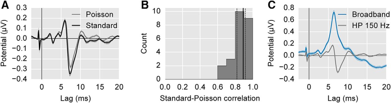
    Comparison of ABR to standard periodic click trains and Poisson click trains. A, The average ABR waveform evoked by the standard, periodic click train at 44.1 clicks/s (black) and the pseudorandom Poisson click train (gray; 44.1 clicks/s overall rate). Areas show ±1 SEM. Both responses are high-pass filtered at 150 Hz. The spike at −1 ms is a stimulus artifact, and occurs before 0 ms to compensate for the 1 ms tube delay of the earphones. B, The histogram of per-subject correlation coefficients between the standard and Poisson click-evoked ABRs. Solid/dotted black lines show median/quartiles. C, Comparison of the Poisson click-evoked ABR with 150-Hz high-pass filtering (gray) and without (i.e., broadband; blue). The latter is used as the benchmark response for the remainder of the study.

  • Figure 3.
    • Download figure
    • Open in new tab
    • Download powerpoint
    Figure 3.

    Comparison of click-evoked responses (blue) with speech-derived responses (red). A, The average waveform across subjects (areas show ±1 SEM). B, The histogram of correlation coefficients between the click-evoked and speech-derived stimuli for each subject. Solid/dotted black lines show median/quartiles. C, Individual subject responses, sorted by descending correlation coefficient. The correlation is shown in the upper right corner.

  • Figure 4.
    • Download figure
    • Open in new tab
    • Download powerpoint
    Figure 4.

    Correlation of speech-derived and click-evoked Wave V latencies (A) and amplitudes (B) across subjects. Because the click-evoked Wave V is known to be subcortical, the strong correlations across subjects point to brainstem neural generators for the speech-derived response as well. Points have been jittered slightly to prevent visual overlap. Regression lines are shown with the 95% confidence interval shaded.

  • Figure 5.
    • Download figure
    • Open in new tab
    • Download powerpoint
    Figure 5.

    Comparison of female-narrated responses (green) with male-narrated responses (purple). A, The average waveforms across subjects (areas show ±1 SEM). B, The histogram of correlation coefficients between the female-evoked and male-evoked stimuli for each subject. Solid/dotted black lines show median/quartiles. The speech-derived ABRs from the male and female narrators show strong similarities, but are not identical, indicating some talker dependence.

  • Figure 6.
    • Download figure
    • Open in new tab
    • Download powerpoint
    Figure 6.

    SNR as a function of data accumulation. SNR was calculated for each subject using the mean of all the data up to each recording epoch by computing the variance in the ABR time interval 0–20 ms, and in the prestimulus noise interval −125 to −10 ms.

  • Figure 7.
    • Download figure
    • Open in new tab
    • Download powerpoint
    Figure 7.

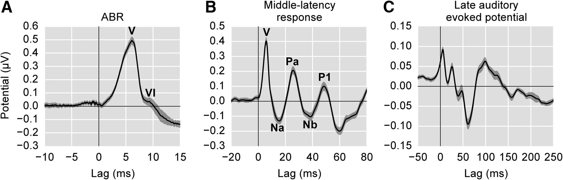
    Changes to the range of lags and filtering parameters allow early, middle, and late responses to be analyzed from the same recording. A, The average speech-derived ABR with Canonical Waves V and VI labeled. B, The middle latency response with its canonical waves labeled (low-pass frequency: 200 Hz). C, The late auditory evoked potential (low-pass frequency: 20 Hz). Due to adaptation, the amplitudes for the later waves are much smaller than typically seen in the event-related potential literature. These peaks are also not given canonical labels as in A, B because their latencies do not directly correspond to the standard N1 and P2 peaks. Shaded areas show ±1 SEM.

Back to top

In this issue

eneuro: 5 (1)
eNeuro
Vol. 5, Issue 1
January/February 2018
  • Table of Contents
  • Index by author
Email

Thank you for sharing this eNeuro article.

NOTE: We request your email address only to inform the recipient that it was you who recommended this article, and that it is not junk mail. We do not retain these email addresses.

Enter multiple addresses on separate lines or separate them with commas.
Auditory Brainstem Responses to Continuous Natural Speech in Human Listeners
(Your Name) has forwarded a page to you from eNeuro
(Your Name) thought you would be interested in this article in eNeuro.
CAPTCHA
This question is for testing whether or not you are a human visitor and to prevent automated spam submissions.
Print
View Full Page PDF
Citation Tools
Auditory Brainstem Responses to Continuous Natural Speech in Human Listeners
Ross K. Maddox, Adrian K. C. Lee
eNeuro 31 January 2018, 5 (1) ENEURO.0441-17.2018; DOI: 10.1523/ENEURO.0441-17.2018

Citation Manager Formats

  • BibTeX
  • Bookends
  • EasyBib
  • EndNote (tagged)
  • EndNote 8 (xml)
  • Medlars
  • Mendeley
  • Papers
  • RefWorks Tagged
  • Ref Manager
  • RIS
  • Zotero
Respond to this article
Share
Auditory Brainstem Responses to Continuous Natural Speech in Human Listeners
Ross K. Maddox, Adrian K. C. Lee
eNeuro 31 January 2018, 5 (1) ENEURO.0441-17.2018; DOI: 10.1523/ENEURO.0441-17.2018
Twitter logo Facebook logo Mendeley logo
  • Tweet Widget
  • Facebook Like
  • Google Plus One

Jump to section

  • Article
    • Visual Abstract
    • Abstract
    • Significance Statement
    • Introduction
    • Materials and Methods
    • Results
    • Discussion
    • Acknowledgments
    • Footnotes
    • References
    • Synthesis
    • Author Response
  • Figures & Data
  • Info & Metrics
  • eLetters
  • PDF

Keywords

  • auditory brainstem response
  • deconvolution
  • electroencephalography
  • encoding model
  • speech

Responses to this article

Respond to this article

Jump to comment:

No eLetters have been published for this article.

Related Articles

Cited By...

More in this TOC Section

Methods/New Tools

  • Superficial Bound of the Depth Limit of Two-Photon Imaging in Mouse Brain
  • A Toolbox of Criteria for Distinguishing Cajal–Retzius Cells from Other Neuronal Types in the Postnatal Mouse Hippocampus
  • Assessment of Spontaneous Neuronal Activity In Vitro Using Multi-Well Multi-Electrode Arrays: Implications for Assay Development
Show more Methods/New Tools

Novel Tools and Methods

  • Superficial Bound of the Depth Limit of Two-Photon Imaging in Mouse Brain
  • A Toolbox of Criteria for Distinguishing Cajal–Retzius Cells from Other Neuronal Types in the Postnatal Mouse Hippocampus
  • Assessment of Spontaneous Neuronal Activity In Vitro Using Multi-Well Multi-Electrode Arrays: Implications for Assay Development
Show more Novel Tools and Methods

Subjects

  • Novel Tools and Methods
  • Home
  • Alerts
  • Follow SFN on BlueSky
  • Visit Society for Neuroscience on Facebook
  • Follow Society for Neuroscience on Twitter
  • Follow Society for Neuroscience on LinkedIn
  • Visit Society for Neuroscience on Youtube
  • Follow our RSS feeds

Content

  • Early Release
  • Current Issue
  • Latest Articles
  • Issue Archive
  • Blog
  • Browse by Topic

Information

  • For Authors
  • For the Media

About

  • About the Journal
  • Editorial Board
  • Privacy Notice
  • Contact
  • Feedback
(eNeuro logo)
(SfN logo)

Copyright © 2025 by the Society for Neuroscience.
eNeuro eISSN: 2373-2822

The ideas and opinions expressed in eNeuro do not necessarily reflect those of SfN or the eNeuro Editorial Board. Publication of an advertisement or other product mention in eNeuro should not be construed as an endorsement of the manufacturer’s claims. SfN does not assume any responsibility for any injury and/or damage to persons or property arising from or related to any use of any material contained in eNeuro.