

Article Figures & Data
Figures
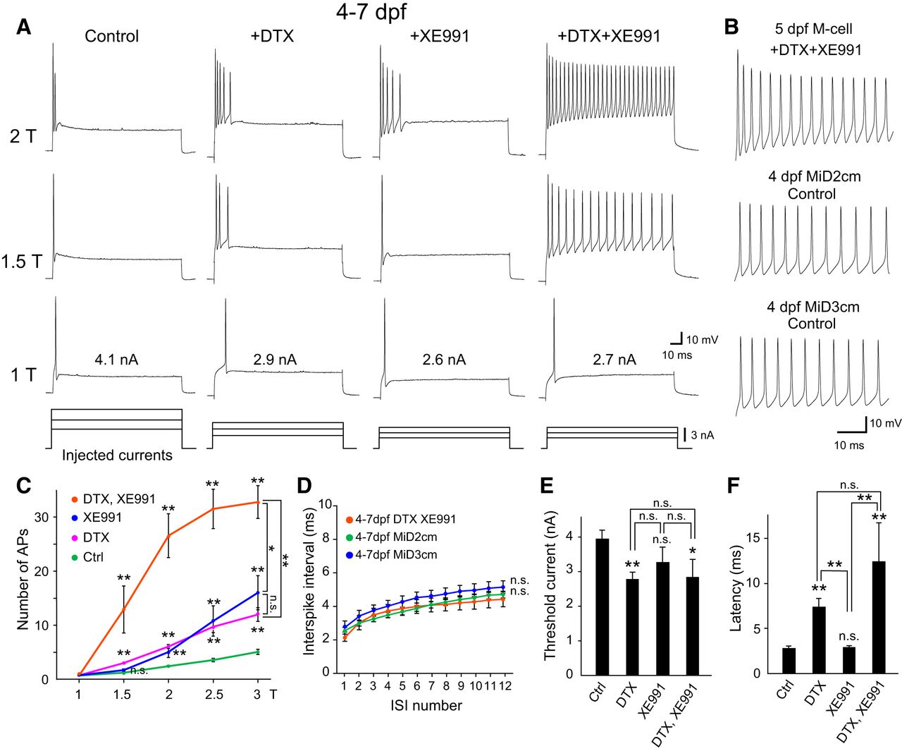
- Figure 1.
Effect of DTX and/or XE991 on single spiking in developed M cells. A, Firing response of M cells at 4–7 dpf [upper three rows of traces elicited by step depolarizing currents (lower traces) of 1×, 1.5×, and 2× threshold intensity (T), as represented in nA on 1T traces] before and after bath application of either 100 nM DTX, 10 μM XE991, or both. B, Initial phase of repetitive firing in response to 2T current injection recorded from M cells with DTX and XE991 treatment, and untreated (control) MiD2cm and MiD3cm cells at 4 dpf. C, Number of APs elicited during a depolarizing current pulse (100 ms) plotted against current intensity (T). The number of APs significantly increased after combined DTX and XE991 treatment compared with each separately. D, Summary data for the ISI of repetitive firing in response to 2T current injection against the interval number. After combined DTX and XE991 treatment, M cells at 4–7 dpf exhibited ISIs that were similar to untreated MiD2cm and MiD3cm cells at the same developmental stage. E, F, The effect of DTX and/or XE991 on threshold current (E) and onset latency of the first spike at 1T (F). Treatment of DTX, but not XE991, reduced the threshold current and increased spiking latency of M cells. n.s., not significant. *p < 0.05, **p < 0.01, Mann–Whitney U test (control, n = 14; +DTX, n = 8; +XE991, n = 7; +DTX + XE991, n = 6). Two-way repeated measures ANOVA was used for ISI (MiD2cm, n = 8; MiD3cm, n = 7).
- Figure 2.
Effect of DTX and/or XE991 on phasic bursting of immature M cells. A, Firing response from M cells at 2 dpf. XE991 treatment caused phasic bursting M cells (control) to fire tonically (+XE991, +DTX + XE991), whereas DTX treatment did not (except for a slight delay of spike onset). B, Summary graph of the blocker effect at 2 dpf showing that the number of APs elicited during 100-ms current pulses significantly increased after XE991 treatment. C, In controls, M cells at 2 dpf showed spike accommodation with a gradual increase in ISI, whereas regular spiking with constant ISI was observed after XE991 treatment. Note that DTX application had no additional effect. D, The threshold current was significantly reduced after combined DTX and XE991 treatment. E, Spike latency greatly increased after XE991 treatment. n.s., not significant. *p < 0.05, **p < 0.01, Mann–Whitney U test (control, n = 6; +DTX, n = 6; +XE991, n = 10; +DTX + XE991, n = 7). Two-way repeated measures ANOVA was used for ISI.
- Figure 3.
Expression of Kv7.4/Kcnq4 mRNA in M cells during development. A, Phylogenetic tree of α-subunit proteins of XE991-sensitive voltage-gated K+ channel Kv7/KCNQ family members in zebrafish, Xenopus, chick, mouse, and human (z, x, c, m, and h as the gene prefix, respectively), and showing identification of zebrafish Kv7.4 (magenta). B, Dorsal views of rhombomeres 4, 5, and 6 (r4, r5, and r6) in the hindbrain at 2 dpf (top) and 5 dpf (bottom) after in situ hybridization using a Kv7.4 antisense probe (green). Kv7.4 mRNA was expressed in M cells (arrowhead) at 2 and 5 dpf, but not in MiD2cm and MiD3cm cells, labeled (magenta) immunohistochemically (with 3A10 antibody, at 2 dpf) or retrogradely (at 5 dpf). Scale bar, 20 μm.
- Figure 4.
Gating properties of zebrafish Kv7.4 and Kv1.1 with/without Kvβ2 in Xenopus oocytes. A, Voltage-gated outward currents recorded from Xenopus oocytes expressing zebrafish Kv7.4 (top), Kv1.1a alone (middle), or Kv1.1a with Kvβ2b subunit (bottom). Command voltages (V) ranging from −80 to +50 mV in 10 mV steps were applied for 200 ms. B, Conductance (G, μs) at peak current amplitude was plotted as a function of the command voltage. C, The G-V relationship normalized to maximum value (Gmax) shows that currents elicited above −60 mV had different open probabilities. D, E, Rise time to half-maximum activation (t1/2) of Kv7.4 (D) above −60 mV was significantly different from Kv1.1a with and without Kvβ2b (E; p < 0.01). **p < 0.01, Kruskal-Wallis test with post hoc Bonferroni correction (Kv7.4, n = 20; Kv1.1a, n = 32; Kv1.1a + Kvβ2b, n = 26).
- Figure 5.
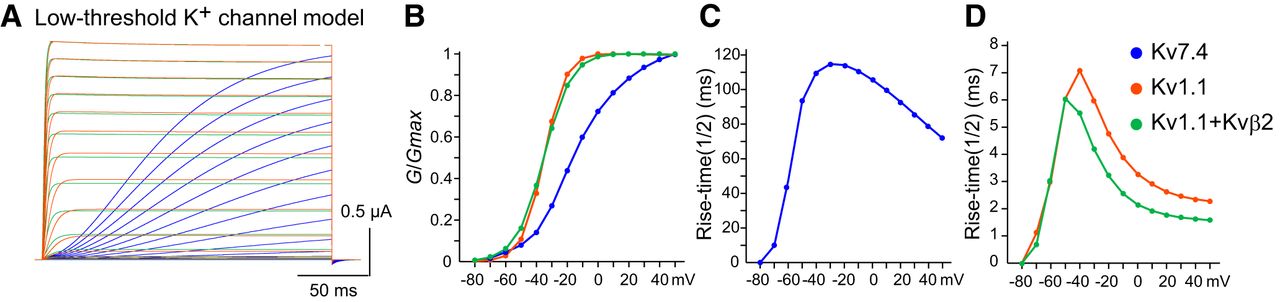
Computational model of low-threshold K+ channels. A, Voltage-gated outward currents of model zebrafish Kv7.4 (blue), Kv1.1 (orange), and Kv1.1 channels coexpressed with Kvβ2 (green) represented by a Hodgkin-Huxley equation. B–D, Normalized G-V plots (B), and rise time (t1/2) of model Kv7.4 (C), Kv1.1 and Kv1.1 + Kvβ2 (D) resemble those obtained in Xenopus oocytes (Fig. 4).
- Figure 6.
M-cell firing model integrating low-threshold K+ channels in response to long step currents. A, B, Representative current-clamp simulation of a model neuron integrating two types of low-threshold K+ channels associated with Kvβ2 subunits into a basic tonic-firing model. Long step currents of 1T, 1.5T, and 2T were injected. Maximum conductances (ḡKv1.1, ḡKv7.4; A) or (ḡKv1.1+Kvβ2, ḡKv7.4; B) are indicated in parentheses. Bold italic numbers represent conductances associated with Kvβ2. Expression of low-threshold K+ channels alters the tonic-firing model to exhibit phasic bursting with long or short firing duration and single spiking, as observed with in vivo M-cell recordings (Figs. 1, 2).
- Figure 7.
Comprehensive analysis of firing parameters in the M-cell model. A–E, Firing parameters of low-threshold K+ channel-integrated model cells with different combinations of (ḡKv1.1, ḡKv7.4; left) or (ḡKv1.1+Kvβ2, ḡKv7.4; right), with respect to threshold current (T; A), onset latency of first AP at 1T (B), number of elicited APs during 100 ms at 1.5T (C) and 2T (D), and ISIlast/ISI1st at 2T (E). In each panel, 1071 firing patterns (21 × 51 combinations) were produced by increments of 100 ns for ḡKv7.4 and 20 ns for ḡKv1.1 or ḡKv1.1+Kvβ2, ranging from 0-2000 ns (40 ms/cm2) and 0-1000 ns (20 ms/cm2), respectively. The Kv1.1 model (ḡKv1.1, ḡKv7.4: 480, 0) and Kv1.1 + Kvβ2 model (ḡKv1.1+Kvβ2, ḡKv7.4: 240, 0) had a similar threshold current (around 500 pA; A). The Kv1.1 model (ḡKv1.1, ḡKv7.4: 920, 0) showed two spikes at 1.5T, and this firing rate was also seen in the Kv1.1 + Kvβ2 model (ḡKv1.1+Kvβ2, ḡKv7.4: 540, 0; C), indicating that faster activation of Kv1.1 by Kvβ2 efficiently suppressed repetitive firing.
- Figure 8.
Na+ and K+ conductance mechanisms for producing characteristic phasic properties. A–D, Representative dynamics of voltage-gated Na+ and K+ channel conductance for gNav (black), ghigh (green), gA (gray), gKv7.4 (blue), and gKv1.1 or gKv1.1+Kvβ2 (red) during current-clamp stimulation. These conductances were normalized to maximum values of Na+ or among K+ conductance at 1.5T using a 2-dpf (A, B) and 4-dpf (C, D) M-cell model, shown in Figure 7A,B, respectively. Conductance dynamics at the beginning of current injection in B, D show the first AP generation of A, C, respectively. E–H, Changes in membrane potential at subthreshold (solid line, below 1–2 pA of 1T), and 1T (dashed line) in the 4-dpf M-cell model (black), XE991-treated model (red), and DTX-treated model (blue; E). The corresponding Na+ and K+ conductance in each model during subthreshold firing (F–H). Na+ conductance became much larger after fast activation of K+ conductance (ḡKv1.1+Kvβ2; F, G) compared with slow activation of K+ conductance (ḡKv7.4; H).
- Figure 9.
Simulation of M-cell firing in response to repetitive short-pulse currents. A, B, Current-clamp simulation of each model neuron (ḡKv1.1, ḡKv7.4; A) or (ḡKv1.1+Kvβ2, ḡKv7.4; B), which was the same as in Figure 6 except that repetitive short-pulse currents (0.5 ms) of 1T, 2 nA, and 3 nA with 500 Hz were given for 100 ms. C, D, Number of APs simulated in (ḡKv1.1, ḡKv7.4; C) or (ḡKv1.1+Kvβ2, ḡKv7.4; D) are plotted during the current injection in 20-pA increments up to 3 nA. E, F, Threshold current (E) and onset latency of the first spike at 1T (F) for each model neuron (ḡKv1.1, ḡKv7.4) or (ḡKv1.1+Kvβ2, ḡKv7.4).


Tables
- Table 1.
Comparison of the parameters of the Boltzmann equation in Xenopus oocytes system is shown
n V1/2 (mV) k Kv7.4 20 −10.1 ± 1.7 15.4 ± 0.7 Kv1.1a 32 −35.4 ± 1.4** 7.4 ± 0.3** Kv1.1a + Kvβ2b 26 −36.6 ± 1.1** 9.4 ± 0.5** All values are represented as mean ± SEM. **p < 0.01, Mann–Whitney U tests against the value of Kv7.4.
Extended Data 1
The model channel generator and firing simulator on NEURON simulator. The NEURON model files encode the channel generator and firing simulator for simulating development and differentiation of the M-cell excitability. The channel generator enables us to generate arbitrary Na+ and K+ channels by changing parameters of a Hodgkin-Huxley model under emulation of two-electrode voltage-clamp recordings in Xenopus oocyte system. The firing simulator simulates current-clamp recordings to generate firing pattern of the model M cell, which are implemented with arbitrary-generated Na+ and K+ conductances and low-threshold K+ channels Kv7.4/KCNQ4 and sole Kv1.1 or Kv1.1 coexpressed with Kvβ2. Download Extended data, ZIP file.
In this issue


