

Article Figures & Data
Figures
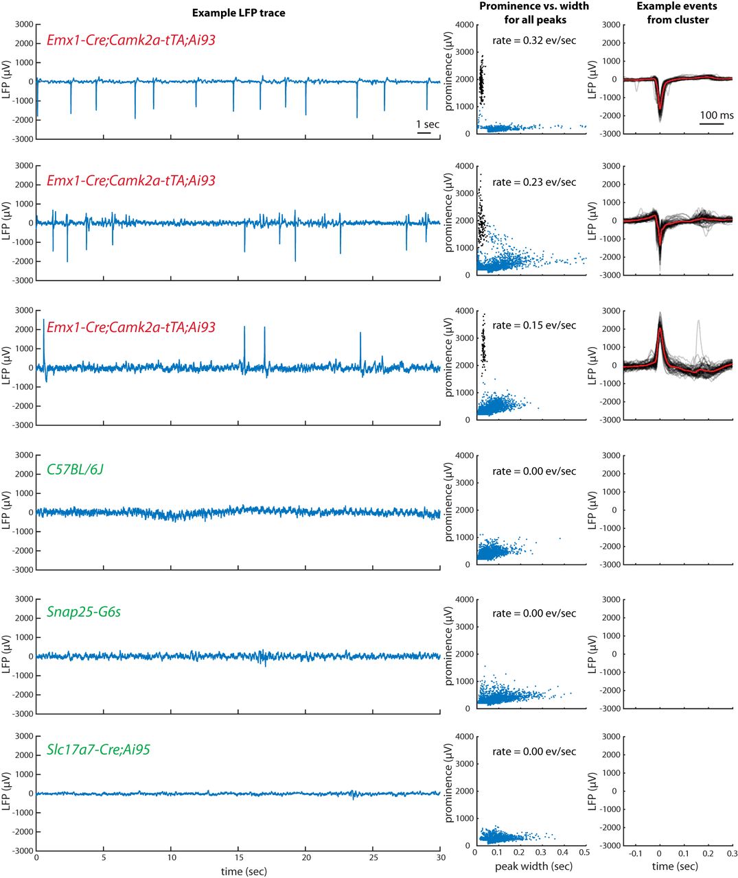
- Figure 1.
Epileptiform electrical activity in frontal cortex of some GCaMP6-expressing mice but not others. Left, each row contains an example segment of raw LFP data from each of six mice, with genotype identified in colored text. Middle, a plot of the prominence versus width of all peaks (see Methods) in the full LFP traces. Points highlighted in black were identified as a distinct cluster and included in the computation of event rate and the example events plotted at right. Right, 50 example events (gray) and the mean of all events (red). For the recording in row C, positive peaks rather than negative were analyzed.
- Figure 2.
Comparison of events observed in LFP and widefield calcium imaging in the same mouse. Format as in Fig. 1. The two LFP recordings were made simultaneously with each other, but not simultaneously with the widefield imaging. In the frontal LFP recording, the two clusters in prominence versus width arise because of the double-peaked shape of the events in this mouse at this site: for some events, the width includes only the first peak; for some, it includes both.
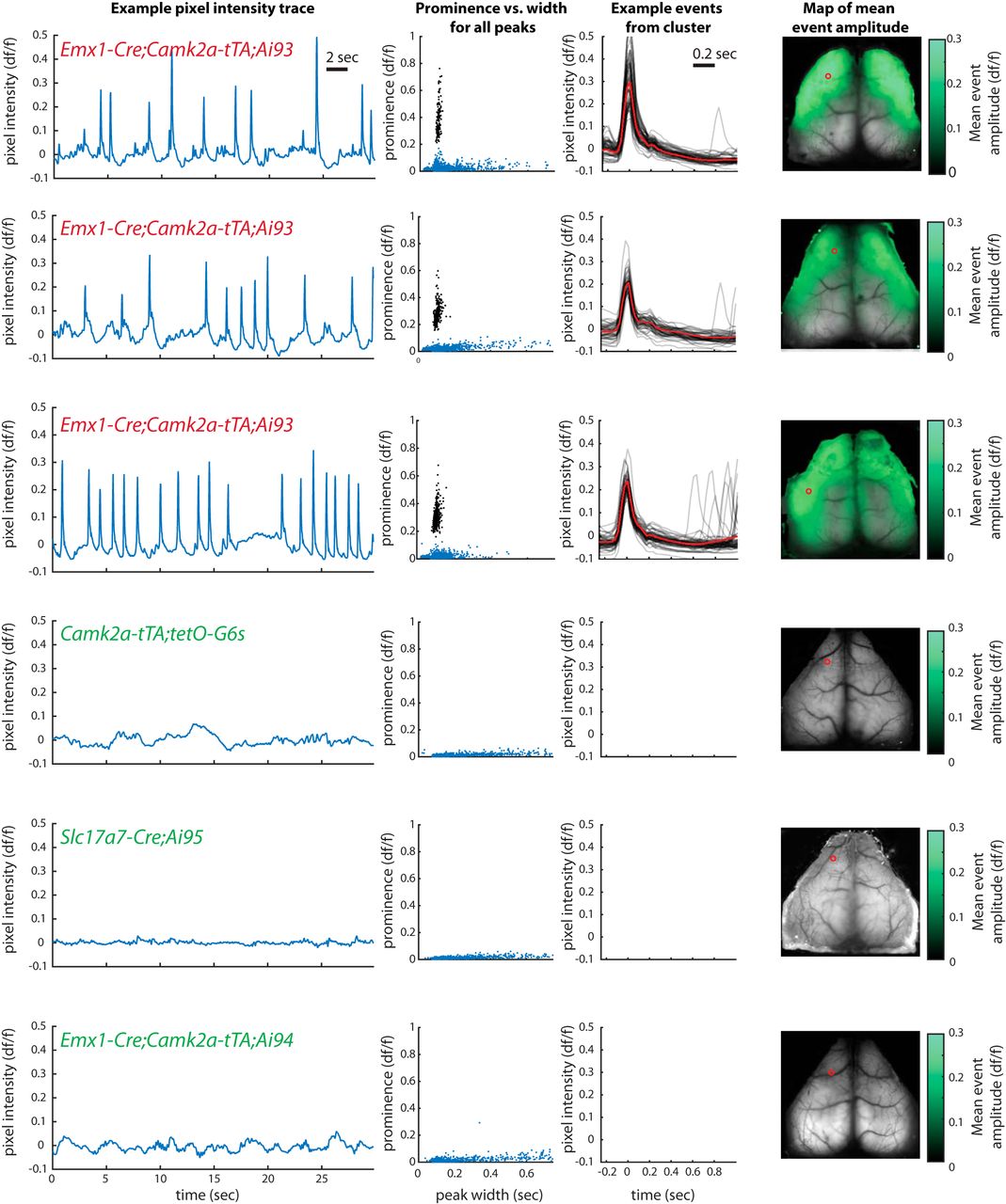
- Figure 3.
Incidence and spatial extent of epileptiform events observed in widefield calcium imaging. Format as in Fig. 1. The intensity trace and detected peaks come from the pixel indicated by the red circle on the brain image at right. Green coloration in the rightmost panel, overlaid on the mean image of the brain, represents the amplitude of the mean event at each point across the brain. See Videos 1, 2, and 3 for examples.
- Figure 4.
Germline Cre recombination results in widespread GCaMP expression. A, B, Native GCaMP6 fluorescence obtained using two-photon serial tomography from two Rbp4-Cre/wt;Camk2a-tTA/wt;Ai93 STOP+/Ai93 STOP+ mice, showing expression of GCaMP only in restricted populations of L5 cortical neurons and moderate expression in hippocampus. C, D, Similar images with matched intensity scale but from a Rbp4-Cre/wt;Camk2a-tTA/wt;Ai93 STOP+/Ai93 STOPdel mouse that had germline Cre recombination, showing high, widespread expression across cortex and hippocampus.
- Figure 5.
Epileptiform events observed in LFP, two-photon calcium imaging, and widefield calcium imaging in one individual mouse, but not simultaneously. Format as in Fig. 1. The genotype of the mouse was Emx1-Cre;Camk2a-tTA;Ai94 (expressing GCaMP6s). Two-photon trace was generated as the mean intensity of each frame across the entire field of view; widefield trace was generated as the mean within an ROI approximating the two-photon field of view.
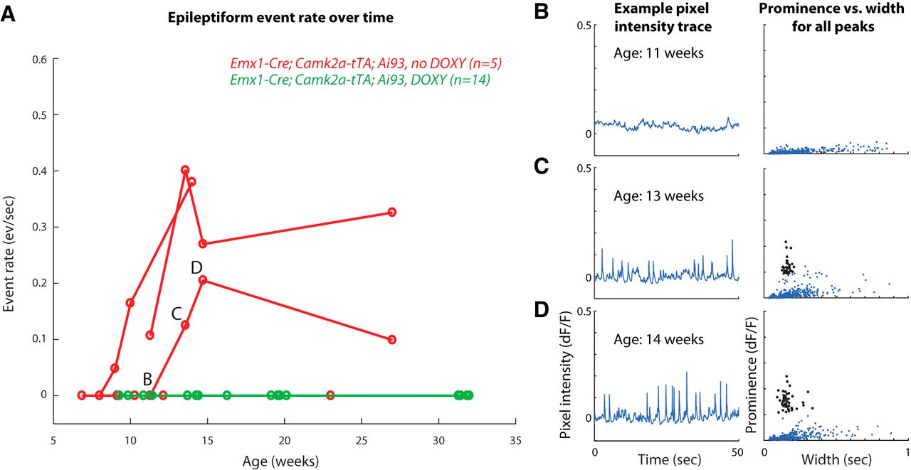
- Figure 6.
Epileptiform events fail to develop over time in mice treated with doxycycline until age 7 weeks. A, Measured rates of epileptiform events in untreated (red) and doxycycline treated (green) mice by age at time of measurement. Connected points indicate observations from the same mouse. All doxycycline-treated mice failed to develop events over the measured time period. Note that the mice represented in this figure are a small subset of all measured mice, and we do not intend to claim that the time courses followed here are representative of all mice developing events. B, C, and D refer to the measurements corresponding to the figure panels at right. B–D, Example traces and prominence versus width plots for an example untreated mouse showing development of events between 11 and 14 weeks.


Tables
- Table 1.
Incidence of epileptiform events in electrophysiological recordings of local field potentials in cortex
Genotype Incidence of events (mice) Emx1-Cre;Camk2a-tTA;Ai93 4/4 Emx1-Cre;Camk2a-tTA;Ai94 1/1 a Slc17a7-Cre;Ai95 0/1 Camk2a-tTA;tetO-G6s 0/2 Snap25-G6s 0/1 Pvalb-Cre;Ai32 0/2 wildtype (C57BL/6J) 0/4 Each count represents one mouse recorded electrophysiologically.
aThe Emx1-Cre;Camk2a-tTA;Ai94 mouse was selected for electrophysiology on the basis of epileptiform events observed in imaging.
Institute/laboratory Cre genotype tTA genotype GCaMP genotype Incidence of events in any size imaging window (mice) Incidence of events in full-hemisphere imaging (mice) High incidence Carandini/Harris, UCL Emx1 Camk2a Ai93 11/11 11/11 Allen Institute Emx1 Camk2a Ai93 9/18 5/7 Margolis, Rutgers Emx1 Camk2a Ai93 3/3 3/3 Margolis, Rutgers Emx1 Rosa26 Ai93 7/7 7/7 Häusser, UCL Emx1-Kess Camk2a Ai93 7/12 – Low incidence Allen Institute Slc17a7 Camk2a Ai93 1/5 1/5 Allen Institute Cux2 Camk2a Ai93 1/7 – Carandini/Harris, UCL Emx1 Camk2a Ai94 1/4 0/3 No events observed Allen Institute Rbp4 Camk2a Ai93 0/11 – Allen Institute Rorb Camk2a Ai93 0/3 – Allen Institute Scnn1a Camk2a Ai93 0/7 – Häusser, UCL Emx1-Kess Camk2a* Ai93 0/11 – Carandini/Harris, UCL Slc17a7 Ai95 0/7 0/7 Allen Institute Emx1 Ai95 0/1 0/1 Allen Institute Emx1 Ai96 0/3 0/3 Carandini/Harris, UCL Camk2a tetO-G6s 0/9 0/9 Allen Institute Camk2a tetO-G6s 0/6 – Carandini/Harris, UCL Snap25-G6s 0/4 0/4 Allen Institute Snap25-G6s 0/2 – Margolis, Rutgers GP4.3 0/2 0/2 Epileptiform events were judged to occur by manually inspecting raw imaging videos, traces, and prominence-versus-width plots for peaks of the traces, as shown in Fig. 3. See Methods for details of mouse lines and imaging preparations. All mice in this table had either intact Cre conditionality or unknown conditionality. Camk2a* indicates that these mice were treated with doxycycline until 7 weeks (see below). See Table 2-1 for further details on these observations.
Cre genotype tTA genotype GCaMP genotype Incidence with germline Cre recombination Incidence without germline Cre recombination Emx1 Camk2a Ai93 9/10 30/44 Ntsr1 Camk2a Ai93 5/9 Rbp4 Camk2a Ai93 4/4 0/11 Rorb Camk2a Ai93 5/8 0/3 “With germline Cre recombination”, mice that had lost Cre conditionality; “without”, mice that were normal, i.e. expressed GCaMP only in subsets of neurons according to the Cre driver line used. In this table, all mice were imaged with widefield calcium imaging of any size window.
Movies
- Movie 1.
Emx1-Cre;Camk2a-tTA;Ai93 mouse. A, The fluorescence signal imaged across the dorsal surface of the mouse brain (df/f). B, at top, velocity of a rubber wheel under the forepaws of the mouse; below, traces of df/f over time from the four identified pixels (matching color points in the first panel). Red points indicate detected epileptiform events. C, Two videos of the mouse. All movies play at half real time. Note that df/f scaling differs between movies for clarity of visualization. For further details of methodology, see Methods of widefield imaging at Carandini/Harris Laboratory.
- Movie 2.
Emx1-Cre;Camk2a-tTA;Ai93 mouse. A, The fluorescence signal imaged across the dorsal surface of the mouse brain (df/f). B, at top, velocity of a rubber wheel under the forepaws of the mouse; below, traces of df/f over time from the four identified pixels (matching color points in the first panel). Red points indicate detected epileptiform events. C, Two videos of the mouse. All movies play at half real time. Note that df/f scaling differs between movies for clarity of visualization. For further details of methodology, see Methods of widefield imaging at Carandini/Harris Laboratory.
- Movie 3.
Slc17a7-Cre;Ai95 mouse. A, The fluorescence signal imaged across the dorsal surface of the mouse brain (df/f). B, at top, velocity of a rubber wheel under the forepaws of the mouse; below, traces of df/f over time from the four identified pixels (matching color points in the first panel). Red points indicate detected epileptiform events. C, Two videos of the mouse. All movies play at half real time. Note that df/f scaling differs between movies for clarity of visualization. For further details of methodology, see Methods of widefield imaging at Carandini/Harris Laboratory.
Table 2-1
Details for all examined mice. The genotype for each transgene is indicated, alleles separated by a slash, with “wt” for wildtype. –, unknown information; NA, not applicable (e.g., for germline Cre-mediated recombination status of transgenes that cannot be subject to Cre-mediated recombination). Germline Cre recombination status is given as all alleles have intact LSL cassette (STOP+); all alleles have recombined LSL (STOPdel+); or one allele recombined and one intact (STOP+/STOPdel+; see Methods). Ages indicate the oldest age observed when no epileptiform events were reported or the youngest age at which epileptiform events were observed when they were. Download Table 2.1, XLS file.
In this issue


