

Article Figures & Data
Figures
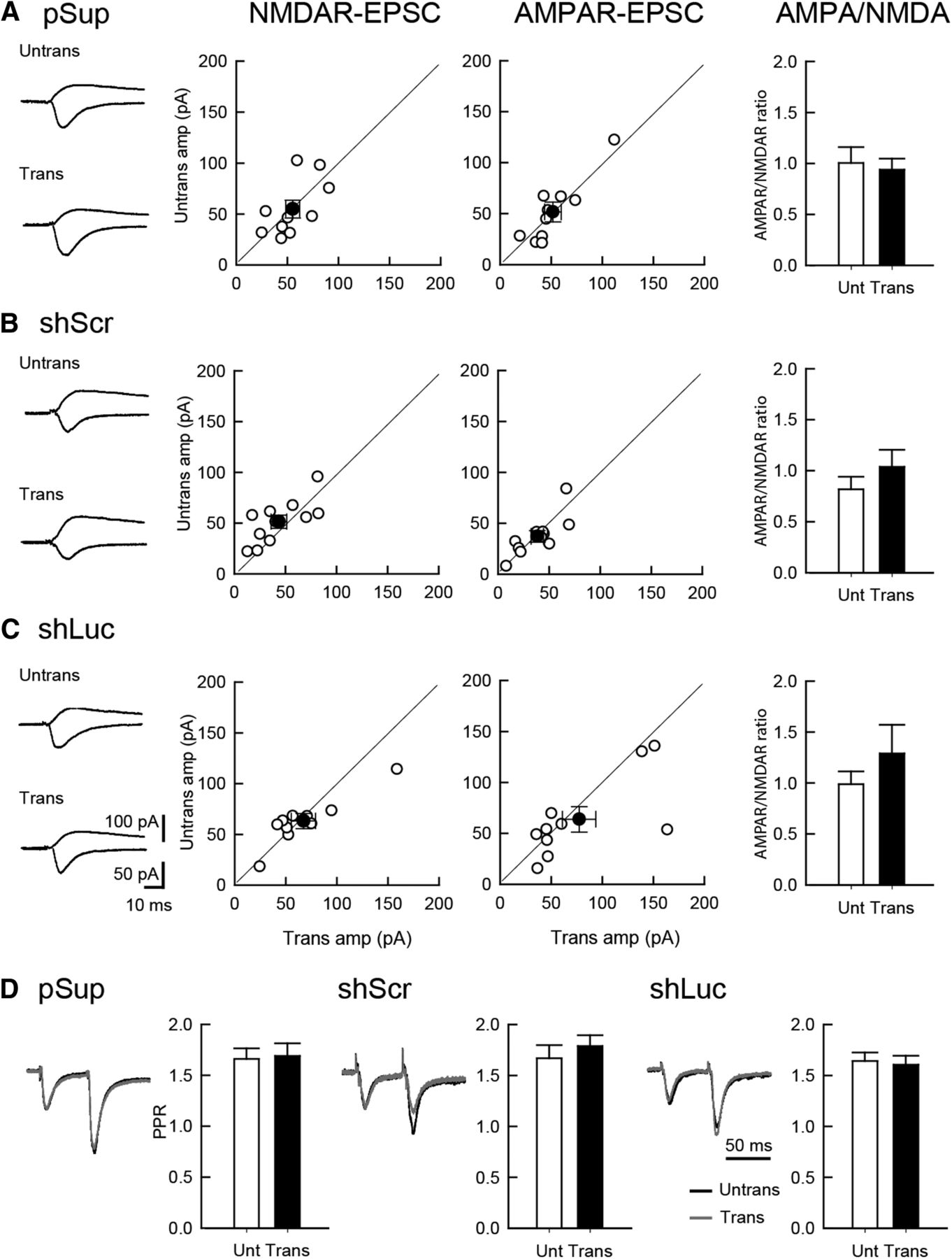
- Figure 1.
Comparable levels of excitatory synaptic transmission between shRNA-transfected and untransfected neurons. Effect of overexpression of three different plasmid transfections on excitatory synaptic transmission in hippocampal CA1 pyramidal cells. An empty vector (pSup; A), shScr (B), and shLuc (C) were transfected together with pCAG-EGFP. Recordings were conducted 5 d following transfection. Left column, Sample EPSC traces mediated by AMPARs (downward) and NMDARs (upward) from pairs of transfected neurons (Trans) and neighboring untransfected neurons (Untrans). Stimulus artifacts were truncated. Middle columns, Scattered plots of NMDAR- (left) and AMPAR- (right) EPSC amplitude (amp). Each pair of transfected and neighboring untransfected cells are presented as open symbols while filled symbols indicate the mean. Right column, Bar graphs of AMPAR/NMDAR ratios. D, PPR of AMPAR-EPSCs recorded from trans- and untransfected neurons, as indicated. Left, Sample traces. Normalized EPSCs to the first EPSC amplitude from trans- (gray) and untransfected neurons (black) are superimposed. Right, Summary graphs of PPR. The PPR was calculated by dividing the average amplitude of the second EPSC by that of the first EPSC. Number of cell pairs tested: pSup, 10 cells/6 mice; shLuc, 10/6; shScr, 11/6 (for AMPAR-EPSC, NMDAR-EPSC, and PPR, respectively).
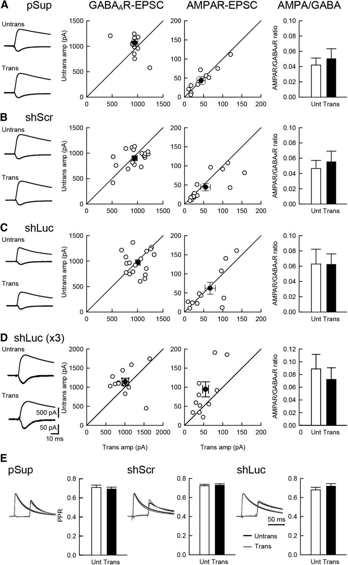
- Figure 2.
Comparable level of inhibitory synaptic transmission between shRNA-transfected and untransfected neurons. Effect of overexpression of the three different plasmid on excitatory and inhibitory synaptic transmission in hippocampal CA1 pyramidal cells. An empty vector (pSup; A), shScr (B), shLuc (C) and shLuc x3 (triple the amount of shLuc) (D) were transfected together with pCAG-EGFP. Left column, Sample AMPAR-EPSC and GABAAR-IPSC traces mediated by AMPARs (downward) and GABAARs (upward) from pairs of transfected neurons (Trans) and neighboring untransfected neurons (Untrans). Stimulus artifacts were truncated. Middle columns, Scattered plots of GABAAR- (left) and AMPAR- (right) EPSC amplitude (amp). Each pair of transfected and neighboring untransfected cells are presented as open symbols while filled symbols indicate the mean. Right column, Bar graphs of AMPAR/GABAAR ratios. E, PPR of GABAAR-IPSCs recorded from trans- and untransfected neurons, as indicated. Left, Sample traces. Normalized IPSCs to the first IPSC amplitude from trans- (gray) and untransfected neurons (black) were superimposed. The First GABAAR-IPSC overlaps with the second IPSC. Therefore, the first EPSC was cancelled by subtracting the traces receiving a single pulse from those receiving a paired pulse, both normalized to the first response. Right, Summary graphs of PPR. The PPR was calculated by dividing the average amplitude of the second IPSC by that of the first IPSC. Number of cell pairs: pSup, 13, 11, and 13 cells/7 mice (13, 11, 13/7); shLuc (18, 13, 18/8); shScr (20, 15, 20/8); shLuc x3 (13, 12/6), for GABAAR-IPSC, AMPAR-EPSC, and PPR, respectively.
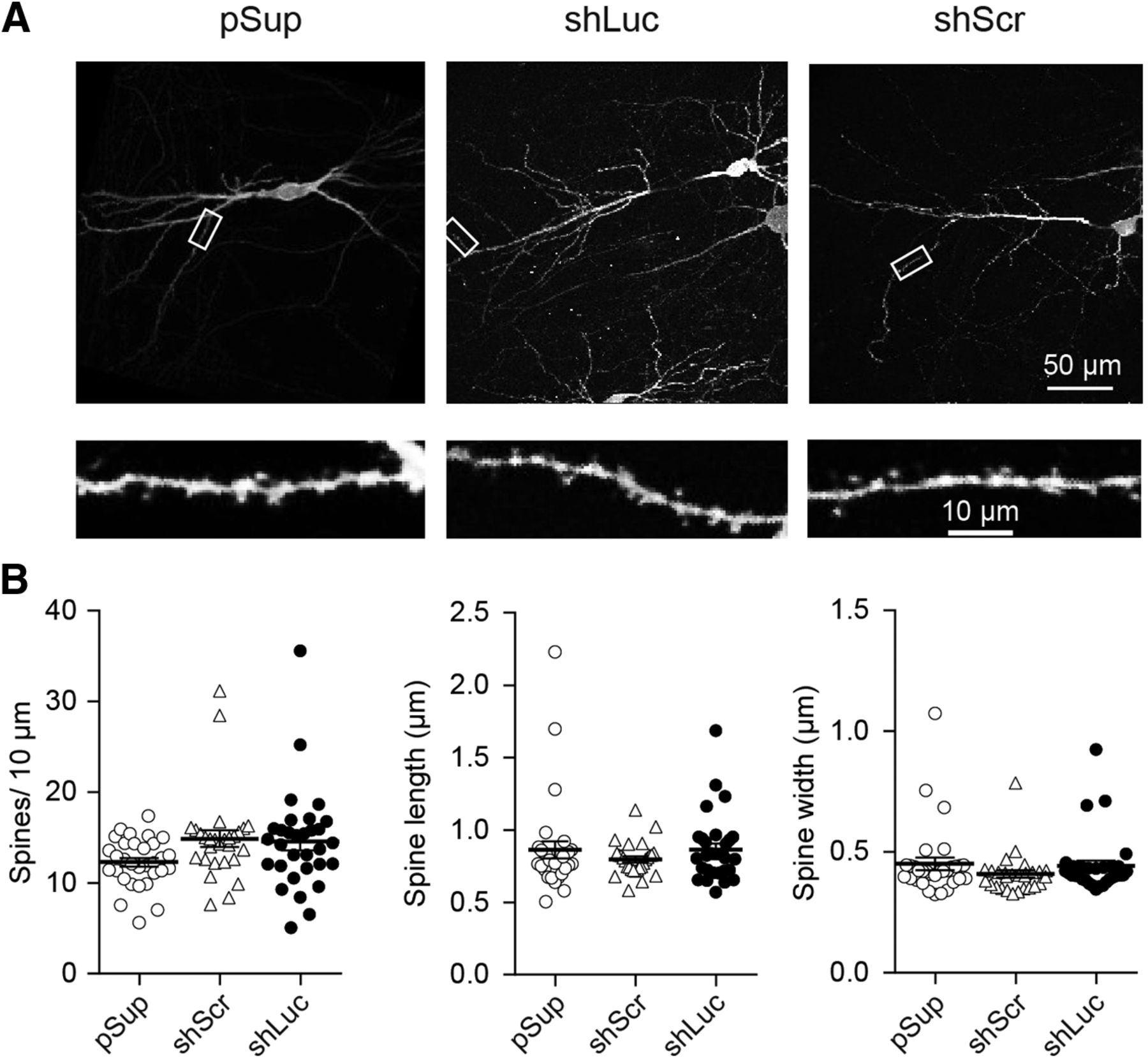
- Figure 3.
Comparable level of dendritic structure between different shRNA-transfected neurons. Effect of overexpression of three different plasmid transfections on dendritic morphology in hippocampal CA1 pyramidal neurons. A, Low (top) and high (bottom) magnification images obtained from empty vector, pSup (left), shLuc (middle), and shScr (right) transfected neurons. Each plasmid was cotransfected with pCAG-EGFP. Fixed slices were immunostained against GFP and neuronal images were obtained by two-photon microscopy. B, Scatter plots of spine length (left), width (middle), and density (right). Error bars indicate SEMs. Note that none of these parameters displayed statistical significance by gene transfection. Number of dendritic segments/cells/mice: pSup, 31/5/5; shLuc, 31/5/5; and shScr, 28/5/5.
- Figure 4.
Reduced membrane excitabilities in shLuc-transfected CA1 pyramidal neurons. A, Effect of shRNA overexpression on neuronal excitability. An empty vector (pSup), shLuc, or shScr was transfected together with pCAG-EGFP. Top, Sample traces from untransfected and transfected CA1 pyramidal neurons in organotypic hippocampal slice cultures. The superimposed traces were elicited by current injections of 0, 100, and 500 pA for 200 ms. Bottom, Summary graph of the frequency of APs in untransfected and transfected neurons. The input–output relationship [number of spikes elicited versus amount of current injection (200-ms duration)] was plotted for untransfected and transfected neurons. Neurons were held at resting membrane potentials (−56.8 mV ± 0.57, n = 100 cells, 9 mice). Number of cells tested: untrans, 45 cells from eight mice (45/8); pSup, 13/7; shScr, 20/5; and shLuc, 22/9. B, Effect of shRNA overexpression on AP kinetics. Top, Sample traces from CA1 pyramidal neurons untransfected and transfected with the three different plasmids. Single APs were induced by current injection (100 pA for 4 ms) and threshold (horizontal arrows) and half width of AP determined by vertical double arrow heads in trans- and untransfected neurons were measured. Bottom, Summary graph of the threshold (left) and half width (right) of single AP in untransfected and transfected neurons. Number of cells tested: untrans, 45 cells/8 mice (45/8); pSup, 13/7; shScr, 20/5; shLuc, 22/9. C, top, Sample traces of sodium currents recorded from untransfected and transfected CA1 pyramidal neurons. Note that these currents were completely blocked by TTX (right). Bottom, Summary graph of the sodium currents in untransfected and transfected neurons. Neurons were voltage-clamped at –80 mV and depolarized from –80 to 30 mV (10-ms duration). Number of cells: untrans, 16 cells/5 mice; pSup, 10/5; shScr, 10/5; shLuc, 14/6. D, top, Sample traces of potassium currents recorded from untransfected and transfected CA1 pyramidal neurons. Note that these upward currents were completely blocked by TEA and 4-AP (right). Bottom, Summary graph of the potassium currents in untransfected and transfected neurons. Neurons were voltage-clamped at –80 mV and depolarized from –80 to 30 mV (10-ms duration). Number of cells tested: untrans, 15 cells/4 mice; pSup, 13/3; shScr, 7/3; shLuc, 9/3.
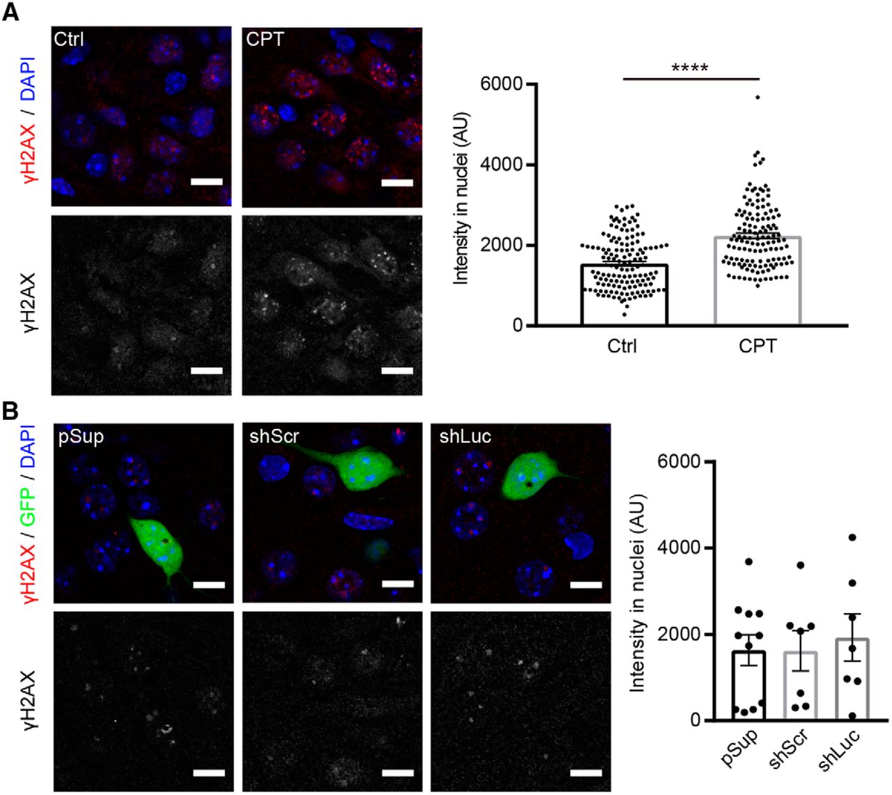
- Figure 5.
Comparable levels of H2A-X phosphorylation in neurons transfected with different shRNA vectors. γ-H2AX immunoreactivity in shRNA-transfected hippocampal CA1 neurons. A, left, Representative confocal images of immunofluorescence staining against γ-H2AX (red) and DAPI (blue) in CA1 pyramidal neurons. Right, Quantification of γ-H2AX signal intensity in nuclei. Treatment with 10 μM camptothecin (CPT) for 6 h increased the signal of γ-H2AX in nuclei of neurons in organotypic slice cultures, confirming the specificity of the anti-γ-H2AX antibody. AU, arbitrary units. Number of cells/mice: mock control (Ctrl), 140/3; CPT, 133/3. B, left, Representative confocal images of immunofluorescence staining against γ-H2AX (red), GFP epifluorescence (green) and DAPI (blue) in pSup-, shLuc-, and shScr-transfected neurons. Right, Quantification of γ-H2AX signal intensity in nuclei. All transfected neurons exhibited comparable levels of γ-H2AX signal. Number of cells/mice: pSup, 11/3; shScr, 7/4; shLuc, 7/3. Scale bars: 10 μm.


Tables
- Table 1.
The basic membrane properties of untransfected and gene transfected hippocampal CA1 neurons
Cell types Peak amplitude of AP (mV) Resting membrane potential (mV) Input resistance (MΩ) Series resistance (MΩ) Number of cells/mice untrans 106.1 ± 1.331 −55.87 ± 0.900 105.6 ± 8.452 8.127 ± 0.2768 45/8 pSup 108.5 ± 2.452 −58.51 ± 1.604 122.9 ± 18.62 8.869 ± 0.6093 13/7 shScr 109.3 ± 1.456 −57.47 ± 1.104 143.1 ± 18.03 8.456 ± 0.3902 20/5 shLuc 106.0 ± 2.224 −57.07 ± 1.054 131.0 ± 11.29 9.349 ± 0.3110 22/9 Statistics p value shLuc vs shScr 0.7606 shLuc vs shScr >0.9999 shLuc vs shScr 0.9893 shLuc vs shScr 0.4875 shLuc vs pSup 0.9657 shLuc vs pSup 0.9777 shLuc vs pSup 0.9996 shLuc vs pSup 0.9755 shLuc vs untrans >0.9999 shLuc vs untrans 0.9604 shLuc vs untrans 0.567 shLuc vs untrans 0.0681 shScr vs untrans 0.6744 shScr vs pSup 0.9958 shScr vs pSup 0.9434 shScr vs pSup 0.988 shScr vs pSup >0.9999 shScr vs untrans 0.8504 shScr vs untrans 0.1487 shScr vs untrans 0.9813 pSup vs untrans 0.9603 pSup vs untrans 0.5991 pSup vs untrans 0.9558 pSup vs untrans 0.7507 Note that all of these parameters were not statistically different between four cell groups. Statistics were done by one-way ANOVA with post hoc Tukey.
Graph Data structure Type of test Dataset p value Fig. 1A Nonparametric Mann-Whitney test AMPAR-EPSC 0.97 NMDAR-EPSC 0.51 AMPAR/NMDAR ratio 0.97 Fig. 1B Nonparametric Mann-Whitney test AMPAR-EPSC 0.69 NMDAR-EPSC 0.47 A/N ratio 0.33 Fig. 1C Nonparametric Mann-Whitney test AMPAR-EPSC 0.79 NMDAR-EPSC 0.79 A/N ratio 0.47 Fig. 1D Parametric Student’s t test PPR of pSup 0.86 PPR of shScr 0.48 PPR of shLuc 0.76 Fig. 2A Nonparametric Mann-Whitney test GABAAR-IPSC 0.21 AMPAR-EPSC 1 AMPAR/GABAAR ratio 0.55 Fig. 2B Nonparametric Mann-Whitney test GABAAR-IPSC 0.43 AMPAR-EPSC 0.41 A/G ratio 0.56 Fig. 2C Nonparametric Mann-Whitney test GABAAR-IPSC 0.69 AMPAR-EPSC 0.92 A/G ratio 0.89 Fig. 2D Nonparametric Mann-Whitney test GABAAR-IPSC 0.15 AMPAR-EPSC 0.24 A/G ratio 0.58 Fig. 2E Parametric Student’s t test PPR of pSup 0.63 PPR of shScr 0.74 PPR of shLuc 0.31 Fig. 3B Parametric One-way ANOVA post hoc Tukey Spine density: pSup vs shLuc 0.45 pSup vs shScr 0.09 shScr vs shLuc 0.65 Spine length: pSup vs shLuc 0.99 pSup vs shScr 0.68 shScr vs shLuc 0.63 Spine width: pSup vs shLuc 0.96 pSup vs shScr 0.37 shScr vs shLuc 0.53 Fig. 4A Parametric Two-way ANOVA post hoc Tukey −100 pA untrans vs shLuc >0.9999 untrans vs shScr >0.9999 untrans vs pSup >0.9999 shLuc vs shScr >0.9999 shLuc vs pSup >0.9999 shScr vs pSup >0.9999 −50 pA untrans vs shLuc >0.9999 untrans vs shScr >0.9999 untrans vs pSup >0.9999 shLuc vs shScr >0.9999 shLuc vs pSup >0.9999 shScr vs pSup >0.9999 0 pA untrans vs shLuc >0.9999 untrans vs shScr >0.9999 untrans vs pSup >0.9999 shLuc vs shScr >0.9999 shLuc vs pSup >0.9999 shScr vs pSup >0.9999 50 pA untrans vs shLuc >0.9999 untrans vs shScr >0.9999 untrans vs pSup 0.9993 shLuc vs shScr >0.9999 shLuc vs pSup 0.9995 shScr vs pSup 0.9996 100 pA untrans vs shLuc 0.9997 untrans vs shScr >0.9999 untrans vs pSup 0.9923 shLuc vs shScr 0.9995 shLuc vs pSup 0.9978 shScr vs pSup 0.9928 150 pA untrans vs shLuc 0.9909 untrans vs shScr >0.9999 untrans vs pSup 0.9964 shLuc vs shScr 0.9959 shLuc vs pSup >0.9999 shScr vs pSup 0.9982 200 pA untrans vs shLuc 0.9818 untrans vs shScr >0.9999 untrans vs pSup 0.8433 shLuc vs shScr 0.9915 shLuc vs pSup 0.9674 shScr vs pSup 0.8983 250 pA untrans vs shLuc 0.7984 untrans vs shScr 0.9421 untrans vs pSup 0.4089 shLuc vs shScr 0.9945 shLuc vs pSup 0.8912 shScr vs pSup 0.8019 300 pA untrans vs shLuc 0.6045 untrans vs shScr 0.8776 untrans vs pSup 0.124 shLuc vs shScr 0.9847 shLuc vs pSup 0.714 shScr vs pSup 0.5448 350 pA untrans vs shLuc 0.2035 untrans vs shScr 0.8942 untrans vs pSup 0.1894 shLuc vs shScr 0.7588 shLuc vs pSup 0.9865 shScr vs pSup 0.6361 400 pA untrans vs shLuc 0.1775 untrans vs shScr 0.7432 untrans vs pSup 0.2523 shLuc vs shScr 0.8662 shLuc vs pSup 0.9987 shScr vs pSup 0.844 450 pA untrans vs shLuc 0.0599 untrans vs shScr 0.8942 untrans vs pSup 0.2917 shLuc vs shScr 0.4825 shLuc vs pSup 0.9928 shScr vs pSup 0.7592 500 pA untrans vs shLuc 0.0477 untrans vs shScr 0.9921 untrans vs pSup 0.6175 shLuc vs shScr 0.2411 shLuc vs pSup 0.8394 shScr vs pSup 0.8383 550 pA untrans vs shLuc 0.0101 untrans vs shScr 0.9971 untrans vs pSup 0.8145 shLuc vs shScr 0.0831 shLuc vs pSup 0.4248 shScr vs pSup 0.9297 600 pA untrans vs shLuc 0.0043 untrans vs shScr >0.9999 untrans vs pSup 0.68 shLuc vs shScr 0.0299 shLuc vs pSup 0.437 shScr vs pSup 0.7694 650 pA untrans vs shLuc 0.0014 untrans vs shScr 0.995 untrans vs pSup 0.8638 shLuc vs shScr 0.0266 shLuc vs pSup 0.176 shScr vs pSup 0.9617 700 pA untrans vs shLuc 0.0003 untrans vs shScr 0.9998 untrans vs pSup 0.9417 shLuc vs shScr 0.0061 shLuc vs pSup 0.0571 shScr vs pSup 0.9735 750 pA untrans vs shLuc 0.0002 untrans vs shScr 0.7664 untrans vs pSup 0.9643 shLuc vs shScr 0.0001 shLuc vs pSup 0.038 shScr vs pSup 0.6549 800 pA untrans vs shLuc 0.0007 untrans vs shScr 0.5283 untrans vs pSup 0.9959 shLuc vs shScr <0.0001 shLuc vs pSup 0.038 shScr vs pSup 0.6058 850 pA untrans vs shLuc 0.0041 untrans vs shScr 0.2197 untrans vs pSup >0.9999 shLuc vs shScr <0.0001 shLuc vs pSup 0.0601 shScr vs pSup 0.4478 900 pA untrans vs shLuc 0.0005 untrans vs shScr 0.4401 untrans vs pSup >0.9999 shLuc vs shScr <0.0001 shLuc vs pSup 0.0174 shScr vs pSup 0.6624 950 pA untrans vs shLuc 0.0001 untrans vs shScr 0.1954 untrans vs pSup 0.9999 shLuc vs shScr <0.0001 shLuc vs pSup 0.006 shScr vs pSup 0.466 1000 pA untrans vs shLuc 0.0004 untrans vs shScr 0.1282 untrans vs pSup 0.9992 shLuc vs shScr <0.0001 shLuc vs pSup 0.0108 shScr vs pSup 0.4014 1050 pA untrans vs shLuc 0.0001 untrans vs shScr 0.2644 untrans vs pSup 0.9762 shLuc vs shScr <0.0001 shLuc vs pSup 0.0025 shScr vs pSup 0.728 1100 pA untrans vs shLuc 0.0003 untrans vs shScr 0.2803 untrans vs pSup 0.999 shLuc vs shScr <0.0001 shLuc vs pSup 0.0087 shScr vs pSup 0.5944 Fig. 4B Parametric One-way ANOVA post hoc Tukey Threshhold: shLuc vs shScr 0.0249 shLuc vs pSup 0.0421 shLuc vs untrans 0.0094 shScr vs pSup 0.9958 shScr vs untrans 0.9991 pSup vs untrans 0.9837 Half width: shLuc vs shScr 0.0002 shLuc vs pSup 0.0165 shLuc vs untrans <0.0001 shScr vs pSup 0.9231 shScr vs untrans 0.9846 pSup vs untrans 0.7637 Fig. 4C Parametric Two-way ANOVA post hoc Tukey -80 mV shLuc vs shScr >0.9999 shLuc vs pSup >0.9999 shLuc vs untrans >0.9999 shScr vs pSup >0.9999 shScr vs untrans >0.9999 pSup vs untrans >0.9999 -70 mV shLuc vs shScr >0.9999 shLuc vs pSup 0.9998 shLuc vs untrans >0.9999 shScr vs pSup >0.9999 shScr vs untrans >0.9999 pSup vs untrans >0.9999 -60 mV shLuc vs shScr >0.9999 shLuc vs pSup 0.9982 shLuc vs untrans 0.9994 shScr vs pSup 0.9995 shScr vs untrans >0.9999 pSup vs untrans 0.9999 −50 mV shLuc vs shScr 0.9996 shLuc vs pSup 0.9956 shLuc vs untrans 0.998 shScr vs pSup 0.9992 shScr vs untrans 0.9999 pSup vs untrans 0.9999 −40 mV shLuc vs shScr 0.999 shLuc vs pSup 0.2815 shLuc vs untrans 0.9959 shScr vs pSup 0.3548 shScr vs untrans 0.9999 pSup vs untrans 0.3237 −30 mV shLuc vs shScr 0.0008 shLuc vs pSup 0.0272 shLuc vs untrans 0.0755 shScr vs pSup 0.7329 shScr vs untrans 0.315 pSup vs untrans 0.9295 −20 mV shLuc vs shScr 0.0007 shLuc vs pSup 0.0009 shLuc vs untrans 0.0235 shScr vs pSup 0.9999 shScr vs untrans 0.5293 pSup vs untrans 0.5778 −10 mV shLuc vs shScr 0.0007 shLuc vs pSup 0.0013 shLuc vs untrans 0.0183 shScr vs pSup 0.9983 shScr vs untrans 0.5872 pSup vs untrans 0.7019 0 mV shLuc vs shScr 0.001 shLuc vs pSup 0.0014 shLuc vs untrans 0.015 shScr vs pSup 0.9998 shScr vs untrans 0.7013 pSup vs untrans 0.7536 10 mV shLuc vs shScr 0.0005 shLuc vs pSup 0.0008 shLuc vs untrans 0.0083 shScr vs pSup 0.9996 shScr vs untrans 0.6986 pSup vs untrans 0.7654 20 mV shLuc vs shScr 0.0011 shLuc vs pSup 0.0017 shLuc vs untrans 0.0064 shScr vs pSup 0.9994 shScr vs untrans 0.856 pSup vs untrans 0.9091 30 mV shLuc vs shScr 0.0015 shLuc vs pSup 0.0017 shLuc vs untrans 0.0042 shScr vs pSup >0.9999 shScr vs untrans 0.9547 pSup vs untrans 0.9613 Fig. 4D Parametric Two-way ANOVA post hoc Tukey −80 mV shLuc vs shScr >0.9999 shLuc vs pSup >0.9999 shLuc vs untrans >0.9999 shScr vs pSup >0.9999 shScr vs untrans >0.9999 pSup vs untrans >0.9999 −70 mV shLuc vs shScr >0.9999 shLuc vs pSup 0.9998 shLuc vs untrans >0.9999 shScr vs pSup >0.9999 shScr vs untrans >0.9999 pSup vs untrans >0.9999 −60 mV shLuc vs shScr >0.9999 shLuc vs pSup 0.9982 shLuc vs untrans 0.9994 shScr vs pSup 0.9995 shScr vs untrans >0.9999 pSup vs untrans 0.9999 −50 mV shLuc vs shScr 0.9996 shLuc vs pSup 0.9956 shLuc vs untrans 0.998 shScr vs pSup 0.9992 shScr vs untrans 0.9999 pSup vs untrans 0.9999 −40 mV shLuc vs shScr 0.999 shLuc vs pSup 0.2815 shLuc vs untrans 0.9959 shScr vs pSup 0.3548 shScr vs untrans 0.9999 pSup vs untrans 0.3237 −30 mV shLuc vs shScr 0.0008 shLuc vs pSup 0.0272 shLuc vs untrans 0.0755 shScr vs pSup 0.7329 shScr vs untrans 0.315 pSup vs untrans 0.9295 −20 mV shLuc vs shScr 0.0007 shLuc vs pSup 0.0009 shLuc vs untrans 0.0235 shScr vs pSup 0.9999 shScr vs untrans 0.5293 pSup vs untrans 0.5778 −10 mV shLuc vs shScr 0.0007 shLuc vs pSup 0.0013 shLuc vs untrans 0.0183 shScr vs pSup 0.9983 shScr vs untrans 0.5872 pSup vs untrans 0.7019 0 mV shLuc vs shScr 0.001 shLuc vs pSup 0.0014 shLuc vs untrans 0.015 shScr vs pSup 0.9998 shScr vs untrans 0.7013 pSup vs untrans 0.7536 10 mV shLuc vs shScr 0.0005 shLuc vs pSup 0.0008 shLuc vs untrans 0.0083 shScr vs pSup 0.9996 shScr vs untrans 0.6986 pSup vs untrans 0.7654 20 mV shLuc vs shScr 0.0011 shLuc vs pSup 0.0017 shLuc vs untrans 0.0064 shScr vs pSup 0.9994 shScr vs untrans 0.856 pSup vs untrans 0.9091 30 mV shLuc vs shScr 0.0015 shLuc vs pSup 0.0017 shLuc vs untrans 0.0042 shScr vs pSup >0.9999 shScr vs untrans 0.9547 pSup vs untrans 0.9613 Fig. 5A Parametric Student’s t test Ctrl vs CPT <0.00001 Fig. 5B Parametric One-way ANOVA post hoc Tukey shLuc vs shScr 0.8937 shLuc vs pSup 0.885 shScr vs pSup 0.9996
In this issue


