
Umbrella menu

  • SfN.org
  • eNeuro
  • The Journal of Neuroscience
  • Neuronline
  • BrainFacts.org

Main menu

  • HOME
  • CONTENT
    • Early Release
    • Featured
    • Latest Articles
    • Issue Archive
    • Editorials
    • Research Highlights
  • TOPICS
    • Cognition and Behavior
    • Development
    • Disorders of the Nervous System
    • History, Teaching and Public Awareness
    • Integrative Systems
    • Neuronal Excitability
    • Novel Tools and Methods
    • Sensory and Motor Systems
  • ALERTS
  • FOR AUTHORS
  • EDITORIAL BOARD
  • BLOG
  • ABOUT
    • Overview
    • For the Media
    • Privacy Policy
    • Contact Us
    • Feedback
  • SfN.org
  • eNeuro
  • The Journal of Neuroscience
  • Neuronline
  • BrainFacts.org

User menu

  • My alerts

Search

  • Advanced search
eNeuro
  • My alerts

eNeuro

Advanced Search

Submit a Manuscript
  • HOME
  • CONTENT
    • Early Release
    • Featured
    • Latest Articles
    • Issue Archive
    • Editorials
    • Research Highlights
  • TOPICS
    • Cognition and Behavior
    • Development
    • Disorders of the Nervous System
    • History, Teaching and Public Awareness
    • Integrative Systems
    • Neuronal Excitability
    • Novel Tools and Methods
    • Sensory and Motor Systems
  • ALERTS
  • FOR AUTHORS
  • EDITORIAL BOARD
  • BLOG
  • ABOUT
    • Overview
    • For the Media
    • Privacy Policy
    • Contact Us
    • Feedback
PreviousNext
Research ArticleNew Research, Neuronal Excitability

Neuregulin and Dopamine D4 Receptors Contribute Independently to Depotentiation of Schaffer Collateral LTP by Temperoammonic Path Stimulation

Yukitoshi Izumi and Charles F. Zorumski
eNeuro 15 August 2017, 4 (4) ENEURO.0176-17.2017; DOI: https://doi.org/10.1523/ENEURO.0176-17.2017
Yukitoshi Izumi
1Department of Psychiatry, Washington University School of Medicine, St. Louis, MO 63110
3Taylor Institute for Innovative Psychiatric Research, Washington University School of Medicine, St. Louis, MO 63110
4Center for Brain Research in Mood Disorders, Washington University School of Medicine, St. Louis, MO 63110
  • Find this author on Google Scholar
  • Find this author on PubMed
  • Search for this author on this site
  • ORCID record for Yukitoshi Izumi
Charles F. Zorumski
1Department of Psychiatry, Washington University School of Medicine, St. Louis, MO 63110
2Department of Neuroscience, Washington University School of Medicine, St. Louis, MO 63110
3Taylor Institute for Innovative Psychiatric Research, Washington University School of Medicine, St. Louis, MO 63110
4Center for Brain Research in Mood Disorders, Washington University School of Medicine, St. Louis, MO 63110
  • Find this author on Google Scholar
  • Find this author on PubMed
  • Search for this author on this site
  • Article
  • Figures & Data
  • Info & Metrics
  • eLetters
  • PDF
Loading

Article Figures & Data

Figures

  • Figure 1.
    • Download figure
    • Open in new tab
    • Download powerpoint
    Figure 1.

    TA-induced LTP-D is blocked by the DA receptor antagonist, clozapine. A, The left graph shows the time course of changes in EPSPs following SC HFS (arrow) and depotentiation by TA/perforant path LFS (PLFS, bar). Note that data in A include control slices done in our prior manuscript (Izumi and Zorumski, 2016) with additional slices added. The right graph in A depicts an analysis from a single slice based on changes in the IO curve. Black circles are baseline IO results, while red circles depict changes 60 min after SC HFS, and red squares show reversal of LTP following PLFS. B, The ability of PLFS to depotentiate SC LTP is completely blocked by 1 μM clozapine (white bar). Upper traces to the right show representative EPSPs at baseline (black lines) and 60 min following SC HFS (redlines) while lower traces show baseline (black lines) compared to 60 min following PLFS (red lines). Calibration: 1 mV, 5 ms.

  • Figure 2.
    • Download figure
    • Open in new tab
    • Download powerpoint
    Figure 2.

    TA-induced LTP-D involves D4Rs. A, The graph shows the inability of a selective D2R antagonist (0.2 μM L-741,626) to block PLFS induced depotentiation of SC LTP. SC HFS was delivered at the arrow; PLFS was administered during the hatched bar. B, In contrast, a selective D4R antagonist (0.1 μM L-745,870) completely inhibited depotentiation. Traces to the right show representative EPSPs as in Figure 1. Calibration: 1 mV, 5 ms.

  • Figure 3.
    • Download figure
    • Open in new tab
    • Download powerpoint
    Figure 3.

    A D4R antagonist blocks homosynaptic SC depotentiation. A, The graph shows the ability of SC LFS [SLFS (1 Hz × 15 min), hatched bar] to depotentiate previously established SC LTP. SC HFS was administered at the arrow. B, The D4R antagonist, 0.1 μM L-745,870, blocked homosynaptic SC depotentiation. For reasons that are uncertain, we observed an increase in variance of EPSPs during perfusion of the D4R antagonist in this set of studies but not in the studies shown in Figure 2. Traces to the right show representative EPSPs as in Figure 1. Calibration: 1 mV, 5 ms.

  • Figure 4.
    • Download figure
    • Open in new tab
    • Download powerpoint
    Figure 4.

    Exogenously administered NRG1β, but not a D4R agonist, depotentiates SC LTP. A, A selective D4R agonist,0.2 μM PD-168,077 (black bar), failed to depotentiate SC LTP when administered for 15 min 60 min following LTP induction. SC HFS was administered at the arrow. B, In contrast to the D4R agonist, 1 nM NRG1β (black bar) induced chemical depotentiation of SC LTP. Traces to the right show EPSPs as in Figure 1. Calibration: 1 mV, 5 ms.
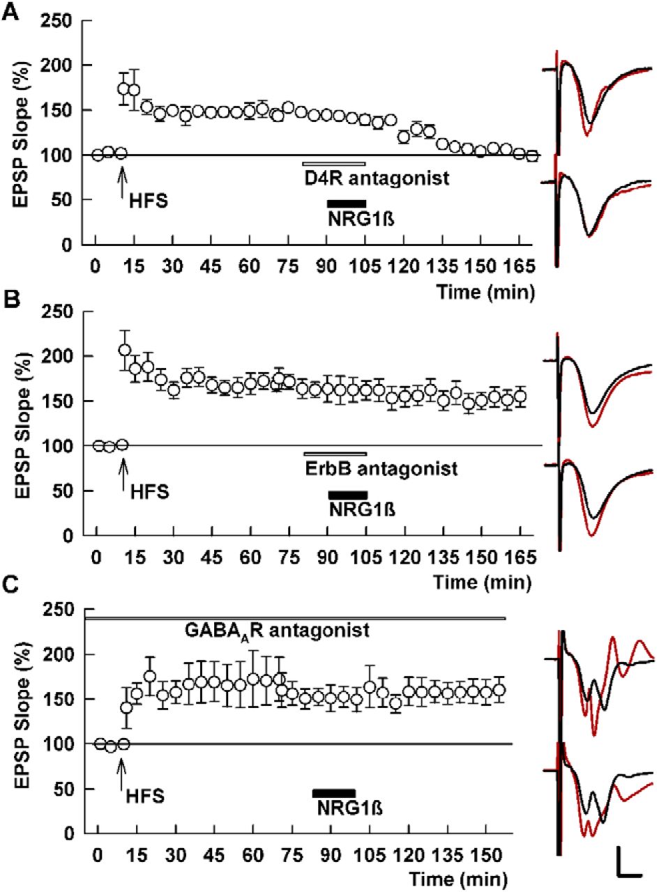
  • Figure 5.
    • Download figure
    • Open in new tab
    • Download powerpoint
    Figure 5.

    Depotentiation by NRG1β is insensitive to D4R antagonism but blocked by an ErbB antagonist and PTX. A, In the presence of 0.1 μM L-745,870 (white bar), 1 nM NRG1β (black bar) induces SC depotentiation. SC HFS was administered at the arrow. B, C, In contrast to the D4R antagonist, a pan-ErbB antagonist (10 μM PD-158,780, white bar) blocks NRG1β-induced depotentiation (B), as does the GABAAR antagonist, 1 μM PTX (C). Traces show representative EPSPs as in Figure 1. Calibration: 1 mV, 5 ms.

  • Figure 6.
    • Download figure
    • Open in new tab
    • Download powerpoint
    Figure 6.

    An ErbB antagonist blocks TA-induced, but not homosynaptic SC depotentiation. A, In the presence of 10 μM PD-158,780, TA stimulation (PLFS, hatched bar) fails to induce persistent SC depotentiation. SC HFS was administered at the arrow. B, In contrast, the ErbB antagonist fails to block depotentiation induced by homosynaptic SC LFS (SLFS, hatched bar). Traces show representative EPSPs as in Figure 1. Calibration: 1 mV, 5 ms.
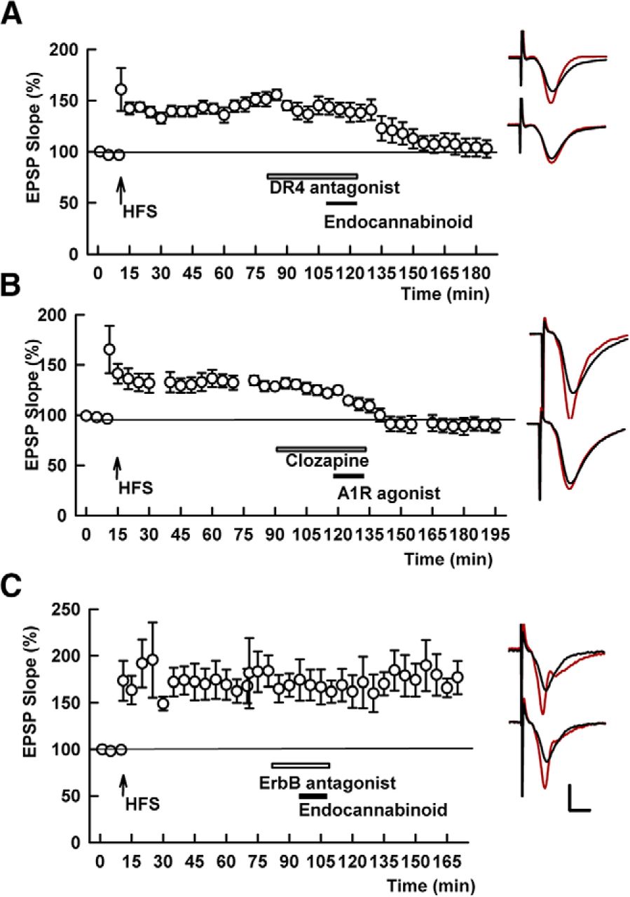
  • Figure 7.
    • Download figure
    • Open in new tab
    • Download powerpoint
    Figure 7.

    D4R antagonism does not block chemical depotentiation by CB1R or adenosine A1R activation, but ErbB antagonism blocks the effects of a CB1R agonist. A, In the presence of the D4R antagonist, L-745,870 (white bar), the endocannabinoid, 20 μM 2-AG (black bar), reversed SC LTP. SC HFS was administered at the arrow. B, Similarly, clozapine, a DAR antagonist with selectivity for D4Rs (white bar), failed to block depotentiation by 10 nM CPA, a selective A1R agonist (black bar). C, The effects of 2AG on SC STP were blocked by the ErbB antagonist, PD-158,780. Traces show representative EPSPs as in Figure 1. Calibration: 1 mV, 5 ms.
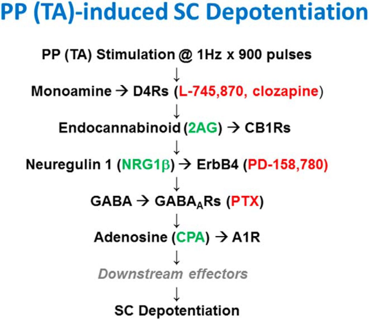
  • Figure 8.
    • Download figure
    • Open in new tab
    • Download powerpoint
    Figure 8.

    The diagram depicts a current scheme for TA-induced SC LTP-D based on prior studies (Izumi and Zorumski, 2008, 2016) and the present results. Agents that promote chemical depotentiation are shown in green while agents that inhibit TA-induced SC depotentiation are shown in red.

Back to top

In this issue

eneuro: 4 (4)
eNeuro
Vol. 4, Issue 4
July/August 2017
  • Table of Contents
  • Index by author
Email

Thank you for sharing this eNeuro article.

NOTE: We request your email address only to inform the recipient that it was you who recommended this article, and that it is not junk mail. We do not retain these email addresses.

Enter multiple addresses on separate lines or separate them with commas.
Neuregulin and Dopamine D4 Receptors Contribute Independently to Depotentiation of Schaffer Collateral LTP by Temperoammonic Path Stimulation
(Your Name) has forwarded a page to you from eNeuro
(Your Name) thought you would be interested in this article in eNeuro.
CAPTCHA
This question is for testing whether or not you are a human visitor and to prevent automated spam submissions.
Print
View Full Page PDF
Citation Tools
Neuregulin and Dopamine D4 Receptors Contribute Independently to Depotentiation of Schaffer Collateral LTP by Temperoammonic Path Stimulation
Yukitoshi Izumi, Charles F. Zorumski
eNeuro 15 August 2017, 4 (4) ENEURO.0176-17.2017; DOI: 10.1523/ENEURO.0176-17.2017

Citation Manager Formats

  • BibTeX
  • Bookends
  • EasyBib
  • EndNote (tagged)
  • EndNote 8 (xml)
  • Medlars
  • Mendeley
  • Papers
  • RefWorks Tagged
  • Ref Manager
  • RIS
  • Zotero
Respond to this article
Share
Neuregulin and Dopamine D4 Receptors Contribute Independently to Depotentiation of Schaffer Collateral LTP by Temperoammonic Path Stimulation
Yukitoshi Izumi, Charles F. Zorumski
eNeuro 15 August 2017, 4 (4) ENEURO.0176-17.2017; DOI: 10.1523/ENEURO.0176-17.2017
del.icio.us logo Digg logo Reddit logo Twitter logo CiteULike logo Facebook logo Google logo Mendeley logo
  • Tweet Widget
  • Facebook Like
  • Google Plus One

Jump to section

  • Article
    • Abstract
    • Significance Statement
    • Introduction
    • Materials and Methods
    • Results
    • Discussion
    • Acknowledgments
    • Footnotes
    • References
    • Synthesis
  • Figures & Data
  • Info & Metrics
  • eLetters
  • PDF

Keywords

  • adenosine
  • Endocannabinoids
  • ErbB
  • GABA
  • hippocampus
  • perforant path

Responses to this article

Respond to this article

Jump to comment:

No eLetters have been published for this article.

Related Articles

Cited By...

More in this TOC Section

New Research

  • Essential Role of Somatic Kv2 Channels in High-Frequency Firing in Cartwheel Cells of the Dorsal Cochlear Nucleus
  • Phenotypic differences between the Alzheimer’s disease-related hAPP-J20 model and heterozygous Zbtb20 knockout mice
  • Distinct basal metabolism in three mouse models of neurodevelopmental disorders
Show more New Research

Neuronal Excitability

  • Essential Role of Somatic Kv2 Channels in High-Frequency Firing in Cartwheel Cells of the Dorsal Cochlear Nucleus
  • Angiotensin-II modulates GABAergic neurotransmission in the mouse substantia nigra
  • Differential Impact of Inhibitory G-Protein Signaling Pathways in Ventral Tegmental Area Dopamine Neurons on Behavioral Sensitivity to Cocaine and Morphine
Show more Neuronal Excitability

Subjects

  • Neuronal Excitability
  • Home
  • Alerts
  • Visit Society for Neuroscience on Facebook
  • Follow Society for Neuroscience on Twitter
  • Follow Society for Neuroscience on LinkedIn
  • Visit Society for Neuroscience on Youtube
  • Follow our RSS feeds

Content

  • Early Release
  • Current Issue
  • Latest Articles
  • Issue Archive
  • Blog
  • Browse by Topic

Information

  • For Authors
  • For the Media

About

  • About the Journal
  • Editorial Board
  • Privacy Policy
  • Contact
  • Feedback
(eNeuro logo)
(SfN logo)

Copyright © 2021 by the Society for Neuroscience.
eNeuro eISSN: 2373-2822

The ideas and opinions expressed in eNeuro do not necessarily reflect those of SfN or the eNeuro Editorial Board. Publication of an advertisement or other product mention in eNeuro should not be construed as an endorsement of the manufacturer’s claims. SfN does not assume any responsibility for any injury and/or damage to persons or property arising from or related to any use of any material contained in eNeuro.