

Article Figures & Data
Figures
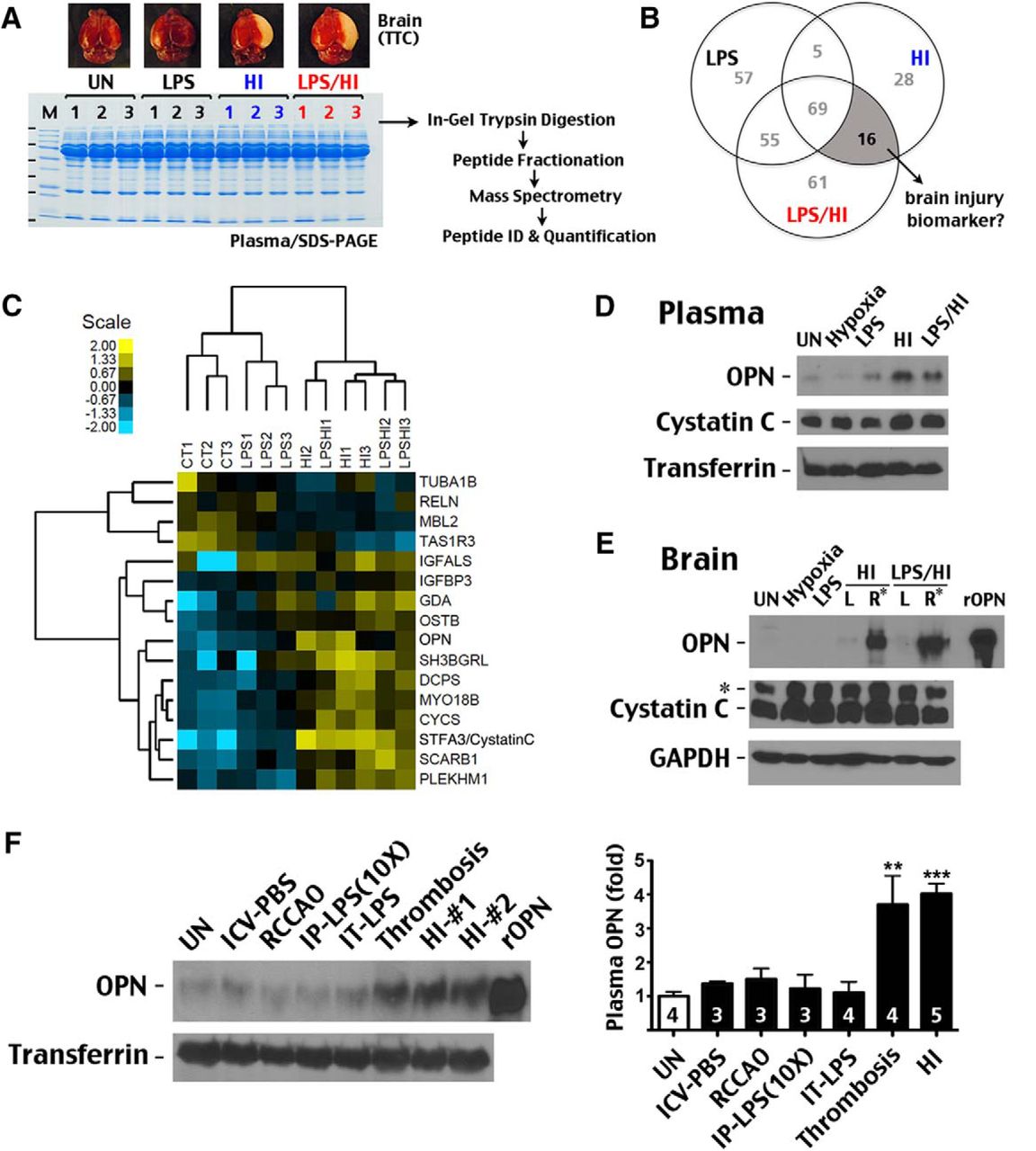
- Figure 1.
Proteomic analysis of plasma biomarkers for murine neonatal HI brain injury. A, Plasma proteins collected from unchallenged (UN), low-dose LPS (0.3 mg/kg, IP)-exposed, and HI- or LPS/HI-injured P11 mice were separated by SDS-PAGE and analyzed as depicted (n = 3 for each). Also shown are representative tetrazolium chloride–stained brains in each group, showing cerebral infarction (pale color) in only HI- or LPS/HI-injured animals. B, Venn diagram showing the number of unique proteins in the depicted conditions compared with the unchallenged animals. C, Heatmap and hierarchical clustering of the 12 examined animals based on the signal intensity of HI- and LPS/HI-associated proteins. Shown is the log2 scale of fold change. D, E, Immunoblot showing specific OPN induction in plasma and the ipsilateral hemisphere (right, R*) of mice after HI or LPS/HI insult. In contrast, although cystatin C showed increased plasma levels after HI and LPS/HI injury, it was not elevated in the brain after these injuries. *, Nonspecific anti–cystatin C band in the brain. rOPN, recombinant mouse OPN used as positive control. F, Immunoblotting showing that cerebral stroke (thrombosis) also induced high levels of plasma OPN, whereas ICV injection of phosphate saline (ICV-PBS), unilateral carotid artery ligation (RCCAO), i.p. injection of 3 mg/kg LPS (IP-LPS[10×]), and intratracheal application of 0/3 mg/kg LPS (IT-LPS) lacked this effect. The numbers of mouse pups examined in each condition are indicated. **, p < 0.01; ***, p < 0.001 by t-test compared with unchallenged animals.
- Figure 2.
Source and specificity of plasma OPN as a biomarker. A, RT-PCR showed that HI triggered a 20- to 30-fold increase of Opn mRNA in the brain of P10 mice and P7 rats at 24-h recovery, but only insignificant changes in PBMCs (n = 4–5 for each group). B, Immunolabeling showed widespread OPN expression by Iba1+ microglia and occasional expression in NG2+ glial progenitors, but rare expression in GFAP+ astrocytes after HI injury (n > 4). Scale bar: 40 μm. C, Application of LPS (1 ng/ml) to microglial SM826 cells in vitro led to induction of OPN protein. D, Similarly, ICV injection of LPS (1 μg) induced Opn mRNA in brain as well as plasma OPN protein at 24-h recovery.
- Figure 3.
Sensitivity and prognostic value of plasma OPN as a biomarker for HI brain injury. A, B, Comparison of OPN and two other candidate biomarkers (MMP-9 and GFAP) in brain and blood after HI and LPS/HI injury. In brain extracts, elevation of MMP-9 or OPN expression and multiple GFAP bands were all observed after injury, but in plasma, only OPN induction was detectable at 24 h post-insult. C, D, Multiplex ELISA was used to compare brain and plasma OPN or MMP-9 levels in P10 mice after HI. Plasma OPN levels showed strong correlation with brain OPN (r = 0.82) and brain MMP-9 levels (r = 0.75, n = 10) 24 h after HI injury. E, HI-injured P7 rats were used to assess the time course of plasma OPN induction after HI injury (n = 5). Strong (>5-fold) induction of plasma OPN occurred only 24 h post-HI. *, Nonspecific signal or hyperphosphorylated OPN. F, Comparison of plasma OPN levels at 48 h post-HI to severity of brain injury at 7 d. Shown are representative subjects for the low and high plasma OPN groups. *, HI-injured hemisphere. G, Quantification showed significant elevation of plasma OPN levels in the severe damage group compared with unchallenged mice (n = 5 and 6 per group for mild and severe brain injury, respectively; p = 0.048 by t-test).


Tables
- Table 1.
Plasma proteins that are uniquely associated with HI and LPS/HI brain injury at 24-h recovery
Symbol Description (all in Mus musculus) Peptide feature counts CT/CT log2(ratio) CT/CT SD, n = 3 LPS/CT log2(ratio) LPS/CT SD, n = 9 HI/CT log2(ratio) HI/CT SD, n = 9 LPS/HI/CT log2(ratio) LPS/HI/CT SD, n = 9 TUBA1B Tubulin alpha-1B chain 58 –0.6 0.33 –0.78 0.5 –0.94 0.47 –1.11 0.41 RELN Reelin precursor 18 0.01 0.22 –0.57 0.35 –0.96 0.27 –1.05 0.14 MBL2 Mannos-binding protein C precursor 86 0.23 0.28 –0.51 0.43 –1.17 0.3 –1.55 0.3 TAS1R3 Taste receptor type 1 member 3 precursor 5 –0.35 0.17 –0.77 0.43 –1.22 0.53 –1.38 0.56 IGFALS Insulin-like growth factor-binding protein complex acid labile subunit precursor 171 –0.11 0.26 –0.56 0.18 –1.78 0.34 –2.06 0.31 IGFBP3 Insulin-like growth factor-binding protein 3 precursor 27 –0.19 0.16 0.13 0.16 –0.92 0.25 –1.28 0.35 GDA Guanine deaminase 15 0.25 0.36 0.62 0.34 1.03 0.45 1.05 0.49 OSTB Organic solute transporter subunit beta 8 0.42 0.14 0.79 0.29 0.9 0.31 1.05 0.33 OPN Osteopontin 11 0.12 0.4 0.69 0.28 1.68 0.6 1.31 0.43 STFA3 Stefin-3 (cystatin C) 14 –0.48 0.99 0.62 0.68 2.63 0.71 1.67 0.6 MYO18B Myosin XVIIIb 2 –0.25 0.15 0.8 0.25 1.51 0.31 1.3 0.2 SH3BGRL SH3 domain-binding glutamic acid-rich like protein 3 0.33 1.21 –0.09 1.98 1.86 0.84 1.7 0.76 DCPS m7GpppX diphosphatase 12 0.2 0.35 0.34 0.45 1.55 0.61 1.47 0.32 PLEKHM1 Pleckstrin homology domain-containing family M member 1 2 –0.05 0.49 –0.12 0.45 0.92 0.42 1.06 0.44 CYCS Cytochrome c, somatic 50 –0.35 0.14 0.64 0.21 1.59 0.23 1.44 0.28 SCARB1 Scavenger receptor class B member 1 isoform 1 2 –0.25 0.1 0.71 0.25 1.3 0.4 1.75 0.46 Plasma proteins associated with HI and LPS/HI brain injury at 24-h recovery in P10 mice. Proteomics detection and statistical analysis were performed as described in the text. Sixteen high-confidence proteins changing in this category with ≥2 MS/MS-identified peptide feature counts used for quantitation across the 12 samples are reported.
In this issue


