
Main menu

  • HOME
  • CONTENT
    • Early Release
    • Featured
    • Current Issue
    • Issue Archive
    • Blog
    • Collections
    • Podcast
  • TOPICS
    • Cognition and Behavior
    • Development
    • Disorders of the Nervous System
    • History, Teaching and Public Awareness
    • Integrative Systems
    • Neuronal Excitability
    • Novel Tools and Methods
    • Sensory and Motor Systems
  • ALERTS
  • FOR AUTHORS
  • ABOUT
    • Overview
    • Editorial Board
    • For the Media
    • Privacy Policy
    • Contact Us
    • Feedback
  • SUBMIT

User menu

Search

  • Advanced search
eNeuro

eNeuro

Advanced Search


  • HOME
  • CONTENT
    • Early Release
    • Featured
    • Current Issue
    • Issue Archive
    • Blog
    • Collections
    • Podcast
  • TOPICS
    • Cognition and Behavior
    • Development
    • Disorders of the Nervous System
    • History, Teaching and Public Awareness
    • Integrative Systems
    • Neuronal Excitability
    • Novel Tools and Methods
    • Sensory and Motor Systems
  • ALERTS
  • FOR AUTHORS
  • ABOUT
    • Overview
    • Editorial Board
    • For the Media
    • Privacy Policy
    • Contact Us
    • Feedback
  • SUBMIT
PreviousNext
Research ArticleNew Research, Development

Neural Progenitor Cells Promote Axonal Growth and Alter Axonal mRNA Localization in Adult Neurons

Tanuja T. Merianda, Ying Jin, Ashley L. Kalinski, Pabitra K. Sahoo, Itzhak Fischer and Jeffery L. Twiss
eNeuro 24 January 2017, 4 (1) ENEURO.0171-16.2017; DOI: https://doi.org/10.1523/ENEURO.0171-16.2017
Tanuja T. Merianda
1Department of Anatomy and Neurobiology, Drexel University College of Medicine, Philadelphia, Pennsylvania 19129
  • Find this author on Google Scholar
  • Find this author on PubMed
  • Search for this author on this site
Ying Jin
1Department of Anatomy and Neurobiology, Drexel University College of Medicine, Philadelphia, Pennsylvania 19129
  • Find this author on Google Scholar
  • Find this author on PubMed
  • Search for this author on this site
Ashley L. Kalinski
2Department of Biological Sciences, University of South Carolina, Columbia, South Carolina 29208
  • Find this author on Google Scholar
  • Find this author on PubMed
  • Search for this author on this site
  • ORCID record for Ashley L. Kalinski
Pabitra K. Sahoo
2Department of Biological Sciences, University of South Carolina, Columbia, South Carolina 29208
  • Find this author on Google Scholar
  • Find this author on PubMed
  • Search for this author on this site
  • ORCID record for Pabitra K. Sahoo
Itzhak Fischer
1Department of Anatomy and Neurobiology, Drexel University College of Medicine, Philadelphia, Pennsylvania 19129
  • Find this author on Google Scholar
  • Find this author on PubMed
  • Search for this author on this site
  • ORCID record for Itzhak Fischer
Jeffery L. Twiss
2Department of Biological Sciences, University of South Carolina, Columbia, South Carolina 29208
  • Find this author on Google Scholar
  • Find this author on PubMed
  • Search for this author on this site
  • ORCID record for Jeffery L. Twiss
  • Article
  • Figures & Data
  • Info & Metrics
  • eLetters
  • PDF
Loading

Article Figures & Data

Figures

  • Figure 1.
    • Download figure
    • Open in new tab
    • Download powerpoint
    Figure 1.

    GRPs enhance axonal growth in DRGs in vitro. A–C, Representative montage images are shown for control dissociated DRG culture (A), dissociated DRGs cocultured with rat GRPs (B), and dissociated DRGs cultures exposed to conditioned medium from alkaline phosphatase-expressing rat GRPs (C). βIII-tubulin (red) immunofluorescence highlights the neurons; GRPs are visualized by immunostaining for alkaline phosphatase (green). D shows quantification for the average length of the longest axon per neuron ± SEM (n ≥ 30 neurons in three separate experiments; **p ≤ 0.01 by Student’s t test). Scale bar, 250 μm.
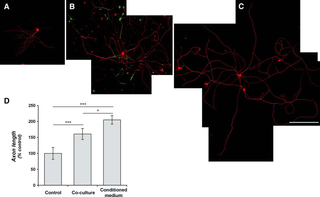
  • Figure 2.
    • Download figure
    • Open in new tab
    • Download powerpoint
    Figure 2.

    Coculture with neural and glial progenitor cells increases axonal outgrowth from adult DRG neurons. A–C, Representative montage images of control-dissociated DRG cultures (A), dissociated DRGs cocultured with rat GRPs/NRPs (B), and dissociated DRG cultures exposed to conditioned medium from parallel rat GRP/NRP cultures (C) are shown. Immunofluorescence for βIII-tubulin (red) and nestin (green) highlight DRG neurons and GRP/NRP nuclei, respectively. D, Quantitation of the average lengths of the longest axon per neuron (±SEM) for the above conditions is shown. Coculture with GRPs/NRPs significantly increases in axon length compared with the standard DRG culture; exposure to conditioned medium from GRP/NRP cultures showed a further increase in axon length (n ≥ 30 neurons in three separate experiments; *p ≤ 0.05 and ***p ≤ 0.001 by Student’s t test). Scale bar, 250 μm. E, Quantitation of axon growth parameters for 7 d injury-conditioned DRG neurons cultured on coverslips laid over a bed of GRP/NRP cells (coculture) or control conditions is shown (n ≥ 200 neurons analyzed in three separate experiments; p values represent ANOVA with Tukey post hoc analyses).

  • Figure 3.
    • Download figure
    • Open in new tab
    • Download powerpoint
    Figure 3.

    Approach for DRG/progenitor cell coculture to isolate axonal processes. A, Schematic of modified Boyden chamber used for culture system for isolation of axons from DRG neurons is shown. For this, DRG explants were plated onto the upper surface of a porous PET membrane, as previously used for dissociated DRGs (Zheng et al., 2001), and GRPs/NRPs were cultured on the surface of the plate well. B–D, Representative confocal projection images of upper membrane surface with DRGs (B), corresponding image stacks for lower membrane surface showing a dense array of axons (C), and GRPs/NRPs along the plate surface (D) are shown. DRGs were stained for βIII-tubulin (green) and DAPI (blue). The GRPs were visualized with DiO stain (cyan). The insets in B and C show mCherry signals (red) of AAV5-mChMYR3'amph-transduced DRGs in cell bodies (B) extending into axons along the lower membrane surface (C). Scale bars: B, C, 100 µm; D, 50 µm. E, Representative RT-PCR from cell body and axonal isolates for DRGs cultured under control conditions or over a bed of GRPs/NRPs. The axonal isolates are depleted of cell body (MAP2 and c-Jun) and glial (GFAP) mRNAs, but contain the known axonal transcript β-actin.
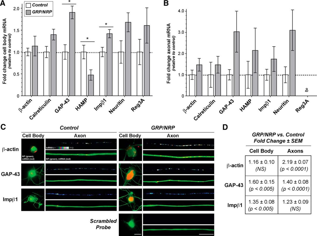
  • Figure 4.
    • Download figure
    • Open in new tab
    • Download powerpoint
    Figure 4.

    Exposure to GRPs/NRPs alters the levels of growth-associated mRNA cell bodies and axons of adult DRG neurons. A, B, The modified coculture system from Figure 3 was used for analyses of RNA levels from the explanted ganglia (“cell body RNA”) and their extended axons (“axonal RNA”). Data from RT-ddPCR analyses with cell body and axonal RNA preparations for known axonal mRNAs are shown in A and B, respectively. Values are shown for the NRP/GRP coculture relative to control samples, as indicated, ±SD for biological replicates (N = 4; p ≤ 0.05 for indicated columns; &no positive droplets were detected for Reg3a amplification despite highest template input levels). C, Representative FISH/IF images of cell bodies and axon shafts are shown for indicated mRNAs for dissociated DRGs cultured under control or GRP/NRP-exposed conditions. For these studies, DRGs were cultured on coverslips over a bed of GRP/NRP cells. Image pairs for control and GRP/NRP conditions are exposure matched, and axonal mRNA signals are shown as a spectral intensity in the upper rows of axon image sequences and cell body, and lower axon panels show merged images for mRNA and NF. Scale bars: cell body panels, 25 µm; axon panels, 10 µm. D, Quantification of axonal FISH signal intensities from matched exposure images are shown for GRP/NRP coculture vs control cultures as the fold change ± SEM (n > 50 for axons and n > 25 for cell bodies in three culture preparations; p values are from ANOVA with Tukey post hoc analyses).

  • Figure 5.
    • Download figure
    • Open in new tab
    • Download powerpoint
    Figure 5.

    Alterations in axonal mRNA levels with GRP/NRP coculture is conferred by axonal mRNA UTRs. Aa–i, Representative fluorescent images for eGFP mRNA (red) and βIII-tubulin protein (green) in cell body and distal axon shaft of dissociated DRG cultures transfected with eGFPMYR plasmids with 3'UTRs of rat GAP43 (a, b), β-actin (c, d), CALR (e, f), and γ-actin mRNAs (g, h). i, Images for sense eGFP probe represent the 3'β-actin construct. Left-hand columns of axon and cell body images show control DRG cultures, and right-hand columns show DRGs cocultured with GRPs/NRPs. All image pairs are exposure matched (control vs GRP/NRP coculture axon and control vs GRP/NRP coculture cell body). As previously published, axonal GFP mRNA is not seen for the construct with 3'UTR of γ-actin, but axonal signals are seen for GAP43, calreticulin, and β-actin 3'UTR constructs (Willis et al., 2007; Vuppalanchi et al., 2010; Yoo et al., 2013). B, C, Quantitation of axonal and cell body GFP mRNA intensities across multiple transfection experiments for DRGs with or without GRPs/NRPs are shown as average signal intensities ± SEM (n ≥ 30 processes over three independent transfections and cultures; ***p ≤ 0.001 by Student’s t test). Scale bars, 20 µm.

Back to top

In this issue

eneuro: 4 (1)
eNeuro
Vol. 4, Issue 1
January/February 2017
  • Table of Contents
  • Index by author
Email

Thank you for sharing this eNeuro article.

NOTE: We request your email address only to inform the recipient that it was you who recommended this article, and that it is not junk mail. We do not retain these email addresses.

Enter multiple addresses on separate lines or separate them with commas.
Neural Progenitor Cells Promote Axonal Growth and Alter Axonal mRNA Localization in Adult Neurons
(Your Name) has forwarded a page to you from eNeuro
(Your Name) thought you would be interested in this article in eNeuro.
CAPTCHA
This question is for testing whether or not you are a human visitor and to prevent automated spam submissions.
Print
View Full Page PDF
Citation Tools
Neural Progenitor Cells Promote Axonal Growth and Alter Axonal mRNA Localization in Adult Neurons
Tanuja T. Merianda, Ying Jin, Ashley L. Kalinski, Pabitra K. Sahoo, Itzhak Fischer, Jeffery L. Twiss
eNeuro 24 January 2017, 4 (1) ENEURO.0171-16.2017; DOI: 10.1523/ENEURO.0171-16.2017

Citation Manager Formats

  • BibTeX
  • Bookends
  • EasyBib
  • EndNote (tagged)
  • EndNote 8 (xml)
  • Medlars
  • Mendeley
  • Papers
  • RefWorks Tagged
  • Ref Manager
  • RIS
  • Zotero
Respond to this article
Share
Neural Progenitor Cells Promote Axonal Growth and Alter Axonal mRNA Localization in Adult Neurons
Tanuja T. Merianda, Ying Jin, Ashley L. Kalinski, Pabitra K. Sahoo, Itzhak Fischer, Jeffery L. Twiss
eNeuro 24 January 2017, 4 (1) ENEURO.0171-16.2017; DOI: 10.1523/ENEURO.0171-16.2017
Reddit logo Twitter logo Facebook logo Mendeley logo
  • Tweet Widget
  • Facebook Like
  • Google Plus One

Jump to section

  • Article
    • Abstract
    • Significance Statement
    • Introduction
    • Materials and Methods
    • Results
    • Discussion
    • Acknowledgments
    • Footnotes
    • References
    • Synthesis
  • Figures & Data
  • Info & Metrics
  • eLetters
  • PDF

Keywords

  • axonal transport
  • Glial restricted progenitor
  • mRNA localization
  • neural restricted progenitor
  • stem cell

Responses to this article

Respond to this article

Jump to comment:

No eLetters have been published for this article.

Related Articles

Cited By...

More in this TOC Section

New Research

  • LIQ HD (Lick Instance Quantifier Home cage Device): An open-source tool for recording undisturbed two-bottle drinking behavior in a home cage environment
  • Insulin-like growth factor-1 supplementation promotes brain maturation in preterm pigs
  • SK and Kv4 channels limit spike timing perturbations in pacemaking dopamine neurons
Show more New Research

Development

  • Ontogeny and Trophic Factor Sensitivity of Gastrointestinal Projecting Vagal Sensory Cell Types
  • Heterozygous Dab1 null mutation disrupts neocortical and hippocampal development
  • The Mobility of Neurofilaments in Mature Myelinated Axons of Adult Mice
Show more Development

Subjects

  • Development

  • Home
  • Alerts
  • Visit Society for Neuroscience on Facebook
  • Follow Society for Neuroscience on Twitter
  • Follow Society for Neuroscience on LinkedIn
  • Visit Society for Neuroscience on Youtube
  • Follow our RSS feeds

Content

  • Early Release
  • Current Issue
  • Latest Articles
  • Issue Archive
  • Blog
  • Browse by Topic

Information

  • For Authors
  • For the Media

About

  • About the Journal
  • Editorial Board
  • Privacy Policy
  • Contact
  • Feedback
(eNeuro logo)
(SfN logo)

Copyright © 2023 by the Society for Neuroscience.
eNeuro eISSN: 2373-2822

The ideas and opinions expressed in eNeuro do not necessarily reflect those of SfN or the eNeuro Editorial Board. Publication of an advertisement or other product mention in eNeuro should not be construed as an endorsement of the manufacturer’s claims. SfN does not assume any responsibility for any injury and/or damage to persons or property arising from or related to any use of any material contained in eNeuro.