
Main menu

  • HOME
  • CONTENT
    • Early Release
    • Featured
    • Current Issue
    • Issue Archive
    • Blog
    • Collections
    • Podcast
  • TOPICS
    • Cognition and Behavior
    • Development
    • Disorders of the Nervous System
    • History, Teaching and Public Awareness
    • Integrative Systems
    • Neuronal Excitability
    • Novel Tools and Methods
    • Sensory and Motor Systems
  • ALERTS
  • FOR AUTHORS
  • ABOUT
    • Overview
    • Editorial Board
    • For the Media
    • Privacy Policy
    • Contact Us
    • Feedback
  • SUBMIT

User menu

Search

  • Advanced search
eNeuro

eNeuro

Advanced Search


  • HOME
  • CONTENT
    • Early Release
    • Featured
    • Current Issue
    • Issue Archive
    • Blog
    • Collections
    • Podcast
  • TOPICS
    • Cognition and Behavior
    • Development
    • Disorders of the Nervous System
    • History, Teaching and Public Awareness
    • Integrative Systems
    • Neuronal Excitability
    • Novel Tools and Methods
    • Sensory and Motor Systems
  • ALERTS
  • FOR AUTHORS
  • ABOUT
    • Overview
    • Editorial Board
    • For the Media
    • Privacy Policy
    • Contact Us
    • Feedback
  • SUBMIT
PreviousNext
Research ArticleFailure to Replicate, Sensory and Motor Systems

Levels of Cocaine- and Amphetamine-Regulated Transcript in Vagal Afferents in the Mouse Are Unaltered in Response to Metabolic Challenges

Xuefeng Yuan, Ying Huang, Sarita Shah, Hua Wu and Laurent Gautron
eNeuro 22 September 2016, 3 (5) ENEURO.0174-16.2016; DOI: https://doi.org/10.1523/ENEURO.0174-16.2016
Xuefeng Yuan
1Division of Hypothalamic Research and Department of Internal Medicine, The University of Texas Southwestern Medical Center, 5323 Harry Hines Blvd., Dallas, Texas 75390
2Department of Orthopedics, Tongji Hospital, Tongji Medical College, Huazhong University of Science and Technology, Wuhan, China
  • Find this author on Google Scholar
  • Find this author on PubMed
  • Search for this author on this site
  • ORCID record for Xuefeng Yuan
Ying Huang
1Division of Hypothalamic Research and Department of Internal Medicine, The University of Texas Southwestern Medical Center, 5323 Harry Hines Blvd., Dallas, Texas 75390
  • Find this author on Google Scholar
  • Find this author on PubMed
  • Search for this author on this site
Sarita Shah
1Division of Hypothalamic Research and Department of Internal Medicine, The University of Texas Southwestern Medical Center, 5323 Harry Hines Blvd., Dallas, Texas 75390
  • Find this author on Google Scholar
  • Find this author on PubMed
  • Search for this author on this site
  • ORCID record for Sarita Shah
Hua Wu
2Department of Orthopedics, Tongji Hospital, Tongji Medical College, Huazhong University of Science and Technology, Wuhan, China
  • Find this author on Google Scholar
  • Find this author on PubMed
  • Search for this author on this site
Laurent Gautron
1Division of Hypothalamic Research and Department of Internal Medicine, The University of Texas Southwestern Medical Center, 5323 Harry Hines Blvd., Dallas, Texas 75390
  • Find this author on Google Scholar
  • Find this author on PubMed
  • Search for this author on this site
  • ORCID record for Laurent Gautron
  • Article
  • Figures & Data
  • Info & Metrics
  • eLetters
  • PDF
Loading

Article Figures & Data
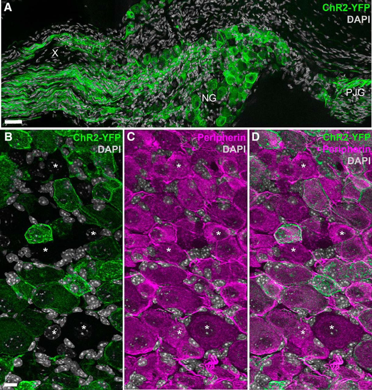
Figures

  • Tables
  • Movies
  • Figure
    • Download figure
    • Open in new tab
    • Download powerpoint
  • Figure 1.
    • Download figure
    • Open in new tab
    • Download powerpoint
    Figure 1.

    Distribution of ChR2-YFP fluorescence in the nodose ganglion of Nav1.8-Cre-ChR2-YFP mice. A, Many neuronal cell bodies and axons were brightly fluorescent in the nodose ganglion (NG) and petrosal-jugular ganglion (PJG) (three images were horizontally stitched together). B–D, Details of NG peripherin-labeled neurons (Alexa Fluor 594). ChR2-YFP is apparent in the membrane and cytoplasm of many NG neurons (but not all) (B). Asterisks are positioned over representative peripherin-positive neurons that were not labeled with ChR2-YFP (B, C). X, cervical vagus nerve. Scale bars, 40 μm in A; 20 μm in B (also applies to C, D).

  • Figure 2.
    • Download figure
    • Open in new tab
    • Download powerpoint
    Figure 2.

    Distribution of ChR2-YFP fluorescence in the dorsovagal complex of Nav1.8-Cre-ChR2-YFP mice. A, Vagal afferents terminating in the AP and NTS were labeled with ChR2-YFP (three horizontally stitched images). B, Highly varicose ChR2-YFP–labeled axons circling the cell body of one TH-positive neuron (Alexa Fluor 594) located in the NTS. C, Single optical section of ChR2-YFP–labeled axons in the AP revealing thin varicose axons of varying sizes. Asterisks are positioned over the presumptive locations of the postsynaptic cell bodies, whereas arrowheads indicate representative vagal varicosities. DMV, dorsal nucleus of the solitary tract; sol, solitary tract; commissural part of the nucleus of the solitary tract; SolDM, dorsomedial part of the nucleus of the solitary tract; SolM, intermediate part of the nucleus of the solitary tract; SubP, subpostrema area. Scale bars, 60 μm in A; 5 μm in B and C.

  • Figure 3.
    • Download figure
    • Open in new tab
    • Download powerpoint
    Figure 3.

    Distribution of ChR2-YFP fluorescence throughout the rostrocaudal extension of the dorsovagal complex of one representative Nav1.8-ChR2-YFP mouse. Several digital images were stitched together (epifluorescence with Apotome filter). The number in the bottom right corner indicates the approximate distance from Bregma according to the Franklin and Paxinos Mouse Brain Atlas (Third Edition). 4v, fourth ventricle; cc, central canal.
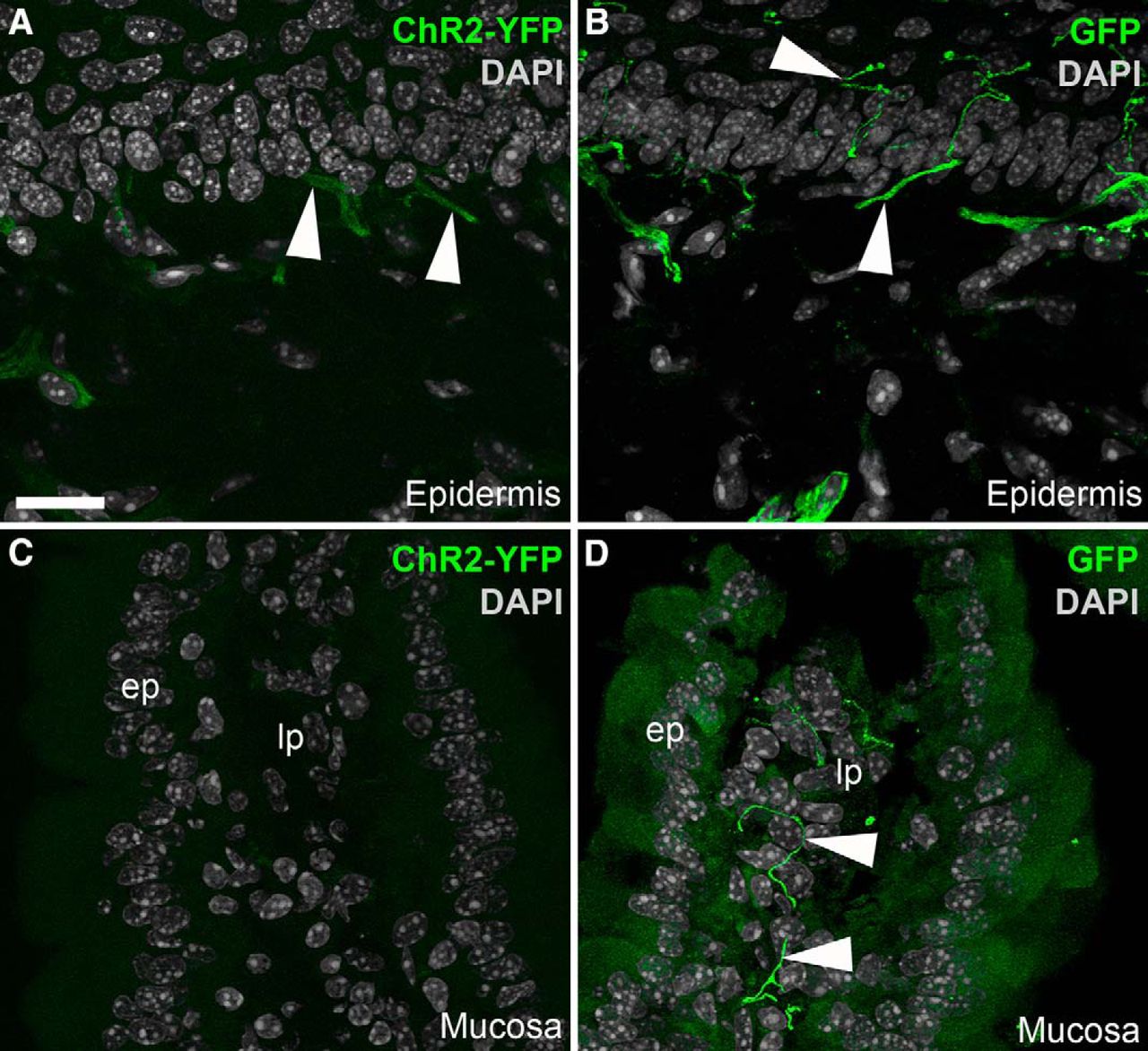
  • Figure 4.
    • Download figure
    • Open in new tab
    • Download powerpoint
    Figure 4.

    Identification of ChR2-YFP–positive fibers in peripheral tissues from Nav1.8-Cre-ChR2-YFP mice. A, Weakly fluorescent structures resembling nerve endings (arrowheads) were observed in the epidermis of the forepaw. B, GFP immunolabeling greatly enhanced the labeling of individual axons ramifying into the epidermis. C, We could not detect endogenous YFP fluorescence in the duodenal mucosa before immunolabeling. D, YFP-labeled axons were evident in the lamina propria (lp) when the tissue was stained for GFP. The antibody produced a small amount of nonspecific background staining in the epithelium (ep). Arrowheads indicate representative YFP-positive fibers. Scale bars, 20 μm in A (also applies to B–D).

  • Figure 5.
    • Download figure
    • Open in new tab
    • Download powerpoint
    Figure 5.

    CART(55–102) detection in the nodose ganglion (NG) of Nav1.8-Cre-ChR2-YFP mice. A, CART-positive cells (Alexa Fluor 594) resembling neurons were observed throughout the NG. The two cells located in the inset are represented at high magnification in C. B, Omission of the primary antibody eliminated CART immunoreactivity. Only a few debris and background could be observed in the NG. C, Two adjacent ChR2-YFP-, CART-positive neurons are shown. CART is detected in vesicle-like structures in the cytoplasm and proximal axon. The larger neuron contained more immunoreactivity than the smaller neuron. Two other YFP-labeled neurons did not contain CART. Scale bars, 50 μm in A and B; 5 μm in C.

  • Figure 6.
    • Download figure
    • Open in new tab
    • Download powerpoint
    Figure 6.

    CART(55–102) immunolabeling of the nodose ganglion (NG) of metabolically challenged Nav1.8-Cre-ChR2-YFP mice. A, C, E, CART-positive perikarya (Alexa Fluor 594) across the feeding groups. B, D, F, CART staining and endogenous ChR2-YFP fluorescence delineate the outline of the vagal afferents. Asterisks are positioned over representative CART-positive cells. Note that CART immunoreactivity is almost always contained within the cell membrane of the YFP-labeled cells, with a few rare exceptions (arrowhead). By visual inspection, the CART distribution pattern and intensity appeared comparable across the feeding groups. Scale bars, 20 μm in A; applies to all images.

  • Figure 7.
    • Download figure
    • Open in new tab
    • Download powerpoint
    Figure 7.

    Estimates of CART-positive neurons and afferents across the feeding groups. A, The percentage of YFP- and CART-positive neurons in the nodose ganglion (NG) of Nav1.8-Cre-ChR2-YFP mice was not influenced by feeding status (n = 7–8 per group). B, The percentage of CART-positive varicosities in the AP was identical in Nav1.8-Cre-ChR2-YFP mice under different feeding conditions. Data are expressed as the average of the mean percentage ± maximum values (n = 7–8 per group). Each circle represents one value from one mouse. C, Frequency of CART immunoreactivity profiles with varying intensity across feeding groups (n = 7 per group). On the left, frequency scattergraph of distributions of individual CART-positive cell profiles with median and interquartile range. Each gray dot is one cell profile. The total number of profiles examined in each group is annotated. On the right, the same data were represented as stacked bars after categorizations of immunoreactivity level. Data are provided as mean percentage ± SEM. D, Frequency of cells expressing pCART mRNA in the nodose ganglia of fed (n = 4) and fasted (n = 3) wild-type mice. Cells were scaled based on the intensity of hybridization signals. The total number of profiles examined in each group is annotated. Stacked bars provide mean percentage ± SEM.

  • Figure 8.
    • Download figure
    • Open in new tab
    • Download powerpoint
    Figure 8.

    A, B, CART(55–102)-positive neurons in the nodose ganglia of lean Zucker rats. Several digital images were stitched together (epifluorescence with Apotome filter). Many CART-positive neurons (Alexa Fluor 594) were observed in the nodose ganglion of fed and fasted rats. White arrows indicate examples of CART neurons. Scale bar, 100 μm in A; applies to B.

  • Figure 9.
    • Download figure
    • Open in new tab
    • Download powerpoint
    Figure 9.

    CART(55–102) immunolabeling of the dorsovagal complex of metabolically challenged Nav1.8-Cre-ChR2-YFP mice. A, B, C, CART-positive (Alexa Fluor 594) perikarya and fibers of various origins were observed throughout the NTS and AP. We noticed a trend toward increased immunoreactivity in the fasted groups. However, our estimates (see Fig. 6) indicate that the levels of CART in vagal afferents remained unchanged across feeding conditions. D, E, F, CART immunoreactivity combined with endogenous ChR2-YFP fluorescence. G, CART was very abundant in the AP. In particular, many YFP-labeled varicosities of vagal origin were enriched for CART. However, we also observed CART immunoreactivity that was not contained in ChR2-YFP–labeled fibers. H, High magnification of the distribution of CART immunoreactivity in the vagal fibers of the AP in a single optical plan. Of note, CART frequently labeled the cytoplasm contained within the YFP-labeled varicosities, which are indicated by arrowheads. Scale bars, 60 μm in A, applies to B–F; 15 μm in G; 5 μm in H.

  • Figure 10.
    • Download figure
    • Open in new tab
    • Download powerpoint
    Figure 10.

    Detection of prepro-CART (pCART) and prepro-MCH (pMCH) mRNAs using chromogenic ISH. A, pCART hybridization signals (brown DAB; bright-field optics) were strong in select hypothalamic nuclei. B, Throughout the nodose ganglion of fed mice, pCART signals of varying intensity were observed in many cell profiles. C, The nodose ganglion of fasted mice also contained pCART hybridization signals. D, Details of the hybridization signal in the nodose ganglion of one fasted mouse. Please note representative cell profiles without signal (*), or with low (l), medium (m), and high (h) signals. E, Hybridization signals for pMCH were very strong in neurons of the lateral hypothalamus. F, G, In contrast to the hypothalamus, pMCH signals were not observed in the nodose ganglia of fed and fasted mice. H, Details of the nodose ganglion of one fasted mouse showing several neuronal profiles completely devoid of signals (*). Tissue was counterstained with hematoxylin. 3V, third ventricle; Arc, arcuate nucleus; DMH, dorsomedial hypothalamus; LHA, lateral hypothalamus; NG, nodose ganglion; VMH, ventromedial hypothalamus; ZI, zona incerta. Scale bars, 500 μm in A and E; 50 μm in B, C, F, and G; 20 μm in D and H.

  • Figure 11.
    • Download figure
    • Open in new tab
    • Download powerpoint
    Figure 11.

    Absence of MCH staining in the vagal afferents of metabolically challenged Nav1.8-Cre-ChR2-YFP mice. A, An antiserum against prepro-MCH (pMCH) labeled perikarya (Alexa Fluor 594) in the lateral hypothalamus (LHA). B, PMCH was undetectable in the nodose ganglion (NG) of fasted Nav1.8-Cre-ChR2-YFP mice. C, D, An antiserum against the MCH peptide labeled very few axons (arrowhead) in the medial NTS (SolM). MCH immunoreactivity was never observed in the YFP-labeled fibers of vagal origin. Arrowheads point to one MCH-positive axon. sol, solitary tract. Scale bars, 40 μm in A and B; 10 μm in C.
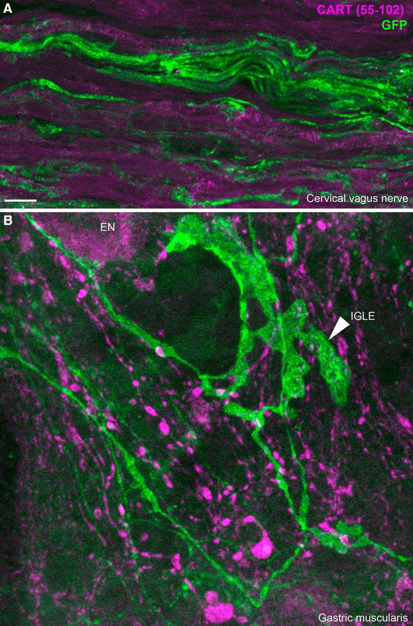
  • Figure 12.
    • Download figure
    • Open in new tab
    • Download powerpoint
    Figure 12.

    CART(55–102) immunolabeling of peripheral ChR2-YFP–labeled fibers. A, ChR2-YFP fibers contained within the cervical vagus nerve. CART was not detected in the vagus nerve. We obtained similar results in the cervical and subdiaphragmatic nerves on both sides. B, In whole mounts of the gastric muscularis, numerous GFP-positive axons and specialized endings can be observed. The leafy structure may correspond to an intraganglionic laminar ending (arrowhead) of vagal origin. CART immunoreactivity was abundant in large varicose fibers and, to a lesser extent, the cell body of enteric neurons (EN). Nonetheless, CART immunoreactivity was not found in GFP-stained fibers. IGLE, intraganglionic laminar ending; EN, enteric neuron. Scale bar, 10 μm in A and B.

Tables

  • Figures
  • Movies
    • View popup
    Table 1.

    Information relative to the primary and secondary antisera used in the study.

    AntibodyManufacturerCatalog no.Lot no.HostWorking dilutionImmunogen
    Primary antibody
        PeripherinEMD MilliporeAB15302446692Rabbit1:500trp-E-peripherin fusion protein containing all but the four N-terminal amino acids of rat peripherin
        CART(55–102)Phoenix PharmaceuticalsH-003-6201251-10; 01251-6Rabbit1:800–1:1000Ile-Pro-Ile-Tyr-Glu-Lys-Lys-Tyr -Gly-Gln-Val-Pro-Met-Cys-Asp-Ala-Gly-Glu-Gln-Cys-Ala-Val-Arg-Lys-Gly-Ala-Arg-Ile-Gly-Lys-Leu-Cys-Asp-Cys-Pro-Arg-Gly-Thr-Ser-Cys-Asn-Ser-Phe-Leu-Leu-Lys-Cys-Leu [disulfide bonds between Cys1-Cys3,Cys2-Cys5,Cys4-Cys6]
        CART 1–39Phoenix PharmaceuticalsH-003-6301102Rabbit1:800–1:1000pGlu-Glu-Asp-Ala-Glu-Leu-Gln-Pro-Arg-Ala-Leu-Asp-Ile-Tyr-Ser-Ala-Val-Asp-Asp-Ala-Ser-His-Glu-Lys-Glu-Leu-Pro-Arg-Arg-Gln-Leu-Arg-Ala-Pro-Gly-Ala-Val-Leu-Gln
        MCHPhoenix PharmaceuticalsH-070-4701629-3Rabbit1:1000Asp-Phe-Asp-Met-Leu-Arg-Cys-Met-Leu-Gly-Arg-Val-Tyr-Arg-Pro-Cys-Trp- Gln-Val
        THAbcamab101853GR120879-17Goat1:1000Synthetic peptide corresponding to human TH(30–100) (N terminal)
        ProMCHSanta Cruz Biotechnologysc-14509B2415Goat1:100–1:100020-amino-acid peptide near the C-terminus of pro-MCH precursor of human origin
        GFPAves LaboratoryGFP-10120GFP697986Chicken1:1000GFP emulsified in Freund’s adjuvant
    Secondary antibody
        Anti-rabbit Alexa Fluor 594Life TechnologiesA212071256153Donkey
        Anti-goat Alexa Fluor 594Life TechnologiesA110581608643Donkey
        Anti-chicken Alexa Fluor 488Life TechnologiesA110391356650Goat
    • View popup
    Table 2.

    List of reagents used for ISH (RNAscope probes from ACD)

    GeneAccession no.Target regionCatalog no., channelChromogenic label
    CARTPTNM_013732.711–860432001-c1DAB, brown
    PMCHNM_029971.24–652478721-c1DAB, brown

Movies

  • Figures
  • Tables
  • Movie 1.

    File compilation of a z-stack of ChR2-YFP–labeled axons (green) around one TH-positive neuron (Alexa Fluor 594) in the AP of the Nav1.8-Cre-ChR2-YFP mouse. Note the varicosities in close apposition to the outline of the TH-positive neuron.

Back to top

In this issue

eneuro: 3 (5)
eNeuro
Vol. 3, Issue 5
September/October 2016
  • Table of Contents
  • Index by author
Email

Thank you for sharing this eNeuro article.

NOTE: We request your email address only to inform the recipient that it was you who recommended this article, and that it is not junk mail. We do not retain these email addresses.

Enter multiple addresses on separate lines or separate them with commas.
Levels of Cocaine- and Amphetamine-Regulated Transcript in Vagal Afferents in the Mouse Are Unaltered in Response to Metabolic Challenges
(Your Name) has forwarded a page to you from eNeuro
(Your Name) thought you would be interested in this article in eNeuro.
CAPTCHA
This question is for testing whether or not you are a human visitor and to prevent automated spam submissions.
Print
View Full Page PDF
Citation Tools
Levels of Cocaine- and Amphetamine-Regulated Transcript in Vagal Afferents in the Mouse Are Unaltered in Response to Metabolic Challenges
Xuefeng Yuan, Ying Huang, Sarita Shah, Hua Wu, Laurent Gautron
eNeuro 22 September 2016, 3 (5) ENEURO.0174-16.2016; DOI: 10.1523/ENEURO.0174-16.2016

Citation Manager Formats

  • BibTeX
  • Bookends
  • EasyBib
  • EndNote (tagged)
  • EndNote 8 (xml)
  • Medlars
  • Mendeley
  • Papers
  • RefWorks Tagged
  • Ref Manager
  • RIS
  • Zotero
Respond to this article
Share
Levels of Cocaine- and Amphetamine-Regulated Transcript in Vagal Afferents in the Mouse Are Unaltered in Response to Metabolic Challenges
Xuefeng Yuan, Ying Huang, Sarita Shah, Hua Wu, Laurent Gautron
eNeuro 22 September 2016, 3 (5) ENEURO.0174-16.2016; DOI: 10.1523/ENEURO.0174-16.2016
del.icio.us logo Digg logo Reddit logo Twitter logo Facebook logo Google logo Mendeley logo
  • Tweet Widget
  • Facebook Like
  • Google Plus One

Jump to section

  • Article
    • Visual Overview
    • Abstract
    • Significance Statement
    • Introduction
    • Materials and Methods
    • Results
    • Discussion
    • Acknowledgments
    • Footnotes
    • References
    • Synthesis
  • Figures & Data
  • Info & Metrics
  • eLetters
  • PDF

Keywords

  • immunohistochemistry
  • metabolism
  • neuropeptide
  • vagus nerve

Responses to this article

Respond to this article

Jump to comment:

  • Food intake rather than physiological state regulates vagal neuropeptide expression
    Guillaume de Lartigue
    Published on: 04 May 2017
  • Published on: (4 May 2017)
    Page navigation anchor for Food intake rather than physiological state regulates vagal neuropeptide expression
    Food intake rather than physiological state regulates vagal neuropeptide expression
    • Guillaume de Lartigue, Assistant Professor, The John B. Pierce Laboratory/Department of Cellular and Molecular Physiology, Yale Medical School

    We have previously demonstrated that vagal afferent neurons switch neuropeptide expression depending on feeding state 1-4. In vivo, vagal afferent neurons of fasted animals express melanin concentrating hormone (MCH) 2, while refeeding reduces MCH expression and increases expression of cocaine and amphetamine regulated transcript (CART) 1,2. Contrary to our previous findings, Yuan et al 5 report constitutive CART expression and a complete absence of MCH expression in vagal afferent neurons in ad libitum fed compared to fasted animal.

    Critically, the feeding paradigm used by Yuan et al 5 was inadequate to observe changes in CART or MCH expression in vagal afferent neurons. In this study mice and rats were either fasted overnight or fed ad libitum and euthanized 3-5 hours into the light phase. Given that fed ad libitum rodents consume most of their calories in the dark phase with almost no intake in the initial period of the light phase, the animals identified as fed are unlikely to have consumed any food for at least 3-5 hours. In contrast, we expose our animals to a longer 24-48h fast on wire-bottom cages to ensure that the stomach is empty, and re-feed half of the fasted animals ad libitum for 2 hours until satiated. This is important since the mechanisms that promote CART and depress MCH expression in vagal afferent neurons requires postprandial release of the gastrointestinal hormone, cholecystokinin (CCK). In support of this, CCK-A receptor encoding nodose gang...

    Show More

    We have previously demonstrated that vagal afferent neurons switch neuropeptide expression depending on feeding state 1-4. In vivo, vagal afferent neurons of fasted animals express melanin concentrating hormone (MCH) 2, while refeeding reduces MCH expression and increases expression of cocaine and amphetamine regulated transcript (CART) 1,2. Contrary to our previous findings, Yuan et al 5 report constitutive CART expression and a complete absence of MCH expression in vagal afferent neurons in ad libitum fed compared to fasted animal.

    Critically, the feeding paradigm used by Yuan et al 5 was inadequate to observe changes in CART or MCH expression in vagal afferent neurons. In this study mice and rats were either fasted overnight or fed ad libitum and euthanized 3-5 hours into the light phase. Given that fed ad libitum rodents consume most of their calories in the dark phase with almost no intake in the initial period of the light phase, the animals identified as fed are unlikely to have consumed any food for at least 3-5 hours. In contrast, we expose our animals to a longer 24-48h fast on wire-bottom cages to ensure that the stomach is empty, and re-feed half of the fasted animals ad libitum for 2 hours until satiated. This is important since the mechanisms that promote CART and depress MCH expression in vagal afferent neurons requires postprandial release of the gastrointestinal hormone, cholecystokinin (CCK). In support of this, CCK-A receptor encoding nodose ganglia neurons co-express with CART6 and MCH2. Furthermore, administration of exogenous CCK in fasted animals increases CART 1 and depresses MCH 2; while, CCK-A receptor antagonist injection in re-fed animals prevents postprandial increase in CART 1 and decrease in MCH 2 expression. In culture, CCK increases CART promoter activation and CART release 7 and within the same cultured vagal afferent neuron MCH expression can be switched to CART within a couple hours following CCK administration 1. Therefore it is likely that in Yuan et al 5 the CART staining in nodose ganglion looked identical in fed, fasted, and obese animals because all the animals were at various points on a fasted spectrum, and perhaps not fasted long enough for MCH to be expressed.

    Yuan et al 5 frequently refer to the fact that their results are in agreement with a previous report by Broberger et al 6 showing no change in CART mRNA in response to food restriction or diet-induced obesity. However Broberger et al did not fast their animals but rather restricted rats to 10g of chow intake per day for 14 days with no indication of when the animals had last eaten prior to euthanasia6. Therefore this neither challenges nor supports the role of feeding and fasting in the control of vagal CART expression. Although Broberger et al found no effect of high fat diet feeding in rats on CART expression in vagal afferent neurons6, it should be noted that these rats were only kept on the diet for 2 weeks. At this time-point rats do not weigh significantly more than lean controls and can therefore not be referred to as diet-induced obese 8-11. Yuan et al quantified CART expression in the nodose ganglia of mice fed a 60% high fat diet for 4 weeks5. No information was provided about the weight gain of these animals in comparison to age matched lean controls. In our previous studies rats were fed an obesigenic diet composed of lower fat content (45% kcal from fat) and higher sucrose content for 8 weeks, at which time these rats weighed significantly more than control littermates maintained on chow 8,12. Therefore even if Yuan et al5 had used a re-feeding paradigm, it is unclear that a blunted CART expression would have been observed in mice maintained on a high-fat low-sugar diet for only 4 weeks.

    It is inaccurate and misleading to refer to the change in CART and MCH expression in vagal afferent neurons as a unique event. Despite being a largely understudied tissue, there have been several examples of stimuli-mediated changes in neuropeptide expression in vagal afferent neurons. Allergic inflammation in guinea pig airways increases the expression of the neuropeptides substance P and CGRP within neurons of the nodose ganglia13. Axonal damage of the vagus nerve increases expression of the neuropeptides galanin, NPY, VIP and CCK, and decreases CCK-1 receptor expression while increasing expression of CCK2 and Y2 receptors in vagal afferent neurons 14-16. Furthermore, metabolic cues from the gut have been extensively reported to alter gene expression in vagal afferent neurons. Expression of clock genes in the nodose ganglia oscillate throughout the day, and are entrained by food intake 17. In lean rats, fasting increases expression of melanin concentrating hormone receptor 12, cannabinoid receptor 1 18, and ghrelin receptor 19. Re-feeding reduces expression of these receptors and promotes expression of Y2 receptor 20,21. Chronic consumption of high fat diets alters mRNA expression of ghrelin receptor 11,19, CB1 receptor 8,22, MCH1 receptor 8, orexin receptor 22, Y2 receptor 8, PPAR-gamma receptor 23, CCK-1 receptor 11,22, GPR4024, GPR41 24, and GPR1 20,24. Given the propensity of vagal afferent neurons to change their neurochemistry in response to peripheral signals, a change in neuropeptide expression in response to nutrient signaling is in keeping with previous findings.

    In summary, the experimental protocol used in Yuan et al 5 is not appropriate to determine neuropeptidergic profile changes in vagal afferent neurons in response to feeding or metabolic state. Crucially the time of last meal and the quantity of food ingested was not controlled in these experiments. However the data by Yuan et al5 suggests that satiation itself is not sufficient to drive the change in neuropeptide expression, since CART and MCH expression are unchanged in animals that chose not to eat despite having access to food during the light phase. A hypothesis that unifies our previous data with those of Yuan et al is that changes in neuropeptide expression is driven by nutrient induced activation of vagal afferent neurons rather than a physiological state that arises from the nutrient consumption.

    1 de Lartigue, G., Dimaline, R., Varro, A. & Dockray, G. J. Cocaine- and amphetamine-regulated transcript: stimulation of expression in rat vagal afferent neurons by cholecystokinin and suppression by ghrelin. J Neurosci 27, 2876-2882, doi:10.1523/JNEUROSCI.5508-06.2007 (2007).
    2 Burdyga, G., Varro, A., Dimaline, R., Thompson, D. G. & Dockray, G. J. Feeding-dependent depression of melanin-concentrating hormone and melanin-concentrating hormone receptor-1 expression in vagal afferent neurones. Neuroscience 137, 1405-1415, doi:10.1016/j.neuroscience.2005.10.057 (2006).
    3 de Lartigue, G., Ronveaux, C. C. & Raybould, H. E. Deletion of leptin signaling in vagal afferent neurons results in hyperphagia and obesity. Mol Metab 3, 595-607, doi:10.1016/j.molmet.2014.06.003 (2014).
    4 de La Serre, C. B., de Lartigue, G. & Raybould, H. E. Chronic exposure to low dose bacterial lipopolysaccharide inhibits leptin signaling in vagal afferent neurons. Physiol Behav 139, 188-194, doi:10.1016/j.physbeh.2014.10.032 (2015).
    5 Yuan, X., Huang, Y., Shah, S., Wu, H. & Gautron, L. Levels of Cocaine- and Amphetamine-Regulated Transcript in Vagal Afferents in the Mouse Are Unaltered in Response to Metabolic Challenges. eNeuro 3, doi:10.1523/ENEURO.0174-16.2016 (2016).
    6 Broberger, C., Holmberg, K., Kuhar, M. J. & Hokfelt, T. Cocaine- and amphetamine-regulated transcript in the rat vagus nerve: A putative mediator of cholecystokinin-induced satiety. Proc Natl Acad Sci U S A 96, 13506-13511 (1999).
    7 De Lartigue, G. et al. Cocaine- and amphetamine-regulated transcript mediates the actions of cholecystokinin on rat vagal afferent neurons. Gastroenterology 138, 1479-1490, doi:10.1053/j.gastro.2009.10.034 (2010).
    8 de Lartigue, G., Barbier de la Serre, C., Espero, E., Lee, J. & Raybould, H. E. Leptin resistance in vagal afferent neurons inhibits cholecystokinin signaling and satiation in diet induced obese rats. PLoS One 7, e32967, doi:10.1371/journal.pone.0032967 (2012).
    9 Mendes, N. F. et al. Knocking down amygdalar PTP1B in diet-induced obese rats improves insulin signaling/action, decreases adiposity and may alter anxiety behavior. Metabolism: clinical and experimental 70, 1-11, doi:10.1016/j.metabol.2017.01.029 (2017).
    10 Boyle, C. N., Rossier, M. M. & Lutz, T. A. Influence of high-fat feeding, diet-induced obesity, and hyperamylinemia on the sensitivity to acute amylin. Physiol Behav 104, 20-28, doi:10.1016/j.physbeh.2011.04.044 (2011).
    11 Paulino, G. et al. Increased expression of receptors for orexigenic factors in nodose ganglion of diet-induced obese rats. Am J Physiol Endocrinol Metab 296, E898-903, doi:10.1152/ajpendo.90796.2008 (2009).
    12 de Lartigue, G., Barbier de la Serre, C., Espero, E., Lee, J. & Raybould, H. E. Diet-induced obesity leads to the development of leptin resistance in vagal afferent neurons. Am J Physiol Endocrinol Metab 301, E187-195, doi:10.1152/ajpendo.00056.2011 (2011).
    13 Myers, A. C., Kajekar, R. & Undem, B. J. Allergic inflammation-induced neuropeptide production in rapidly adapting afferent nerves in guinea pig airways. Am J Physiol Lung Cell Mol Physiol 282, L775-781, doi:10.1152/ajplung.00353.2001 (2002).
    14 Broberger, C., Holmberg, K., Shi, T. J., Dockray, G. & Hokfelt, T. Expression and regulation of cholecystokinin and cholecystokinin receptors in rat nodose and dorsal root ganglia. Brain Res 903, 128-140 (2001).
    15 Zhang, X. et al. Expression of peptides, nitric oxide synthase and NPY receptor in trigeminal and nodose ganglia after nerve lesions. Exp Brain Res 111, 393-404 (1996).
    16 Zhang, X. et al. Expression and regulation of the neuropeptide Y Y2 receptor in sensory and autonomic ganglia. Proc Natl Acad Sci U S A 94, 729-734 (1997).
    17 Kentish, S. et al. in Obesity Week (Los Angeles, CA, 2015).
    18 Burdyga, G. et al. Expression of cannabinoid CB1 receptors by vagal afferent neurons is inhibited by cholecystokinin. J Neurosci 24, 2708-2715, doi:10.1523/jneurosci.5404-03.2004 (2004).
    19 Kentish, S. et al. Diet-induced adaptation of vagal afferent function. J Physiol 590, 209-221, doi:10.1113/jphysiol.2011.222158 (2012).
    20 Dockray, G. J. & Burdyga, G. Plasticity in vagal afferent neurones during feeding and fasting: mechanisms and significance. Acta Physiol (Oxf) 201, 313-321, doi:10.1111/j.1748-1716.2010.02219.x (2011).
    21 Burdyga, G. et al. Cholecystokinin regulates expression of Y2 receptors in vagal afferent neurons serving the stomach. J Neurosci 28, 11583-11592, doi:10.1523/JNEUROSCI.2493-08.2008 (2008).
    22 Nefti, W., Chaumontet, C., Fromentin, G., Tome, D. & Darcel, N. A high-fat diet attenuates the central response to within-meal satiation signals and modifies the receptor expression of vagal afferents in mice. American journal of physiology. Regulatory, integrative and comparative physiology 296, R1681-1686, doi:10.1152/ajpregu.90733.2008 (2009).
    23 Liu, C. et al. PPARgamma in vagal neurons regulates high-fat diet induced thermogenesis. Cell Metab 19, 722-730, doi:10.1016/j.cmet.2014.01.021 (2014).
    24 Duca, F. A., Swartz, T. D., Sakar, Y. & Covasa, M. Decreased intestinal nutrient response in diet-induced obese rats: role of gut peptides and nutrient receptors. Int J Obes (Lond) 37, 375-381, doi:10.1038/ijo.2012.45 (2013).

    Show Less
    Competing Interests: None declared.

Related Articles

Cited By...

More in this TOC Section

Failure to Replicate

  • Experimental Design and Data Analysis Issues Contribute to Inconsistent Results of C-Bouton Changes in Amyotrophic Lateral Sclerosis
  • Limited Efficacy of α-Conopeptides, Vc1.1 and RgIA, To Inhibit Sensory Neuron CaV Current
Show more Failure to Replicate

Sensory and Motor Systems

  • Experimental Design and Data Analysis Issues Contribute to Inconsistent Results of C-Bouton Changes in Amyotrophic Lateral Sclerosis
  • Limited Efficacy of α-Conopeptides, Vc1.1 and RgIA, To Inhibit Sensory Neuron CaV Current
  • Efferent Activity Controls Hair Cell Response to Mechanical Overstimulation
Show more Sensory and Motor Systems

Subjects

  • Sensory and Motor Systems

  • Home
  • Alerts
  • Visit Society for Neuroscience on Facebook
  • Follow Society for Neuroscience on Twitter
  • Follow Society for Neuroscience on LinkedIn
  • Visit Society for Neuroscience on Youtube
  • Follow our RSS feeds

Content

  • Early Release
  • Current Issue
  • Latest Articles
  • Issue Archive
  • Blog
  • Browse by Topic

Information

  • For Authors
  • For the Media

About

  • About the Journal
  • Editorial Board
  • Privacy Policy
  • Contact
  • Feedback
(eNeuro logo)
(SfN logo)

Copyright © 2022 by the Society for Neuroscience.
eNeuro eISSN: 2373-2822

The ideas and opinions expressed in eNeuro do not necessarily reflect those of SfN or the eNeuro Editorial Board. Publication of an advertisement or other product mention in eNeuro should not be construed as an endorsement of the manufacturer’s claims. SfN does not assume any responsibility for any injury and/or damage to persons or property arising from or related to any use of any material contained in eNeuro.