

Article Figures & Data
Figures
- Figure 1.
VTA GABA cells project to the hippocampus. A, Schematic of the viral injection of the Cre-dependent ChR2-EYFP construct in the VTA of GAD65-Cre mice. B, Coronal section of the VTA showing the expression of ChR2-EYFP in GABA neurons. C, D, ChR2-EYFP-expressing axons are observable in the GCL of the DG (C) and in the CA2 region (D). E, Sagittal section of illustrating the anteroposterior distribution of the infected GABA neurons withing the VTA. The ML coordinate from the midline is indicated at the top. F, G, Confocal images at low (F) and high (G) magnification showing the absence of colocalization between VTA GABA fibers and TH in the DG. H, Schematic showing the injection of CTB555 into the dorsal DG and of a DIO-EYFP-expressing viral vector into the VTA of a GAD65-Cre mouse. I, Coronal section of the dDG showing the CTB555 injection site. J, Higher-magnification image detailing the presence of EYFP-expressing axons in the GCL. K, L, Confocal images at low (K) and high (L) magnification showing the retrograde labeling of CTB555 in medial VTA neurons, some of which are EYFP-expressing GABA neurons. M, Schematic representation of the localization of the retrogradely labeled neurons within the VTA and the proportion of neurons colocalizing with EYFP. The AP coordinate from bregma is indicated at the top.
- Figure 2.
DG granule cells receive input from VTA GABA axons. A, Schematic for the whole-cell patch-clamp experiments. B, Sample neuron loaded with biocytin after recording in the GCL. C, Left, Example trace of the current-clamp responses to current steps. Right, Example voltage-clamp trace in a paired-pulse light stimulation protocol. D, PPR of the light-evoked currents in GCL neurons measured at different interpulse intervals. E, Same as in C but for neurons recorded in the hilus. F, Same as in C and E, but for CA2 neurons. G, Mean amplitude (±SEM) of the light-evoked currents in the GCL, hilus, and CA2 plotted against the percentage of connected neurons.
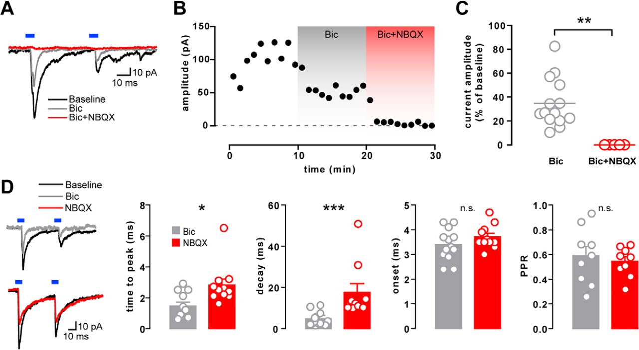
- Figure 3.
Mixed neurotransmission onto DG granule cells. A, Example voltage-clamp traces of a GCL neuron before and after bath application of bicuculline alone (Bic) or in combination with NBQX (Bic+NBQX). B, Time course of the current peak amplitude (1 min bins) from the same example neuron as in A. C, Residual current amplitude following Bic (n = 14) or Bic+NBQX (n = 5) application, shown as a percentage of the baseline current amplitude. D, Sample traces before and after the application of Bic or NBQX alone and the effect on the time to peak (n = 11, 11), decay time (n = 13, 11) onset latency (n = 13, 10), and PPR (n = 9, 9) of the light-evoked currents.
- Figure 4.
VTA glutamatergic neurons release GABA in the DG. A, Schematic showing the injection procedures for retrograde tracing or for whole-cell recordings in VGLUT2-Cre mice (as done in Fig. 1A,H for GAD65-Cre mice). B, CTB555 injection site in the dDG. C, High-magnification image of the retrogradely labeled neurons in the VTA, some of which colocalize with EYFP. D, Schematic localization of retrogradelylabeled cells and the proportion of CTB-positive neurons colocalizing with EYFP in VGLUT2-Cre mice. E, Graph showing the average amplitude (±SEM) of the light-evoked PSCs recorded in the GCL of VGLUT2- or GAD65-Cre mice as a function of their connectivity. F, Example traces showing the light-evoked PSC recorded in a granule cell at baseline and after bicuculline alone (Bic) or together with NBQX (Bic+NBQX). G, Peak amplitude time course from the same example neuron in F. H, Residual current amplitude after Bic (n = 8) or Bic+NBQX (n = 7) application, shown as a percentage of the baseline current amplitude. I, Sample traces before and after the application of Bic or NBQX alone. J, Effect of Bic or NBQX alone on the time to peak (n = 8,9), decay time (n = 7,11), onset latency (n = 8,11), and PPR (n = 8,11) of the light-evoked currents.
- Figure 5.
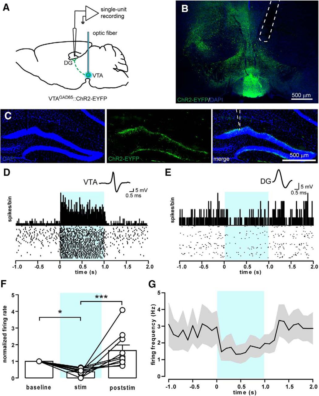
Stimulation of VTA GABA neurons reduces firing activity in the DG in vivo. A, Schematic of the single-unit recordings in the DG of anesthetized mice. An optic fiber connected to a blue laser was implanted above the VTA for the light stimulation of ChR2-expressing GABA neurons (20 5 ms pulses at 20 Hz). B, Sample confocal image showing the optical fiber positioning above the VTA. C, Sample confocal images showing the position of the recording electrode in the upper blade of the DG. D, Spike waveform, PSTH, and raster plot of a sample GABA neuron recorded in the VTA and showing an increase in firing rate upon blue light stimulation (light blue-shaded area). E, Spike waveform, PSTH, and raster plot of a sample responsive neuron in the DG showing a moderate reduction in the firing rate during blue light stimulation. F, Average firing rate of responsive neurons during (stim) or after (poststim) light stimulation normalized to baseline firing (n = 10). G, Average firing frequency of responsive neurons recorded in the DG (n = 10).


Tables
- Table 1:
Basic electrophysiological properties in light-responsive and nonresponsive DG granule cells
Light-responsive neurons (n = 15) Nonresponsive neurons (n = 15) p Value (light-responsive vs nonresponsive) Resting membrane potential −72.9 ± 1.2 mV −70.8 ± 2.1 mV 0.5606 Capacitance 14.2 ± 1.4 pF 13.6 ± 1.4 pF 0.8519 Input resistance 232.5 ± 24.5 MΩ 253.4 ± 25.8 MΩ 0.5336 Quantification of resting membrane potential, capacitance, and input resistance values shows no difference between DG granule cells receiving the VTA GABA input and nonconnected neurons (i.e. light-responsive vs nonresponsive neurons).
Figure Panel Data structure Type of test p Value 2 G Non-normal distribution Kruskal–Wallis test 0.2086 3 C Non-normal distribution Mann–Whitney test 0.0013 D Non-normal distribution Mann–Whitney test (time to peak) 0.0102 Non-normal distribution Mann–Whitney test (decay) 0.0004 Non-normal distribution Mann–Whitney test (onset) 0.2817 Non-normal distribution Mann–Whitney test (PPR) 0.6048 4 E Non-normal distribution Mann–Whitney test 0.1921 H Non-normal distribution Mann–Whitney test 0.0013 J Non-normal distribution Mann–Whitney test (time to peak) 0.0045 Non-normal distribution Mann–Whitney test (decay) 0.0006 Non-normal distribution Mann–Whitney test (onset) 0.2814 Non-normal distribution Mann–Whitney test (PPR) 0.3020 5 C Non-normal distribution Friedman test Friedman statistic: 15.80; <0.0001 Dunn’s multiple comparison post-test (baseline vs stimulation) Rank sum difference: 13.00; 0.0110 Dunn’s multiple comparison post-test (baseline vs poststimulation) Difference in rank sum: -4.000; >0.9999 Dunn’s multiple comparison post-test (stimulation vs poststimulation) Difference in rank sum: -17.00; 0.0004 Table 1 Non-normal distribution Mann–Whitney test (resting membrane potential) 0.5606 Non-normal distribution Mann–Whitney test (capacitance) 0.8519 Non-normal distribution Mann–Whitney test (input resistance) 0.5336
In this issue


