
Main menu

  • HOME
  • CONTENT
    • Early Release
    • Featured
    • Current Issue
    • Issue Archive
    • Blog
    • Collections
    • Podcast
  • TOPICS
    • Cognition and Behavior
    • Development
    • Disorders of the Nervous System
    • History, Teaching and Public Awareness
    • Integrative Systems
    • Neuronal Excitability
    • Novel Tools and Methods
    • Sensory and Motor Systems
  • ALERTS
  • FOR AUTHORS
  • ABOUT
    • Overview
    • Editorial Board
    • For the Media
    • Privacy Policy
    • Contact Us
    • Feedback
  • SUBMIT

User menu

Search

  • Advanced search
eNeuro
eNeuro

Advanced Search


  • HOME
  • CONTENT
    • Early Release
    • Featured
    • Current Issue
    • Issue Archive
    • Blog
    • Collections
    • Podcast
  • TOPICS
    • Cognition and Behavior
    • Development
    • Disorders of the Nervous System
    • History, Teaching and Public Awareness
    • Integrative Systems
    • Neuronal Excitability
    • Novel Tools and Methods
    • Sensory and Motor Systems
  • ALERTS
  • FOR AUTHORS
  • ABOUT
    • Overview
    • Editorial Board
    • For the Media
    • Privacy Policy
    • Contact Us
    • Feedback
  • SUBMIT
PreviousNext
Research ArticleNew Research, Development

Krüppel-Like Factor 4 (KLF4) Is Not Required for Retinal Cell Differentiation

Jiahua Fang, Peter X. Shaw, Yan Wang and Jeffrey L. Goldberg
eNeuro 13 January 2016, 3 (1) ENEURO.0117-15.2016; https://doi.org/10.1523/ENEURO.0117-15.2016
Jiahua Fang
1Department of Ophthalmology and Shiley Eye Institute, University of California San Diego, La Jolla, California 92093
2Department of Ophthalmology, Changsha First People’s Hospital, Changsha 410008, Hunan, China
  • Find this author on Google Scholar
  • Find this author on PubMed
  • Search for this author on this site
Peter X. Shaw
1Department of Ophthalmology and Shiley Eye Institute, University of California San Diego, La Jolla, California 92093
  • Find this author on Google Scholar
  • Find this author on PubMed
  • Search for this author on this site
  • ORCID record for Peter X. Shaw
Yan Wang
1Department of Ophthalmology and Shiley Eye Institute, University of California San Diego, La Jolla, California 92093
  • Find this author on Google Scholar
  • Find this author on PubMed
  • Search for this author on this site
Jeffrey L. Goldberg
1Department of Ophthalmology and Shiley Eye Institute, University of California San Diego, La Jolla, California 92093
3Byers Eye Institute at Stanford, Palo Alto, California 94303
  • Find this author on Google Scholar
  • Find this author on PubMed
  • Search for this author on this site
  • ORCID record for Jeffrey L. Goldberg
  • Article
  • Figures & Data
  • Info & Metrics
  • eLetters
  • PDF
Loading

Abstract

During early vertebrate eye development, a regulatory network of transcription factors regulates retinal cell differentiation and survival into adulthood. Among those factors, Krüppel-like factor 4 (KLF4) plays the dual role of maintaining the stem cell status of retinal progenitors cells and repressing the intrinsic axon regeneration ability in retinal ganglion cells (RGCs) after injury. This study further investigated whether KLF4 plays a role in early retinal cell differentiation or survival into adulthood. We examined different types of retinal neurons, including RGCs, amacrine cells, bipolar cells, Müller cells, and photoreceptor cells, in adult mice in which KLF4 was conditionally deleted in early retinal development using Chx10-promoted Cre by immunohistochemistry. We compared the numbers of retinal neurons and the thickness of photoreceptor and nerve fiber layers between Chx10–Cre-driven KLF4 deletion mice and wild-type mice. There was no significant difference in cell number among any of the retinal cell types or in photoreceptor layer thickness with KLF4 deletion during early development. The thickness of axon bundles in the nerve fiber layer in the Chx10 conditional KLF4 knock-out mice was greater than that in wild-type mice. These results suggest that KLF4 is not required for retinal cell differentiation or survival, but does normally limit retinal ganglion cell axon bundle thickness. These data support a hypothesis that KLF4 suppresses axon growth during development.

  • axon growth
  • Krüppel-like factor 4
  • retinal development
  • retinal ganglion cells

Significance Statement

Although it has been well studied for its role in reprogramming somatic cells into induced pluripotent stem cells and the repression of axon regeneration of optic nerve injury, whether Krüppel-like factor 4 (KLF4) plays any role in retinal neuronal differentiation and survival into adulthood is still poorly understood. This study provides a line of evidence that the absence of KLF4 in early development does not affect retinal neuron differentiation into designated cell types.

Introduction

The neural retina comprises six types of neurons (retinal ganglion cells, amacrine, horizontal, bipolar, and rod and cone photoreceptors) and one type of glial cell (Müller cell). They are generated from a common pool of multipotent retinal progenitor cells (RPCs). During vertebrate retina development, the differentiation of retinal cells occurs in a defined order (Cepko et al., 1996; Livesey and Cepko, 2001; Turner and Cepko, 1987). Retinal ganglion cells (RGCs) are generated first along with cone photoreceptors, amacrine cells, and horizontal cells, followed by bipolar neurons and rod photoreceptors, and ending with Müller glia, with considerable overlap among these phases (Cepko, 1999; Livesey and Cepko, 2001). In the early stages of retinal development, RPCs generate these different cell types under the regulation of a combination of extrinsic and intrinsic influences (Livesey and Cepko, 2001). The ability of RPCs to generate particular progeny is specified by transcription factors that define their competence to differentiate into single or perhaps subsets of retinal cell types (Cepko, 1999). Transcription factors known to regulate these functions include members of the basic helix-loop-helix (Hatakeyama and Kageyama, 2004; Yao et al., 2010), homeodomain (Dorval et al., 2005), and forkhead families (Moose et al., 2009).

Krüppel-like factors (KLFs) are a subfamily of evolutionarily conserved zinc finger DNA-binding transcription factors (Bieker, 2001; Ghaleb et al., 2005). Seventeen mammalian KLFs have been identified, all of which have three highly conserved zinc finger motifs at the C terminus; bind to similar consensus GT-box or CACCC DNA response elements; and function as transcriptional activators, repressors, or both (Dang et al., 2000; Moore et al., 2011; Turner and Crossley, 1999). These factors regulate various biological functions in development and adulthood, including cell growth, apoptosis, proliferation, and differentiation (McConnell and Yang, 2010). Different KLFs have been linked to the differentiation of different cell types, for example, KLF1 in erythropoiesis (Drissen et al., 2005), KLF2 in thymocytes (Carlson et al., 2006), KLF7 in olfactory sensory neurons (Kajimura et al., 2007), and KLF4 in monocytes (Alder et al., 2008) and corneal epithelial cells (Swamynathan et al., 2007).

KLF4 is expressed during murine eye development (Chiambaretta et al., 2004). When the KLF4 gene was selectively deleted in the surface ectoderm-derived structures of the eye with Pax6-driven Cre (le-Cre mice), 8-week-old mice exhibited multiple ocular defects, including corneal epithelial fragility, stromal edema, defective lens, and loss of conjunctival goblet cells (Swamynathan et al., 2007). We previously reported that KLF4 is developmentally regulated in RGCs and suppresses their intrinsic capacity for rapid axon growth (Moore et al., 2009). In that article, we did not see a defect in RGC numbers in adult mice after deleting KLF4 with a thy-1-promoted Cre in early development, but whether KLF4 regulates retinal neurons, including RGC differentiation, when deleted at the beginning of retinal development before Thy1 expression is turned on in postmitotic RGCs, remains to be determined. In this report, we created early retinal KLF4 conditional knock-out mice by breeding Chx10-Cre Tg with KLF4fl/fl mice. Using immunohistochemical methods, we examined retinal architecture compared with age-matched wild-type (WT) controls.

Materials and Methods

Animals

All procedures involving animals were in accordance with the ARVO Statement for the Use of Animals in Ophthalmic and Vision Research and were approved by the Institutional Biosafety Committee and the Institutional Animal Care and Use Committee at the University of California, San Diego. Conditional knock-out mice were generated by breeding KLF4-floxed mice (Moore et al., 2009) and Chx10-Cre mice (Jax mice stock #005105, The Jackson Laboratory); genotyping was confirmed by PCR, as in our prior publication (Moore et al., 2009). Age-matched littermates without the Chx10-cre allele served as controls. For counting and statistical analysis of adult retinas, a minimum of three mice of either sex were included in each group.

Western blot

Adult mice of indicated genotypes were killed, and retinas were dissected and lysed with lysis buffer (Cell Signaling Technology) containing 0.5 mm phenylmethylsulfonyl fluoride (Sigma-Aldrich). Protein concentration was determined by BCA protein assay (Thermo Fisher Scientific). Samples (25 μg) were separated by SDS-PAGE in 4-20% gradient Tris-glycine precast gels (Invitrogen) and transferred to a polyvinylidene difluoride membrane (Millipore). The membrane was incubated for 1 h in blocking solution containing 5% nonfat milk powder and 0.1% Tween-20, pH 7.6. This was followed by overnight incubation at 4°C in the blocking buffer containing rabbit primary antibodies against KLF4 (1:50; Ab72543, Abcam). Subsequently, the labeled proteins were visualized by incubation with a horseradish peroxidase (HRP)-conjugated anti-goat or rabbit IgG (1:2000; Santa Cruz Biotechnology) followed by development with a chemiluminescence substrate for HRP (Thermo Fisher Scientific). The images of Western blots were captured by GE ImageQuant. Relative band intensities were analyzed using ImageJ software and normalized to GAPDH.

Immunohistochemistry

Animals under deep anesthesia underwent cardiac perfusion with 4% paraformaldehyde. Whole adult eyes were isolated, the corneas were incised to facilitate intraocular fixative penetration, and the eyeballs were transferred to PBS containing 4% paraformaldehyde for 1 h at room temperature, followed by 10% sucrose in PBS and, finally, 30% sucrose in PBS. Eyes were then mounted in optimal cutting temperature compound (Sakura Finetek USA), frozen, and cryosectioned at 5 μm thickness. At least three sections with an interval of 50 μm were acquired from each eye across the optic nerve to ensure retinal centration, for a total of 18 sections in each group. Cryosections were rinsed with PBS and blocked with 5% goat serum albumin (Thermo Fisher Scientific) and 0.2% Triton X-100 (Sigma-Aldrich) in antibody buffer for 1 h. Tissue was then incubated in antibody buffer containing primary antibody for 24 h at 4°C. The following antibodies were used: for RGCs, mouse anti-Brn3a (1:100; MAB 1585, EMD Millipore); for RGC axons, mouse monoclonal βIII-tubulin (1:100; Tuj1, Convance); for amacrine cells, mouse monoclonal anti-Pax6 (1:100; sc-81649, Santa Cruz Biotechnology); for photoreceptors and outer segment (OS) layer thickness, recoverin (1:100; EMD Millipore) and rhodopsin (1:100; Pierce/Thermo Scientific); for bipolar cells, rabbit polyclonal anti-protein kinase C-α (PKC-α; 1:100; sc-208, Santa Cruz Biotechnology); and for Müller glia rabbit anti-glutamine synthetase (GS; 1:100; Sigma-Aldrich). Sections were then rinsed in PBS and incubated with a fluorescent secondary antibody containing 4',6'-diamidino-2-phenylindole dihydrochloride (DAPI; 1:2000; Molecular Probes) for 24 h at 4°C for nuclei staining. The following two secondary antibodies were used in a dilution of 1:500: Alexa Fluor 488 mouse anti-rabbit Texas red or Alexa Fluor 546 goat anti-mouse (Molecular Probes). After a subsequent three washes in PBS, these sections were mounted with Vectashield H1000 (Vector Laboratories) and photographed with a Leica microscope.

Morphologic analysis

Retinal cell counts were measured in cross sections to calculate cell density at a distance of 100-200 μm (both sides) from the center of the optic disc. The thickness of axon bundles in the ganglion cell layer (GCL) was quantified by measuring the thickness of the βIII-tubulin-immunostained RGC axons in the retinal nerve fiber layer at 200 μm from the center of the optic disc. Thickness of retinal layers containing photoreceptor cells plus outer segments (recoverin+ layer), and containing rod outer segments (rhodopsin+ layer) were also measured at 200 μm from the center of the optic disc. All measurements were collected by masked investigators.

Statistical analysis

Data are presented as the mean ± SD with N = 18 replicates per condition; Student's t test (SPSS for Mac version 20.0) was used to test for significance at a p < 0.05 level.

Results

Early KLF4 deficiency does not affect photoreceptors into adulthood

We generated mice with retinas deficient in KLF4 by crossing a floxed KLF4 allele (Moore et al., 2009) with a cre recombinase-expressing line driven by the pan-retinal progenitor expressing marker CHX10 (Jax mice stock #005105, The Jackson Laboratory). Western blot analysis confirmed that the level of KLF4 protein expression in retinal tissues of KLF4fl/fl/Chx10-Cre+ mice was significantly decreased compared with that of the wild-type control mice (Fig. 1). To investigate whether the development of photoreceptors is influenced by retinal expression of KLF4, we examined recoverin on retinal cross sections of conditional knock-out (KLF4-cKO) and WT control mice. Recoverin is a calcium-binding protein that is present in both rod and cone photoreceptors (Polans et al., 1995). Both cKO and WT mice displayed intact photoreceptor cell somata, which showed very high levels of recoverin immunoreactivity throughout the outer nuclear layer (ONL), as well as normal-appearing inner segments (ISs) and OSs (Fig. 2A). We found no significant difference in the thickness of recoverin+ retinal immunostaining in WT versus KLF4-cKO mice (p > 0.1; Fig. 2C).

Rhodopsin is a single-chain integral membrane protein localized to rod outer segment membranes. Similar to recoverin, strong rhodopsin immunoreactivity was found in the photoreceptor outer segments of both WT and KLF4-cKO mice (Fig. 2B), with no significant difference in thickness between the two groups (Fig. 2D). Thus, both IS and OS photoreceptor measures are not dependent on retinal KLF4 expression.

Figure 1.
  • Download figure
  • Open in new tab
  • Download powerpoint
Figure 1.

Western blot quantitative analysis of KLF4 protein expression in the retinae of KLF4 cKO and control mice. A, Representative images of Western blots against KLF4 and GAPDH as marked. B, Quantification of the relative protein expression normalized to total protein and GAPDH as marked (N = 3 for each biological sample; values are reported as the mean ± SD).

Figure 2.
  • Download figure
  • Open in new tab
  • Download powerpoint
Figure 2.

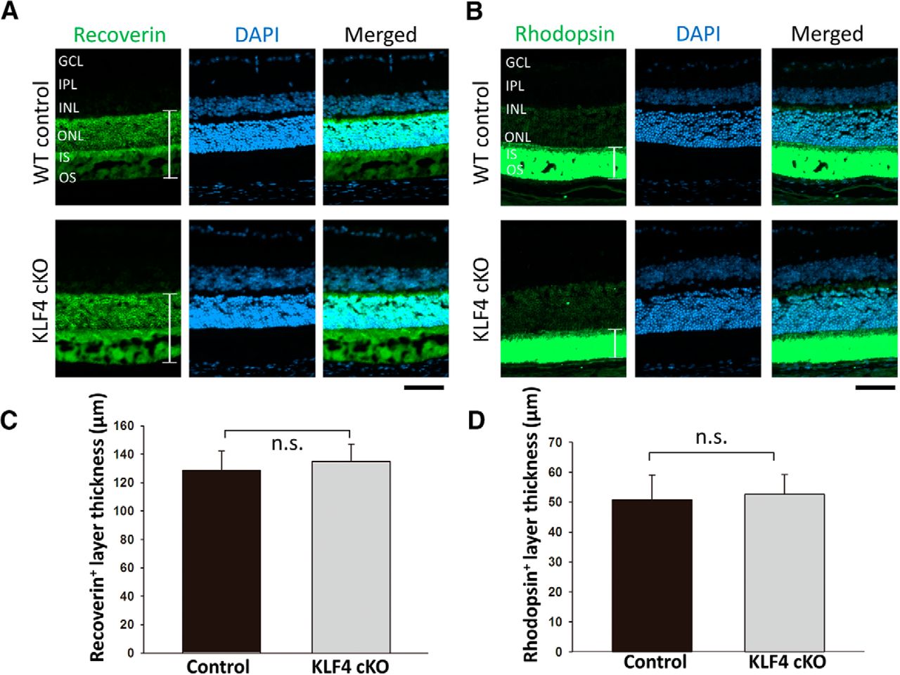
A, B, KLF4 is not required for photoreceptors or outer segments. Immunofluorescence for recoverin (A) and rhodopsin (B; green) was counterstained with DAPI for nuclei (blue) in retina cross sections from wild-type control mice (top) and KLF4-cKO mice (bottom). IPL, Inner plexiform layer. C, D, The thickness of recoverin+ (C) and rhodopsin+ (D) layers (as marked with brackets in A and B) were measured and found to be nonsignificant (n.s.; values are reported as the mean ± SD; N = 18). Scale bar, 50 μm.

KLF4 is not associated with Müller, amacrine, and bipolar cell survival into adulthood

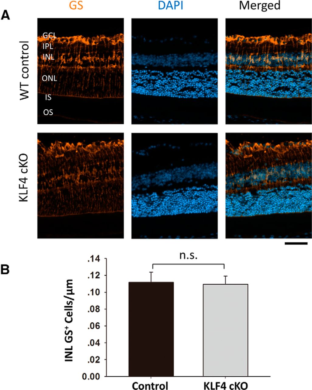
We next examined other retinal cell markers to determine whether early KLF4 deficiency affects differentiation and survival into adulthood. GS, an enzyme that converts neuron-released glutamate to glutamine, is a specific marker for Müller glia (Fischer and Reh, 2001; Jablonski et al., 2001). Immunostaining for GS demonstrated prominent labeling of Müller cell bodies located in the inner nuclear layer (INL) and their processes, extending to the inner and outer limiting membranes (Fig. 3A). GS+ cell bodies were counted only in the INL, not counting cells in the GCL wrapped by GS+ Müller glia end feet. We found no significant difference in Müller cell number between WT and KLF4-cKO mice (Fig. 3B; p > 0.1).

Figure 3.
  • Download figure
  • Open in new tab
  • Download powerpoint
Figure 3.

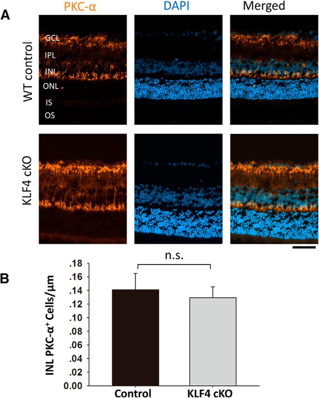
KLF4 does not affect rod bipolar cell numbers. A, Retinal cross sections were immunostained for PKC-α (orange) and counterstained with DAPI (blue). B, PKC-α+ cells in the INL showed no statistically significant difference between WT and KLF4-cKO groups (n.s.; values are reported as the mean ± SD; N = 18). Scale bar, 50 μm.

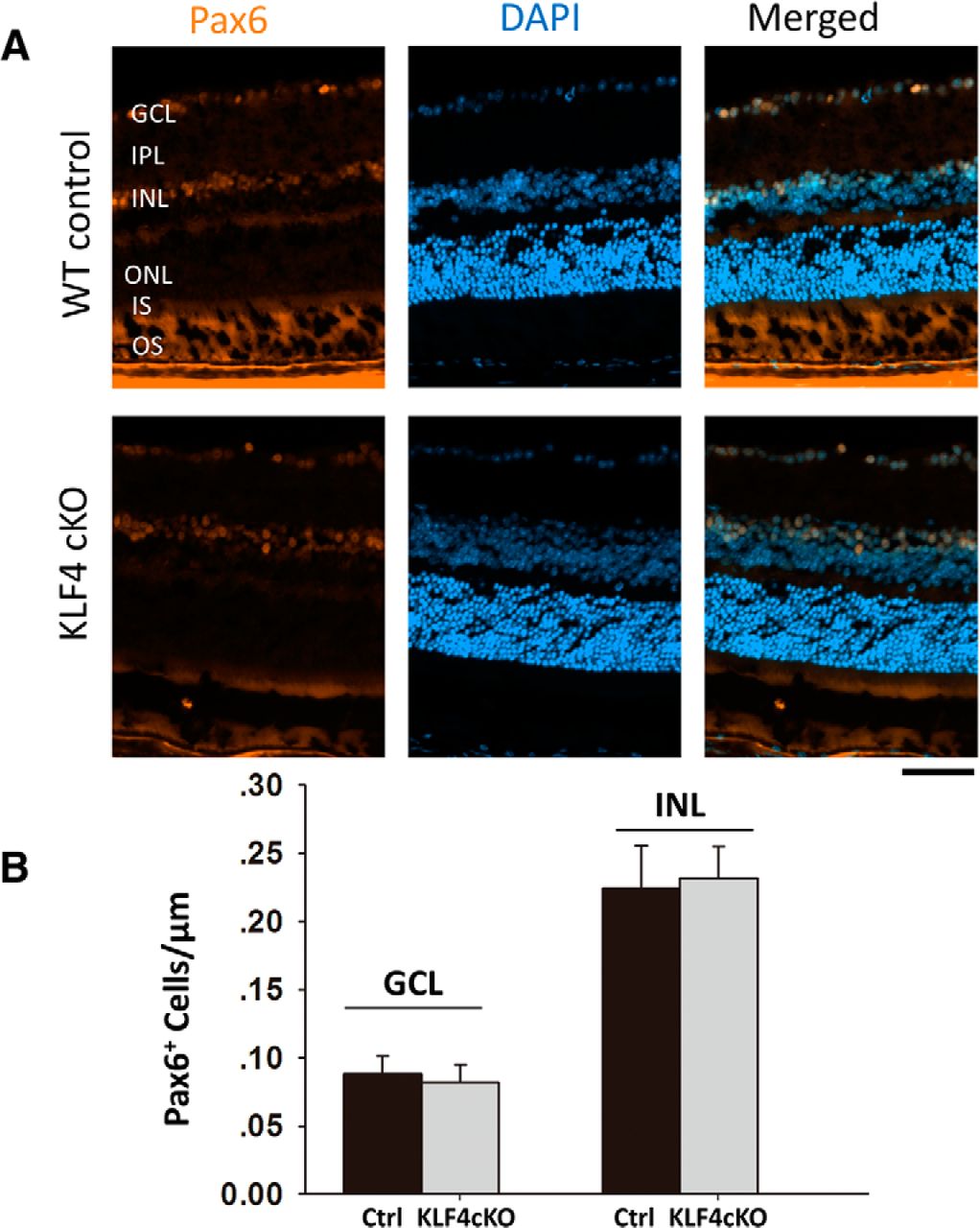
Using Pax6 as a marker for amacrine cells in the INL as well as the displaced amacrine cells in the ganglion cell layer (Fig. 4A), we found no significant difference between WT and KLF4-cKO mice (Fig. 4B). Using PKC-α, which is expressed in cell bodies and processes of ON bipolar cells that mainly connect to rods, we detected PKC-α immunoreactivity in a morphologically distinct population of rod bipolar cells with somata located in the distal row of the inner nuclear layer (Fig. 5A). Again, there was no difference in cell density of bipolar cells in WT versus KLF4-cKO mice (p > 0.05; Fig. 5B). Thus, KLF4 is not required to generate the normal number of bipolar, amacrine, or Müller glial cells in the adult retina.

Figure 4.
  • Download figure
  • Open in new tab
  • Download powerpoint
Figure 4.

KLF4 does not affect Müller glial cell numbers. A, Immunofluorescence staining was performed to detect GS (orange), and cells were counterstained with DAPI (blue). B, The GS+ cells in the INL demonstrated no statistical difference between WT and KLF4-cKO groups (n.s.; values are reported as the mean ± SD; N = 18). Scale bar, 50 μm.

Figure 5.
  • Download figure
  • Open in new tab
  • Download powerpoint
Figure 5.

KLF4 is not required for amacrine cell numbers. A, Immunofluorescence staining for Pax6 (orange) and counterstaining with DAPI (blue). B, Pax6+ cells per unit length showed no statistical difference between WT and KLF4-cKO groups in either the GCL or INL (n.s.; values are reported as the mean ± SD; N = 18). Scale bar, 50 μm.

KLF4 is not associated with RGC differentiation and survival into adulthood

We previously reported that, using a Thy1-Cre driver to disrupt KLF4 expression in approximately half of RGCs, RGC cell numbers into adulthood were not dependent on the postmitotic expression of KLF4 in RGCs themselves (Moore et al., 2009). Here, we extended those studies to determine whether KLF4 deficiency in progenitors of all retinal cells would affect RGC numbers into adulthood using the Chx10-cre allele. We used the RGC-specific marker Brn3a (Fig. 6A; Nadal-Nicolás et al., 2009) and found no significant difference in RGC numbers into adulthood (p > 0.05; Fig. 6B). Thus, for all retinal cell types studied, KLF4 deficiency in early retinal progenitor cells does not affect cell numbers or photoreceptor layer thickness into adulthood.

Figure 6.
  • Download figure
  • Open in new tab
  • Download powerpoint
Figure 6.

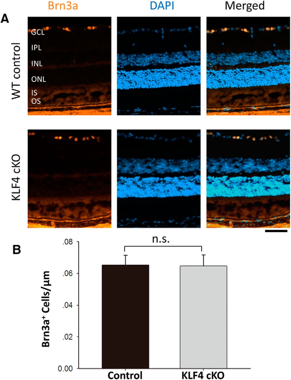
KLF4 is not required for RGC cell numbers. A, Retinal cross sections were immunostained for Brn3a (orange) and counterstained with DAPI (blue). B, Brn3a+ cells showed no statistically significant difference between WT and KLF4-cKO groups (n.s.; values are reported as the mean ± SD; N = 18). Scale bar, 50 μm.

KLF4 is associated with nerve fiber layer thickness

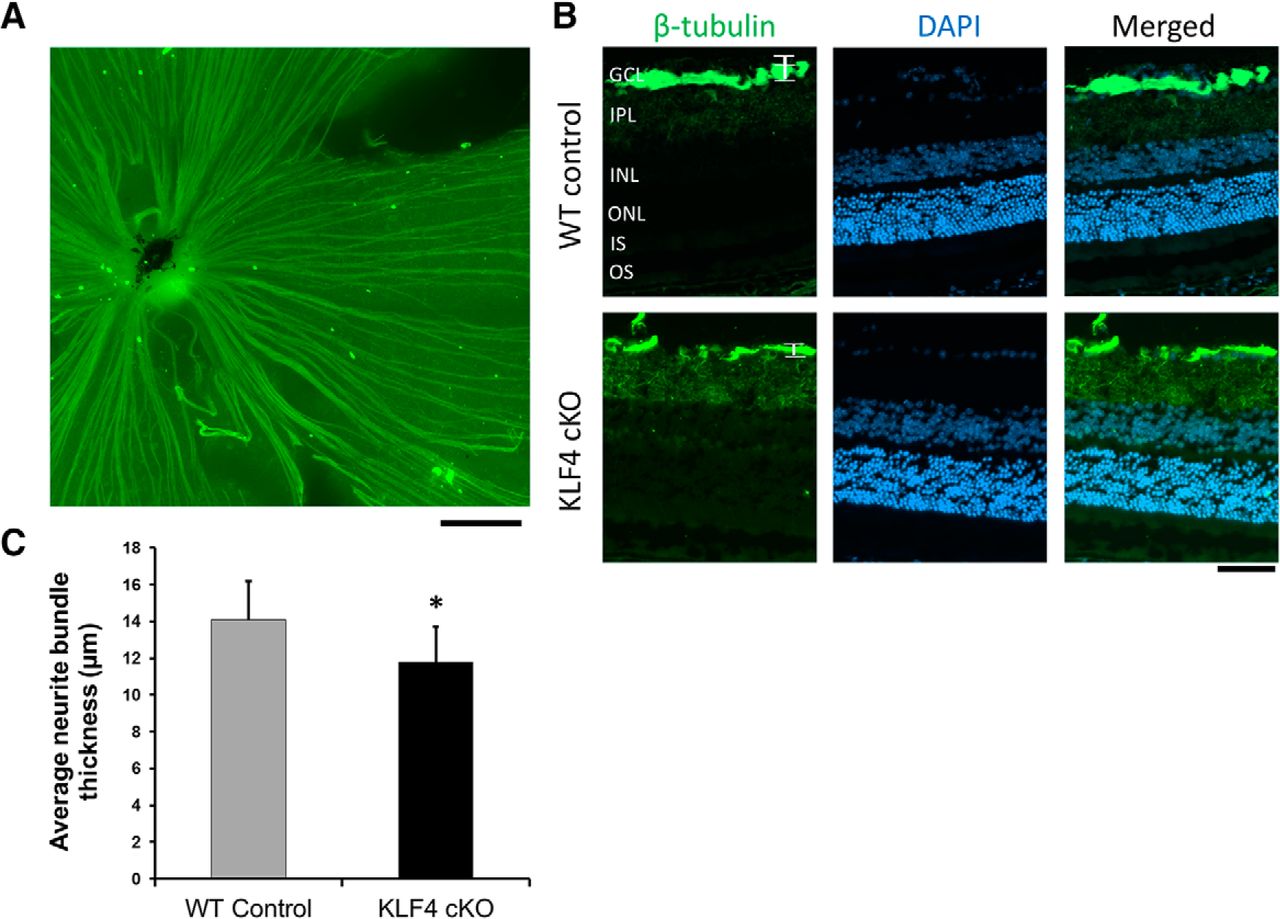
Finally, because we and others previously found that KLF4 profoundly affects the ability of RGCs to regenerate their axons after optic nerve injury (Moore et al., 2009; Qin et al., 2013), we asked whether KLF4 affects RGC axon growth during normal retinal development in vivo. We used βIII-tubulin to stain RGC axons in flat-mounted retinas (Fig. 7A) and cross sections (Fig. 7B). Sections that traversed toward the optic nerve were analyzed. Due to the variability of nerve fiber bundles at the optic nerve head and the sparseness of axon bundles in the peripheral retina, we measured axon bundles 200 μm away from the optic nerve head to maintain consistency. By analyzing βIII-tubulin, which labeled fasciculated RGC axon bundles in the nerve fiber layer of the retina (Fig. 7B), we found that the thickness of axon bundles in KLF4-cKO mice is significantly increased compared to that of WT mice (p < 0.05; Fig. 7C). This result supports a model in which KLF4 expression normally suppresses some measure of axon growth in vivo.

Figure 7.
  • Download figure
  • Open in new tab
  • Download powerpoint
Figure 7.

KLF4 deficiency leads to an increase in RGC axon fascicles in the adult retina. A, Representative immunofluorescent staining of retinal flat mount. Scale bar, 200 μm. B, Retinal cross sections were immunostained for β-tubulin (green) and counterstained with DAPI (blue). C, The thickness of RGC axon fascicles (brackets in B) was measured. There was a significant increase in nerve fiber layer axon bundle thickness in KLF4-cKO compared with WT mice (*p < 0.05; values are reported as the mean ± SD; N = 18). Scale bar, 50 μm.

Discussion

During early vertebrate eye development, a regulatory network of transcription factors regulates retinal cell differentiation (Ohsawa and Kageyama, 2008) and survival into adulthood, including Pax6, Six3, Rx, Chx10, Notch, and Notch pathway components (Badea and Nathans, 2011; Mu and Klein, 2004). KLFs including KLF4 regulate differentiation of other cell types throughout the body (Apara and Goldberg, 2014; Moore et al., 2011), and KLF4 is one of a set of four factors capable of reprogramming somatic cells into induced pluripotent stem cells (Hanna et al., 2008; Takahashi et al., 2007; Takahashi and Yamanaka, 2006; Welstead et al., 2008; Yu et al., 2007). KLF4 plays an important role in cell cycle arrest and growth inhibition (Choi et al., 2006; Wei et al., 2005, 2008), and is highly expressed in the postmitotic cells of both the gut and skin (Shields et al., 1996; Zheng et al., 2009). KLF4 has been found to be downregulated in many types of cancers, indicating its possible contribution to cellular hyperproliferation and malignant transformation (Hu et al., 2009; Huang et al., 2005; Wei et al., 2005). However, in other types of primary tumors, such as breast cancer, the expression of KLF4 is upregulated (Foster et al., 2000; Pandya et al., 2004). An exact mechanism by which KLF4 affects negative or positive regulation in cell cycle control remains largely unknown. Progesterone receptor agonists can induce KLF4 expression and inhibit cyclin D1 expression, resulting in the inhibition of cell proliferation in human endometrial epithelial cells (Shimizu et al., 2010). Downregulation of cyclin D1 also controls the proliferation of retinal progenitor cells (Njaine et al., 2010). In addition, p21Cip1 coexpression may determine the tumor suppressor or oncogenic function of KLF4 (Rowland et al., 2005; Rowland and Peeper, 2006). The promoter region of KLF4 contains typical CpG islands, and, thus, hypermethylation may regulate KLF4 transcription and function in cell cycle control (Song et al., 2013).

In order to characterize the potential roles of KLF4 in retinal neuron differentiation and RGC development, we generated KLF4 conditional knock-out mice driven by Chx10-promoted Cre expression. We performed a Western blot analysis with retinal tissues from cKO and control mice. The results demonstrated the significant reduction (>80%) of KLF4 protein in Chx10 Cre-driven KLF4 cKO. The “escape” of a small amount of KLF4 expression in KLF4 cKO retinae may represent either Chx10-derived retinal cells not adequately excising the gene, or cells not derived from retinal progenitors, such as endothelial cells or nerve fiber layer astrocytes; this is a potential limitation of the cKO strategy. Because we did not find any differences in retinal cell numbers in the KLF4-deficient mouse retina, we hypothesize that KLF4 is dispensable for retinal progenitor cell differentiation and/or survival into adulthood. However, it remains plausible that KLF4 plays a role in both differentiation and survival into adulthood, and that alterations in both of these balance out to normal adult retinal cell numbers. The examination of earlier time points in retinal differentiation, for example at late embryonic and early postnatal ages, could assess this question in future experiments. It is also possible that KLF4 contributes to these processes but that other KLFs compensate in the absence of KLF4 (Moore et al., 2011). Furthermore, although we did not detect a difference in total Pax6+ amacrine cell numbers in the INL, amacrine cell subtype-specific immuno-identification (Kunzevitzky et al., 2013) could reveal changes in the differentiation of specific subpopulations.

RGCs and other CNS neurons in early development demonstrate robust axon growth and regenerative abilities, but their intrinsic capacity for rapid axon growth declines substantially after birth (Goldberg and Barres, 2000; Goldberg et al., 2002). We have recently reported that KLF4 plays a role in regulating intrinsic axon growth ability in RGCs (Moore et al., 2009). Overexpression of KLF4 significantly decreased neurite outgrowth in RGCs, hippocampal and cortical neurons, and RGCs lacking KLF4 showed increased axon growth both in vitro and after optic nerve injury in vivo.

Despite not affecting RGC numbers in the adult animal, in this study, we found that the thickness of RGC axon bundles adjacent to the optic nerve is increased in KLF4-cKO mice compared with WT adult mice. We declined to use optical coherence tomography (OCT), a promising technology particularly for longitudinal assessments of retinal layer thickness, as our data revealed subtle changes and OCT does not provide the resolution this available with histology and immunostaining. βIII-tubulin is a microtubule element expressed exclusively in neurons, and is frequently used to characterize RGCs and their axons in the retina. Other proteins such as neurofilament (NF) can mark RGC axons, but NF staining can vary between the axon hillock and the whole axon, depending on phosphorylation and the relative expression of NF-heavy, NF-medium, and NF-light chains, which vary in expression in different stages of development and after injury. Studying NF and other axon and synapse proteins would be an interesting area for future research in trying to determine how KLF4 suppresses axon growth in vitro and in vivo.

These data do not differentiate between axon number and axon caliber; an increase in either could explain these findings. Future experiments examining this question at the level of electron microscopy could differentiate between these possibilities, as well as to explore whether there are differences in dendrite elaboration in vivo, for example using single-cell imaging techniques. It would be interesting as well to explore whether these differences in nerve fiber layer thickness detected histologically could be detected in the adult mouse in vivo using noninvasive means such as OCT. Nevertheless, the finding of KLF4 function in suppressing some measure of axon development in vivo is consistent with our and others’ previous reports (Moore et al., 2009; Qin et al., 2013) that KLF4 normally acts to inhibit the intrinsic properties of neurite growth.

Footnotes

  • ↵1 J.L.G. is a coinventor on a patent relating to Krüppel-Like Factor 4 in axon regeneration. The authors declare no other competing financial interests.

  • ↵3 This research was supported by National Institutes of Health/National Eye Institute Grants R01-EY-020913, R01-EY-025693, and P30-EY-022589; an unrestricted grant from Research to Prevent Blindness; and a Visiting Scholar Grant from President Li Mengxiong and the First Hospital of Jingzhou to J.F.

This is an open-access article distributed under the terms of the Creative Commons Attribution 4.0 International, which permits unrestricted use, distribution and reproduction in any medium provided that the original work is properly attributed.

References

  1. ↵
    Alder JK, Georgantas RW, Hildreth RL, Kaplan IM, Morisot S, Yu XB, McDevitt M, Civin CI (2008) Kruppel-like factor 4 is essential for inflammatory monocyte differentiation in vivo. J Immunol 180:5645-5652. pmid:18390749
    OpenUrlAbstract/FREE Full Text
  2. ↵
    Apara A, Goldberg JL (2014) Molecular mechanisms of the suppression of axon regeneration by KLF transcription factors. Neural Regen Res 9:1418-1421. doi:10.4103/1673-5374.139454 pmid:25317150
    OpenUrlCrossRefPubMed
  3. ↵
    Badea TC, Nathans J (2011) Morphologies of mouse retinal ganglion cells expressing transcription factors Brn3a, Brn3b, and Brn3c: analysis of wild type and mutant cells using genetically-directed sparse labeling. Vision Res 51:269-279. doi:10.1016/j.visres.2010.08.039
    OpenUrlCrossRefPubMed
  4. ↵
    Bieker J.J. (2001). Krüppel-like factors: three fingers in many pies. J Biol Chem 276:34355-34358. doi:10.1074/jbc.R100043200 pmid:11443140
    OpenUrlFREE Full Text
  5. ↵
    Carlson CM, Endrizzi BT, Wu JH, Ding XJ, Weinreich MA, Walsh ER, Wani MA, Lingrel JB, Hogquist KA, Jameson SC (2006) Kruppel-like factor 2 regulates thymocyte and T-cell migration. Nature 442:299-302. doi:10.1038/nature04882 pmid:16855590
    OpenUrlCrossRefPubMed
  6. ↵
    Cepko CL (1999) The roles of intrinsic and extrinsic cues and bHLH genes in the determination of retinal cell fates. Curr Opin Neurobiol 9:37-46. pmid:10072376
    OpenUrlCrossRefPubMed
  7. ↵
    Cepko CL, Austin CP, Yang X, Alexiades M, Ezzeddine D (1996) Cell fate determination in the vertebrate retina. Proc Natl Acad Sci U S A 93:589-595. pmid:8570600
    OpenUrlAbstract/FREE Full Text
  8. ↵
    Chiambaretta F, De Graeve F, Turet G, Marceau G, Gain P, Dastugue B, Rigal D, Sapin V (2004) Cell and tissue specific expression of human Krüppel-like transcription factors in human ocular surface. Mol Vis 10:901-909. pmid:15570219
    OpenUrlPubMed
  9. ↵
    Choi BJ, Cho YG, Song JW, Kim CJ, Kim SY, Nam SW, Yoo NJ, Lee JY, Park WS (2006) Altered expression of the KLF4 in colorectal cancers. Pathology, research and practice 202:585-589. doi:10.1016/j.prp.2006.05.001
    OpenUrlCrossRefPubMed
  10. ↵
    Dang DT, Pevsner J, Yang VW (2000) The biology of the mammalian Krüppel-like family of transcription factors. Int J Biochem Cell Biol 32:1103-1121. pmid:11137451 [Mismatch]
    OpenUrlCrossRefPubMed
  11. ↵
    Dorval KM, Bobechko BP, Ahmad KF, Bremner R (2005) Transcriptional activity of the paired-like homeodomain proteins CHX10 and VSX1. J Biol Chem 280:10100-10108. doi:10.1074/jbc.M412676200 pmid:15647262
    OpenUrlAbstract/FREE Full Text
  12. ↵
    Drissen R, von Lindern M, Kolbus A, Driegen S, Steinlein P, Beug H, Grosveld F, Philipsen S (2005) The erythroid phenotype of EKLF-null mice: defects in hemoglobin metabolism and membrane stability. Mol Cell Biol 25:5205-5214. doi:10.1128/MCB.25.12.5205-5214.2005 pmid:15923635
    OpenUrlAbstract/FREE Full Text
  13. ↵
    Fischer AJ, Reh TA (2001) Müller glia are a potential source of neural regeneration in the postnatal chicken retina. Nat Neurosci 4:247-252. doi:10.1038/85090
    OpenUrlCrossRefPubMed
  14. ↵
    Foster KW, Frost AR, McKie-Bell P, Lin CY, Engler JA, Grizzle WE, Ruppert JM (2000) Increase of GKLF messenger RNA and protein expression during progression of breast cancer. Cancer research 60:6488-6495. pmid:11103818
    OpenUrlAbstract/FREE Full Text
  15. ↵
    Ghaleb AM, Nandan MO, Chanchevalap S, Dalton WB, Hisamuddin IM, Yang VW (2005) Krüppel-like factors 4 and 5: the yin and yang regulators of cellular proliferation. Cell Res 15:92-96. doi:10.1038/sj.cr.7290271 pmid:15740636
    OpenUrlCrossRefPubMed
  16. ↵
    Goldberg JL, Barres BA (2000) The relationship between neuronal survival and regeneration. Annu Rev Neurosci 23:579-612. doi:10.1146/annurev.neuro.23.1.579 pmid:10845076
    OpenUrlCrossRefPubMed
  17. ↵
    Goldberg JL, Klassen MP, Hua Y, Barres BA (2002) Amacrine-signaled loss of intrinsic axon growth ability by retinal ganglion cells. Science 296:1860-1864. doi:10.1126/science.1068428 pmid:12052959
    OpenUrlAbstract/FREE Full Text
  18. ↵
    Hanna J, Markoulaki S, Schorderet P, Carey BW, Beard C, Wernig M, Creyghton MP, Steine EJ, Cassady JP, Foreman R, Lengner CJ, Dausman JA, Jaenisch R (2008) Direct reprogramming of terminally differentiated mature B lymphocytes to pluripotency. Cell 133:250-264. doi:10.1016/j.cell.2008.03.028 pmid:18423197
    OpenUrlCrossRefPubMed
  19. ↵
    Hatakeyama J, Kageyama R (2004) Retinal cell fate determination and bHLH factors. Semin Cell Dev Biol 15:83-89. doi:10.1016/j.semcdb.2003.09.005 pmid:15036211
    OpenUrlCrossRefPubMed
  20. ↵
    Hu W, Hofstetter WL, Li H, Zhou Y, He Y, Pataer A, Wang L, Xie K, Swisher SG, Fang B (2009) Putative tumor-suppressive function of Kruppel-like factor 4 in primary lung carcinoma. Clinical cancer research : an official journal of the American Association for Cancer Research 15:5688-5695. pmid:19737957
    OpenUrlAbstract/FREE Full Text
  21. ↵
    Huang CC, Liu Z, Li X, Bailey SK, Nail CD, Foster KW, Frost AR, Ruppert JM, Lobo-Ruppert SM (2005) KLF4 and PCNA identify stages of tumor initiation in a conditional model of cutaneous squamous epithelial neoplasia. Cancer biology & therapy 4:1401-1408. pmid:PMC1361751
    OpenUrlPubMed
  22. ↵
    Jablonski MM, Tombran-Tink J, Mrazek DA, Iannaccone A (2001) Pigment epithelium-derived factor supports normal Müller cell development and glutamine synthetase expression after removal of the retinal pigment epithelium. Glia 35:14-25. doi:10.1002/glia.1066
    OpenUrlCrossRefPubMed
  23. ↵
    Kajimura D, Dragomir C, Ramirez F, Laub F (2007) Identification of genes regulated by transcription factor KLF7 in differentiating olfactory sensory neurons. Gene 388:34-42. doi:10.1016/j.gene.2006.09.027 pmid:17123745
    OpenUrlCrossRefPubMed
  24. ↵
    Kunzevitzky NJ, Willeford KT, Feuer WJ, Almeida MV, Goldberg JL (2013) Amacrine cell subtypes differ in their intrinsic neurite growth capacity. Investigative ophthalmology & visual science 54:7603-7613. doi:10.1167/iovs.13-12691 pmid:PMC3832218
    OpenUrlAbstract/FREE Full Text
  25. ↵
    Livesey FJ, Cepko CL (2001) Vertebrate neural cell-fate determination: lessons from the retina. Nat Rev Neurosci 2:109-118. doi:10.1038/35053522 pmid:11252990
    OpenUrlCrossRefPubMed
  26. ↵
    McConnell BB, Yang VW (2010) Mammalian Krüppel-like factors in health and diseases. Physiol Rev 90:1337-1381. doi:10.1152/physrev.00058.2009 pmid:20959618
    OpenUrlAbstract/FREE Full Text
  27. ↵
    Moore DL, Apara A, Goldberg JL (2011) Krüppel-like transcription factors in the nervous system: novel players in neurite outgrowth and axon regeneration. Mol Cell Neurosci 47:233-243. doi:10.1016/j.mcn.2011.05.005 pmid:21635952
    OpenUrlCrossRefPubMed
  28. ↵
    Moore DL, Blackmore MG, Hu Y, Kaestner KH, Bixby JL, Lemmon VP, Goldberg JL (2009) KLF family members regulate intrinsic axon regeneration ability. Science 326:298-301. doi:10.1126/science.1175737 pmid:19815778
    OpenUrlAbstract/FREE Full Text
  29. ↵
    Moose HE, Kelly LE, Nekkalapudi S, El-Hodiri HM (2009) Ocular forkhead transcription factors: seeing eye to eye. Int J Dev Biol 53:29-36. doi:10.1387/ijdb.072505hm pmid:19123124
    OpenUrlCrossRefPubMed
  30. ↵
    Mu X, Klein WH (2004) A gene regulatory hierarchy for retinal ganglion cell specification and differentiation. Semin Cell Dev Biol 15:115-123. doi:10.1016/j.semcdb.2003.09.009 pmid:15036214
    OpenUrlCrossRefPubMed
  31. ↵
    Nadal-Nicolás FM, Jiménez-López M, Sobrado-Calvo P, Nieto-López L, Cánovas-Martínez I, Salinas-Navarro M, Vidal-Sanz M, Agudo M (2009) Brn3a as a marker of retinal ganglion cells: qualitative and quantitative time course studies in naive and optic nerve-injured retinas. Invest Ophthalmol Vis Sci 50:3860-3868. doi:10.1167/iovs.08-3267
    OpenUrlAbstract/FREE Full Text
  32. ↵
    Njaine B, Martins RA, Santiago MF, Linden R, Silveira MS (2010) Pituitary adenylyl cyclase-activating polypeptide controls the proliferation of retinal progenitor cells through downregulation of cyclin D1. The European journal of neuroscience 32:311-321. doi:10.1111/j.1460-9568.2010.07286.x pmid:20646049
    OpenUrlCrossRefPubMed
  33. ↵
    Ohsawa R, Kageyama R (2008) Regulation of retinal cell fate specification by multiple transcription factors. Brain Res 1192:90-98. doi:10.1016/j.brainres.2007.04.014 pmid:17488643
    OpenUrlCrossRefPubMed
  34. ↵
    Pandya AY, Talley LI, Frost AR, Fitzgerald TJ, Trivedi V, Chakravarthy M, Chhieng DC, Grizzle WE, Engler JA, Krontiras H, et al. (2004) Nuclear localization of KLF4 is associated with an aggressive phenotype in early-stage breast cancer. Clinical cancer research: an official journal of the American Association for Cancer Research 10:2709-2719. pmid:15102675
    OpenUrlAbstract/FREE Full Text
  35. ↵
    Polans AS, Witkowska D, Haley TL, Amundson D, Baizer L, Adamus G (1995) Recoverin, a photoreceptor-specific calcium-binding protein, is expressed by the tumor of a patient with cancer-associated retinopathy. Proc Natl Acad Sci U S A 92:9176-9180. doi:10.1073/pnas.92.20.9176
    OpenUrlAbstract/FREE Full Text
  36. ↵
    Rowland BD, Bernards R, Peeper DS (2005) The KLF4 tumour suppressor is a transcriptional repressor of p53 that acts as a context-dependent oncogene. Nature cell biology 7:1074-1082. pmid:16244670
    OpenUrlCrossRefPubMed
  37. ↵
    Rowland BD, Peeper DS (2006) KLF4, p21 and context-dependent opposing forces in cancer. Nature reviews Cancer 6:11-23.
    OpenUrlCrossRefPubMed
  38. ↵
    Qin S, Zou Y, Zhang CL (2013) Cross-talk between KLF4 and STAT3 regulates axon regeneration. Nat Commun 4:2633. doi:10.1038/ncomms3633 pmid:24129709
    OpenUrlCrossRefPubMed
  39. ↵
    Shields JM, Christy RJ, Yang VW (1996) Identification and characterization of a gene encoding a gut-enriched Kruppel-like factor expressed during growth arrest. The Journal of biological chemistry 271:20009-20017. pmid:PMC2330254
    OpenUrlAbstract/FREE Full Text
  40. ↵
    Shimizu Y, Takeuchi T, Mita S, Notsu T, Mizuguchi K, Kyo S (2010) Kruppel-like factor 4 mediates anti-proliferative effects of progesterone with G(0)/G(1) arrest in human endometrial epithelial cells. Journal of endocrinological investigation 33:745-750. doi:10.3275/7044
    OpenUrlCrossRefPubMed
  41. ↵
    Song E, Ma X, Li H, Zhang P, Ni D, Chen W, Gao Y, Fan Y, Pang H, Shi T, et al. (2013) Attenuation of Kruppel-like factor 4 facilitates carcinogenesis by inducing g1/s phase arrest in clear cell renal cell carcinoma. PLoS One 8:e67758. doi:10.1371/journal.pone.0067758
    OpenUrlCrossRef
  42. ↵
    Swamynathan SK, Katz JP, Kaestner KH, Ashery-Padan R, Crawford MA, Piatigorsky J (2007) Conditional deletion of the mouse Klf4 gene results in corneal epithelial fragility, stromal edema, and loss of conjunctival goblet cells. Mol Cell Biol 27:182-194. doi:10.1128/MCB.00846-06 pmid:17060454
    OpenUrlAbstract/FREE Full Text
  43. ↵
    Takahashi K, Yamanaka S (2006) Induction of pluripotent stem cells from mouse embryonic and adult fibroblast cultures by defined factors. Cell 126:663-676. doi:10.1016/j.cell.2006.07.024 pmid:16904174
    OpenUrlCrossRefPubMed
  44. ↵
    Takahashi K, Tanabe K, Ohnuki M, Narita M, Ichisaka T, Tomoda K, Yamanaka S (2007) Induction of pluripotent stem cells from adult human fibroblasts by defined factors. Cell 131:861-872. doi:10.1016/j.cell.2007.11.019
    OpenUrlCrossRefPubMed
  45. ↵
    Turner DL, Cepko CL (1987) A common progenitor for neurons and glia persists in rat retina late in development. Nature 328:131-136. doi:10.1038/328131a0 pmid:3600789
    OpenUrlCrossRefPubMed
  46. ↵
    Turner J, Crossley M (1999) Mammalian Krüppel-like transcription factors: more than just a pretty finger. Trends Biochem Sci 24:236-240. pmid:10366853
    OpenUrlCrossRefPubMed
  47. Wang L, Yao JC, Wu TT, Huang S, Xie K (2005) Drastic down-regulation of Kruppel-like factor 4 expression is critical in human gastric cancer development and progression. Cancer research 65:2746-2754. pmid:PMC1361751
    OpenUrlAbstract/FREE Full Text
  48. ↵
    Wei D, Gong W, Kanai M, Schlunk C, Wang L, Yao JC, Wu TT, Huang S, Xie K (2005) Drastic down-regulation of Kruppel-like factor 4 expression is critical in human gastric cancer development and progression. Cancer research 65:2746-2754.
    OpenUrlAbstract/FREE Full Text
  49. ↵
    Wei D, Kanai M, Jia Z, Le X, Xie K (2008) Kruppel-like factor 4 induces p27Kip1 expression in and suppresses the growth and metastasis of human pancreatic cancer cells. Cancer research 68:4631-4639.
    OpenUrlAbstract/FREE Full Text
  50. ↵
    Welstead GG, Brambrink T, Jaenisch R (2008) Generating iPS cells from MEFS through forced expression of Sox-2, Oct-4, c-Myc, and Klf4. J Vis Exp (14):e734.
  51. ↵
    Yu J, Vodyanik MA, Smuga-Otto K, Antosiewicz-Bourget J, Frane JL, Tian S, Nie J, Jonsdottir GA, Ruotti V, Stewart R, Slukvin II, Thomson JA (2007) Induced pluripotent stem cell lines derived from human somatic cells. Science 318:1917-1920. doi:10.1126/science.1151526 pmid:18029452
    OpenUrlAbstract/FREE Full Text
  52. ↵
    Yao JH, Zhou JY, Liu QL, Lu DR, Wang L, Qiao XJ, Jia W (2010) Atoh8, a bHLH transcription factor, is required for the development of retina and skeletal muscle in zebrafish. PLoS One 5. doi:10.1371/journal.pone.0010945
    OpenUrlCrossRefPubMed
  53. ↵
    Zheng H, Pritchard DM, Yang X, Bennett E, Liu G, Liu C, Ai W (2009) KLF4 gene expression is inhibited by the notch signaling pathway that controls goblet cell differentiation in mouse gastrointestinal tract. American journal of physiology Gastrointestinal and liver physiology 296:G490-G498. pmid:19109406
    OpenUrlAbstract/FREE Full Text

Synthesis

The decision was a result of the Reviewing Editor Elizabeth Powell and the peer reviewers coming together and discussing their recommendations until a consensus was reached. A fact-based synthesis statement explaining their decision and outlining what is needed to prepare a revision is listed below. The following reviewers agreed to reveal their identity: Budd Tucker

The submission describes the role of KLF4 in early retinal cell differentiation and survival. While KLF4 is critical for stem cell growth, the role in retinal cell differentiation is yet unknown. The authors new data extend the field of knowledge and advances the field of retinal development and the overall roles of KLF4. The reviewers are enthusiastic about the manuscript, but they have a few issues that should be addressed. The individual comments are included:

Reviewer 1

In the manuscript titled "Kru¨ppel-like factor 4 (KLF4) is not required for retinal cell differentiation" the authors have investigated the effects of KLF4 in regulating retinal cell differentiation and survival. Their findings imply that KLF4 is not required for retinal progenitor cell differentiation and survival of retinal cells into adulthood. To identify the effects of KLF4, they have utilized CHX10-Cre driven KLF4 conditional knockout (cKO) mice and studied cell counts of retinal neurons: RGCs, amacrine, bipolar, Muller and photoreceptor cells. In addition, the authors have quantified the NFL/RGC axon bundle thickness.

Major revisions:

1.The authors suggest that KLF4 is not required for differentiation of retinal cells and cKO of KLF4 shows no significant difference in retinal cell counts between WT mice and the CHX10-Cre driven KLF4 cKO mice. However, the authors have only shown fluorescent microscopy images (Recoverin, Rhodopsin, GS, PAX6, PKC-a and Brn3a) 200um from the center of the optic disc. Please indicated if this analysis was perfromed in a masked fashion and why only 200um from the center of the optic disc was chosen?

Furthermore a discussion of how the different retinal layers were quantified and why the authors not quantify the size of the retinal fiber layers in vivo using OCT should be provided.

2.The authors should address in detail, the role of KLF4 in cell cycle control, and how it affects negative or positive regulation (Wei et al. 2008, Hu et al. 2009, Shimizu et al. 2010, Chen et al. 2010) and the effect in retinal progenitors (Njaine et al. 2010).

3.To quantify the thickness of the axon bundles, the only staining utilized was for BIII-tubulin. Would a neurofilament staining be better (For e.g. NFL)? The authors should also show cross-sections and traverse sections of the optic nerve to present a better case for axon bundle thickness.

For Figure 6, the authors mention, within the results (page 10) and discussion (page 11) sections that the axon bundles are significantly increased in the KLF4 cKO mice. However, the representative bracket of the GCL in figure 6 is thicker in WT than the KLF4 cKO. Please address.

Minor Comment:

There are several instances where the figures and figure legends throughout the manuscript do not match the results sections, please scan thoroughly and address as needed.

The authors are using PAX6 as a marker for amacrine cells, markers such as HPC1 and Calbindin may be useful.

Reviewer 2

1. I Would like to see confirmation that there is no expression of KLF4 in these animals by rq-PCR of total retinal RNA.

2. In figure 6, the B-tubulin stained bundles of k/o's are actually thinner than those of the WT control, although the graph shows the reverse, and the authors state that the k/o's have thicker bundles. Please replace photo with more representative confocal photo.

3. The authors suggest there may be some differences in mean axon diameter between WT and k/o animals. They suggest analysis of ON cross-sections (by TEM) might show difference in mean axonal diameter, if there is a shift in the type of axons, as well as simply looking at optical coherence tomography. It would enhance the paper's significance greatly if either of these approaches is included in the current report, and can be easily done (particularly with OCT).

Back to top

In this issue

eneuro: 3 (1)
eNeuro
Vol. 3, Issue 1
January/February 2016
  • Table of Contents
  • Index by author
Email

Thank you for sharing this eNeuro article.

NOTE: We request your email address only to inform the recipient that it was you who recommended this article, and that it is not junk mail. We do not retain these email addresses.

Enter multiple addresses on separate lines or separate them with commas.
Krüppel-Like Factor 4 (KLF4) Is Not Required for Retinal Cell Differentiation
(Your Name) has forwarded a page to you from eNeuro
(Your Name) thought you would be interested in this article in eNeuro.
CAPTCHA
This question is for testing whether or not you are a human visitor and to prevent automated spam submissions.
Print
View Full Page PDF
Citation Tools
Krüppel-Like Factor 4 (KLF4) Is Not Required for Retinal Cell Differentiation
Jiahua Fang, Peter X. Shaw, Yan Wang, Jeffrey L. Goldberg
eNeuro 13 January 2016, 3 (1) ENEURO.0117-15.2016; DOI: 10.1523/ENEURO.0117-15.2016

Citation Manager Formats

  • BibTeX
  • Bookends
  • EasyBib
  • EndNote (tagged)
  • EndNote 8 (xml)
  • Medlars
  • Mendeley
  • Papers
  • RefWorks Tagged
  • Ref Manager
  • RIS
  • Zotero
Respond to this article
Share
Krüppel-Like Factor 4 (KLF4) Is Not Required for Retinal Cell Differentiation
Jiahua Fang, Peter X. Shaw, Yan Wang, Jeffrey L. Goldberg
eNeuro 13 January 2016, 3 (1) ENEURO.0117-15.2016; DOI: 10.1523/ENEURO.0117-15.2016
Twitter logo Facebook logo Mendeley logo
  • Tweet Widget
  • Facebook Like
  • Google Plus One

Jump to section

  • Article
    • Abstract
    • Significance Statement
    • Introduction
    • Materials and Methods
    • Results
    • Discussion
    • Footnotes
    • References
    • Synthesis
  • Figures & Data
  • Info & Metrics
  • eLetters
  • PDF

Keywords

  • axon growth
  • Krüppel-like factor 4
  • retinal development
  • retinal ganglion cells

Responses to this article

Respond to this article

Jump to comment:

No eLetters have been published for this article.

Related Articles

Cited By...

More in this TOC Section

New Research

  • A Very Fast Time Scale of Human Motor Adaptation: Within Movement Adjustments of Internal Representations during Reaching
  • Optogenetic Activation of β-Endorphin Terminals in the Medial Preoptic Nucleus Regulates Female Sexual Receptivity
  • Hsc70 Ameliorates the Vesicle Recycling Defects Caused by Excess α-Synuclein at Synapses
Show more New Research

Development

  • Examining Relationships between Functional and Structural Brain Network Architecture, Age, and Attention Skills in Early Childhood
  • Evidence That Dmrta2 Acts through Repression of Pax6 in Cortical Patterning and Identification of a Mutation Impairing DNA Recognition Associated with Microcephaly in Human
  • Deletion of endocannabinoid synthesizing enzyme DAGLα in Pcp2-positive cerebellar Purkinje cells decreases depolarization-induced short-term synaptic plasticity, reduces social preference, and heightens anxiety
Show more Development

Subjects

  • Development
  • Home
  • Alerts
  • Follow SFN on BlueSky
  • Visit Society for Neuroscience on Facebook
  • Follow Society for Neuroscience on Twitter
  • Follow Society for Neuroscience on LinkedIn
  • Visit Society for Neuroscience on Youtube
  • Follow our RSS feeds

Content

  • Early Release
  • Current Issue
  • Latest Articles
  • Issue Archive
  • Blog
  • Browse by Topic

Information

  • For Authors
  • For the Media

About

  • About the Journal
  • Editorial Board
  • Privacy Notice
  • Contact
  • Feedback
(eNeuro logo)
(SfN logo)

Copyright © 2025 by the Society for Neuroscience.
eNeuro eISSN: 2373-2822

The ideas and opinions expressed in eNeuro do not necessarily reflect those of SfN or the eNeuro Editorial Board. Publication of an advertisement or other product mention in eNeuro should not be construed as an endorsement of the manufacturer’s claims. SfN does not assume any responsibility for any injury and/or damage to persons or property arising from or related to any use of any material contained in eNeuro.