

Article Figures & Data
Figures
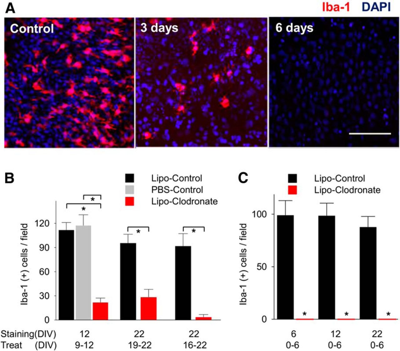
- Figure 1.
Elimination of microglia using liposomal (Lipo) clodronate from organotypic hippocampal slices of rat. A, Iba-1-positive cells (microglia and macrophages) in CA1 were depleted by using liposomal clodronate (0.2 mg/ml). B, Treatment for 3 d (from DIV9 to 12) decreased the number of microglia comparing liposome-control group and saline control group. Six day treatment from DIV16 to DIV22 eliminated more Iba-1(+) cells than 3 d treatment (96.2% vs 70.4%, n = 4 per group, p = 0.07). C, Microglial depletion persisted 16 d after washout. All values are expressed as mean ± SEM. *p < 0.05. Scale bar, 100 µm.
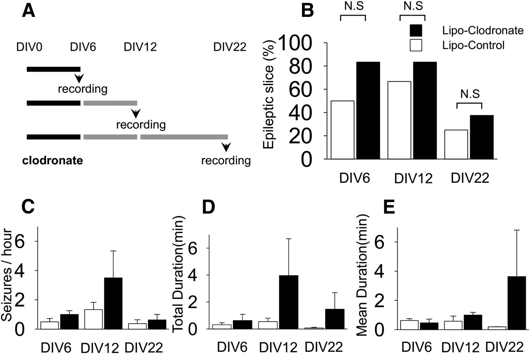
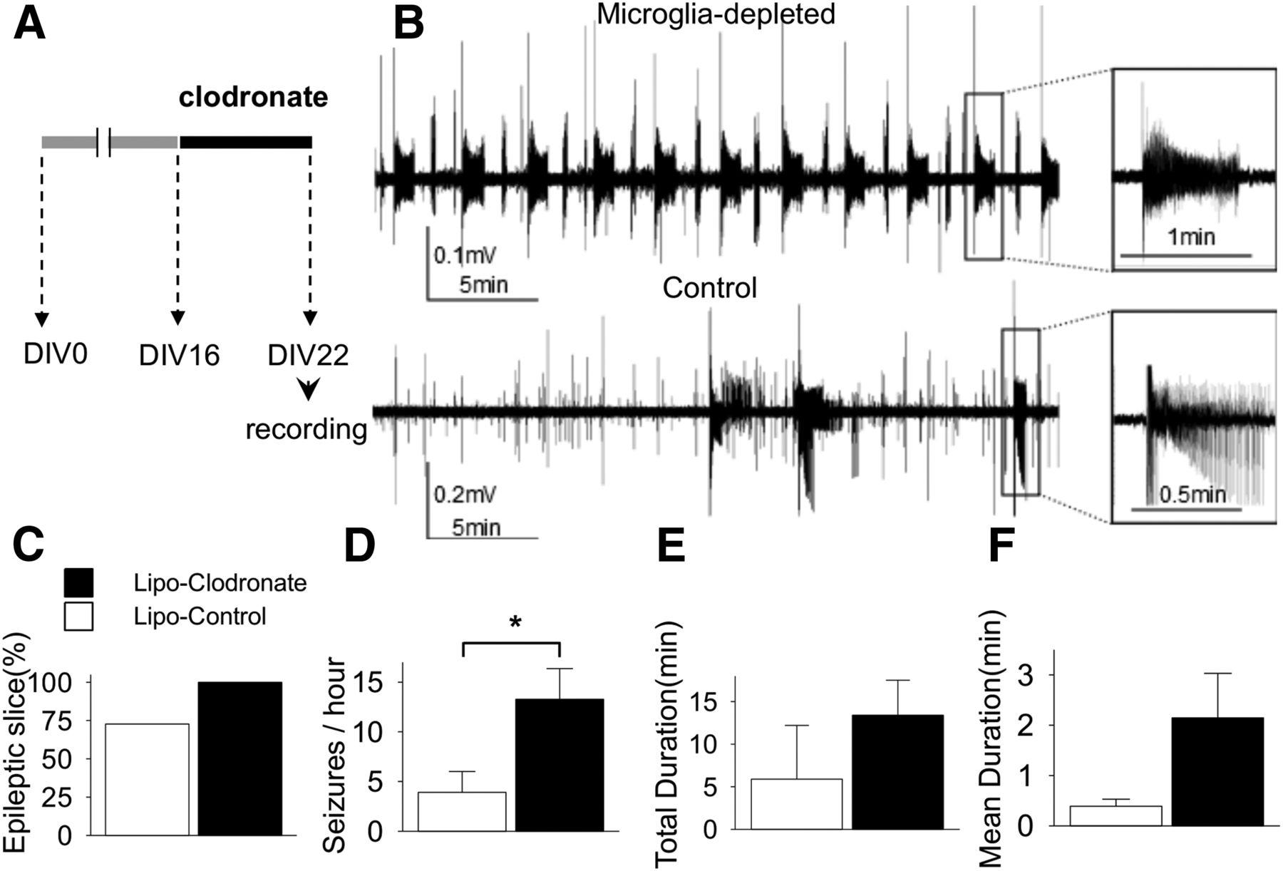
- Figure 2.
Effect of microglial depletion on ictogenesis in cultured rat slices. A, Schematic drawing of experiment protocol. B, Representative traces of field potentials recorded at DIV22 show that the microglia-depleted slice had more frequent and longer seizure-like activities compared to the control slice. C, Bar graphs indicate the percentage of slices with more than one seizure-like activity during observation period in each group. D−F, The frequency of seizure-like activities was significantly greater in the microglia-depleted group (13.3 ± 3.1 vs 3.9 ± 2.1/h, n = 11 per group, p = 0.02). All values are expressed as mean ± SEM, *p < 0.05.
- Figure 3.
Effect of microglial depletion on epileptogenesis in cultured rat slices. A, Liposomal (Lipo) clodronate or liposome-control was exposed to slices from DIV0 to 6 and spontaneous seizure-like activities were recorded at DIV6, 12, or 22 according to the indicated protocols. B, The proportions of slices demonstrating seizure-like activity during recording were not different between microglia-negative group and control group (n = 6-8 per group, p = 0.24, 0.52, and 0.60. respectively). C−E, Seizure frequency, total recorded seizure time, and mean seizure duration tend to be higher in slices depleted of microglia, although none of these differences were statistically different (n = 6-8 per group, p = 0.24, 0.30, and 0.28, respectively). N.S, Not significant. All values are expressed as mean ± SEM.
- Figure 4.
Effect of microglial depletion on epileptogenesis and ictogenesis in wild-type mice slices. A, Data was recorded at DIV6 after exposure of liposome clodronate from DIV0 to 6. B, Data was recorded at DIV12 after exposure of liposomal clodronate from DIV6 to 12. Microglial depletion did not alter the frequency, total duration, or mean duration of seizure-like activities (n = 4-5 per group). C, Representative traces recorded at DIV6 from microglia-depleted and control group, shows similar patterns of spontaneous seizure-like activities. All values are expressed as mean ± SEM. N.S, Not significant.
- Figure 5.
Microglial depletion from slices of nude mouse. A, Comparison of the density of Iba1-positive cells in area CA1 in control slices from different species (n = 5-6 per group) versus the percentage of slices that displayed seizure activity (n = 5-8 per group; different slices used for Iba-1 staining and recording). Across species, there was no significant correlation between microglial density and fraction of epileptic slices (R = −0.10, p = 0.94). B, Examples of hippocampal cultures from nude mice at each time point. Liposomal (Lipo) clodronate (top, 0.02 mg/ml) or liposome-control (bottom) was applied from DIV0 to 6. C, D, Double immunostaining was performed with NeuN and Iba-1 antibodies. Quantification at CA1 reveals that liposomal clodronate did not affect the neuronal populations of nude mouse (p = 0.33), whereas it depleted all microglia (n = 5 per group). E, The proportion of epileptic slices and total duration of seizure-like activities in microglia-depleted slices did not differ significantly from control slices, whereas the frequency of seizure-like activity was somewhat lower in microglia-depleted slices (n = 8-10 per group). All values are expressed as mean ± SEM. *p < 0.05; N.S, not significant. Scale bar, 100 μm.
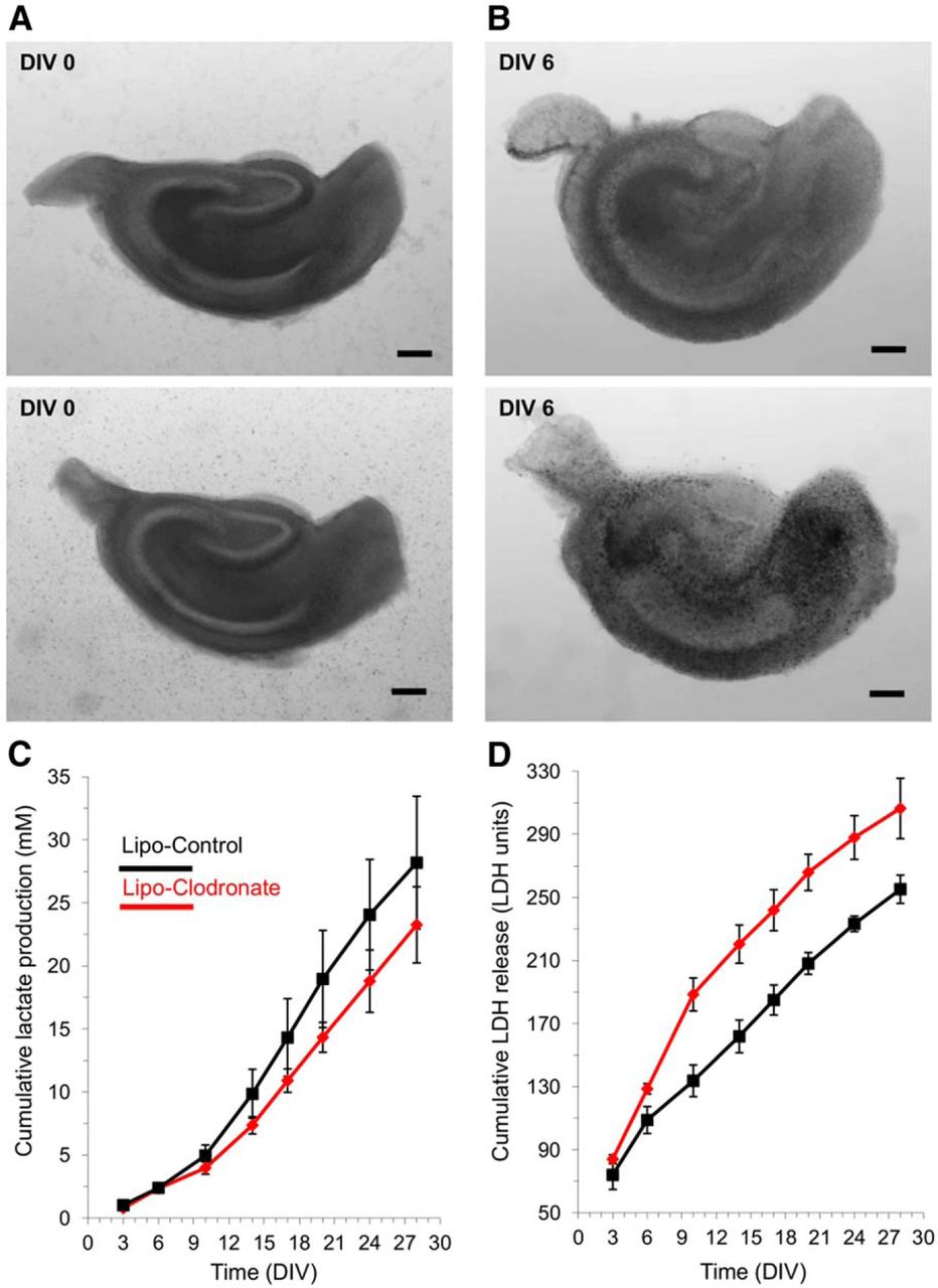
- Figure 6.
Long-term assays of epileptogenesis in microglia-depleted versus control hippocampal slice cultures. A, Examples of slice culture brightfield micrographs at DIV0 prior to clodronate treatment of upper slice. B, Brightfield micrographs of the same slice cultures on DIV6 at the conclusion of clodronate treatment to the upper slice and empty liposome treatment of the lower slice. No deleterious effects of clodronate are evident at this magnification. C, Cumulative group mean lactate production, assayed in the spent culture media at 3-4 daintervals during twice weekly media changes. N = 3 slices each group; all slices from the same animal. D, Cumulative group mean LDH release, assayed in the spent culture media. Same groups slices and media as for lactate assays in panel C. All values are expressed as mean ± SEM. Scale bar, 250 μm.


Tables
Recording timing Slice numbers (per group) Frequency of seizure (/h) Number of animals used Incidence of epilepsy (%) Clodronate Control Clodronate Control Figure 2 DIV12 11 1-36 0-24 3 rats 100 72.7 Figure 3 DIV6 6 0-2 0-2 2 rats 83.3 50 DIV12 6 0-12 0-3 2 rats 83.3 66.7 DIV22 8 0-3 0-2 2 rats 37.5 25.0 Figure 4 DIV6 4-5 9-19 1-16 2 mice 100 100 DIV12 4-6 2-26 2-21 2 mice 100 100 Figure 5 DIV12 4-6 0-2 0-4 3 nude mice 40.0 62.5 Data structure Type of test Power a Normally distributed t test 0.004 b Normally distributed t test 0.001 c Normally distributed t test 0.77 d Categorical Fisher’s exact test 0.07 e Normally distributed One-way ANOVA 0.02 f Normally distributed One-way ANOVA 0.11 g Normal distributed One-way ANOVA 0.14 h Categorical Fisher’s exact test 0.24 i Categorical Fisher’s exact test 0.52 j Categorical Fisher’s exact test 0.60 k Normally distributed Two-way ANOVA 0.39 for frequency, 0.46 for total duration, and 0.30 for mean duration l Normally distributed One-way ANOVA 0.31 m Normally distributed One-way ANOVA 0.36 n Normally distributed One-way ANOVA 0.56 o Normally distributed One-way ANOVA 0.99 p Normally distributed One-way ANOVA 0.89 q Normally distributed One-way ANOVA 0.62 r Normally distributed t test 0.003 s Normally distributed t test 0.01 t Categorical Fisher’s exact test 0.36 u Normally distributed One-way ANOVA 0.54 v Normally distributed One-way ANOVA 0.051 w* Normally distributed t test with Sidak correction 0.02, 0.42, 0.08, 0.07, 0.09, 0.08, 0.08, and 0.13 at each time point x* Normally distributed t test with Sidak correction 0.10, 0.02, 0.001, 0.002, 0.002, 0.002, 0.007, 0.01 at each time point *αSID = 1 − (1 − α)1/ m , α = 0.05, m = 1,2,3,4,5,6,7,8. αSID(MAX) = 0.06
- Table 3.
Comparison of seizure-like activities between microglia-depleted and control slices
Microglia-depleted Control p value Recording at DIV6 Frequency (SLA/h) 1.0 ± 0.3 0.5 ± 0.2 0.17 Total duration (s/h) 37.7 ± 27.7 18.7 ± 9.0 0.53 Mean duration (s) 27.7 ± 15.3 37.3 ± 7.7 0.67 Recording at DIV12 Frequency (SLA/h) 3.5 ± 1.8 1.3 ± 0.5 0.24 Total duration (s/h) 237.7 ± 164.2 32.8 ± 15.3 0.30 Mean duration (s) 59.6 ± 11.1 34.5 ± 20.9 0.28 Recording at DIV22 Frequency (SLA/h) 0.6 ± 0.4 0.4 ± 0.3 0.59 Total duration (s/h) 88.0 ± 73.8 4.4 ± 3.1 0.28 Mean duration (s) 217.8 ± 191.1 11.5 ± 0.5 0.46
In this issue


