
Main menu

  • HOME
  • CONTENT
    • Early Release
    • Featured
    • Current Issue
    • Issue Archive
    • Blog
    • Collections
    • Podcast
  • TOPICS
    • Cognition and Behavior
    • Development
    • Disorders of the Nervous System
    • History, Teaching and Public Awareness
    • Integrative Systems
    • Neuronal Excitability
    • Novel Tools and Methods
    • Sensory and Motor Systems
  • ALERTS
  • FOR AUTHORS
  • ABOUT
    • Overview
    • Editorial Board
    • For the Media
    • Privacy Policy
    • Contact Us
    • Feedback
  • SUBMIT

User menu

Search

  • Advanced search
eNeuro

eNeuro

Advanced Search


  • HOME
  • CONTENT
    • Early Release
    • Featured
    • Current Issue
    • Issue Archive
    • Blog
    • Collections
    • Podcast
  • TOPICS
    • Cognition and Behavior
    • Development
    • Disorders of the Nervous System
    • History, Teaching and Public Awareness
    • Integrative Systems
    • Neuronal Excitability
    • Novel Tools and Methods
    • Sensory and Motor Systems
  • ALERTS
  • FOR AUTHORS
  • ABOUT
    • Overview
    • Editorial Board
    • For the Media
    • Privacy Policy
    • Contact Us
    • Feedback
  • SUBMIT
PreviousNext
Research ArticleResearch Article: Confirmation, Cognition and Behavior

Hippocampal Neuronal Activity Preceding Stimulus Predicts Later Memory Success

Soyeon Jun, June Sic Kim and Chun Kee Chung
eNeuro 31 January 2023, 10 (2) ENEURO.0252-22.2023; DOI: https://doi.org/10.1523/ENEURO.0252-22.2023
Soyeon Jun
1Neuroscience Research Institute, Seoul National University College of Medicine, Seoul, South Korea, 03080
4Department of Neurosurgery, Seoul National University Hospital, Seoul, South Korea, 03080
  • Find this author on Google Scholar
  • Find this author on PubMed
  • Search for this author on this site
June Sic Kim
3Research Institute of Basic Sciences, Seoul National University, Seoul, South Korea, 03080
  • Find this author on Google Scholar
  • Find this author on PubMed
  • Search for this author on this site
Chun Kee Chung
2Department of Brain and Cognitive Sciences, Seoul National University, Seoul, South Korea, 08826
4Department of Neurosurgery, Seoul National University Hospital, Seoul, South Korea, 03080
  • Find this author on Google Scholar
  • Find this author on PubMed
  • Search for this author on this site
  • ORCID record for Chun Kee Chung
  • Article
  • Figures & Data
  • Info & Metrics
  • eLetters
  • PDF
Loading

Article Figures & Data

Figures

  • Tables
  • Figure 1.
    • Download figure
    • Open in new tab
    • Download powerpoint
    Figure 1.

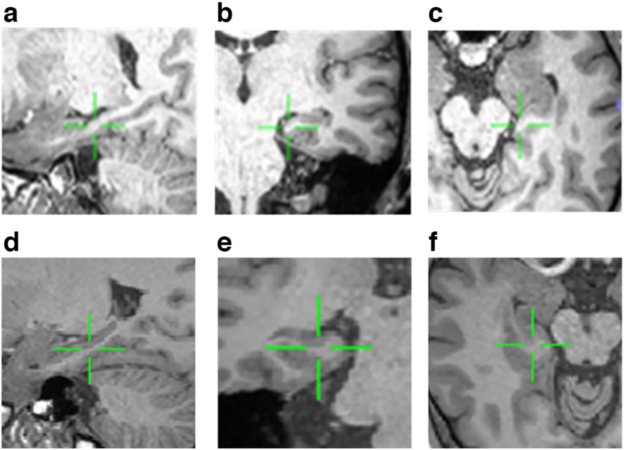
    Electrode localization with structural magnetic resonance images. Examples of hippocampal microelectrodes from two patients are shown (see Table 1 for demographic information). The patient’s left brain is shown on the left side in the axis plane. All images are T1 images recorded using Sigma 3-tesla scanner (GE). Green arrows indicate electrodes inserted in the hippocampal subfield of Cornu Ammonis (CA3 (above) and CA1 (bottom)). a, Sagittal image of the left hippocampal CA3. b, Coronal hippocampal CA3. c, Axial hippocampal CA3. d, Sagittal image of the left hippocampal CA1. e, Coronal CA1 image. f, Axial CA1 image.

  • Figure 2.
    • Download figure
    • Open in new tab
    • Download powerpoint
    Figure 2.

    Word memory tasks. a, Example of the timeline of the visual item memory task and (b) associative memory task. Memory tasks comprised three successive stages encoding, distractor, and retrieval. Each patient sequentially completed words items during encoding, which was followed by a white fixation cue with a black screen for 1 s. The green dashed line box indicates the preceding stimulus interval.

  • Figure 3.
    • Download figure
    • Open in new tab
    • Download powerpoint
    Figure 3.

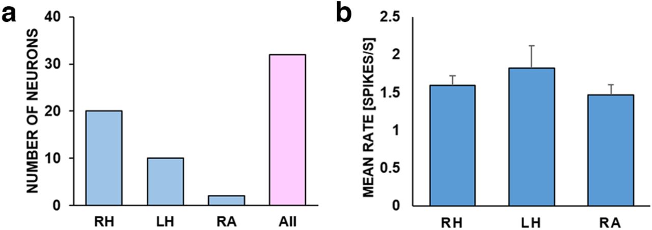
    Numbers of neurons and mean firing rates from different brain areas. a, Units’ numbers from each brain area were as follows, from left to right: 20, 10, and 2. b, The mean firing rates of units, respectively. All units are included, regardless of firing rates. The mean firing rates of the hippocampal and amygdala are 1.71 and 1.47, respectively. Error bars are ±SEM. RH, right hippocampus; LH, left hippocampus; RA, the right amygdala.

  • Figure 4.
    • Download figure
    • Open in new tab
    • Download powerpoint
    Figure 4.

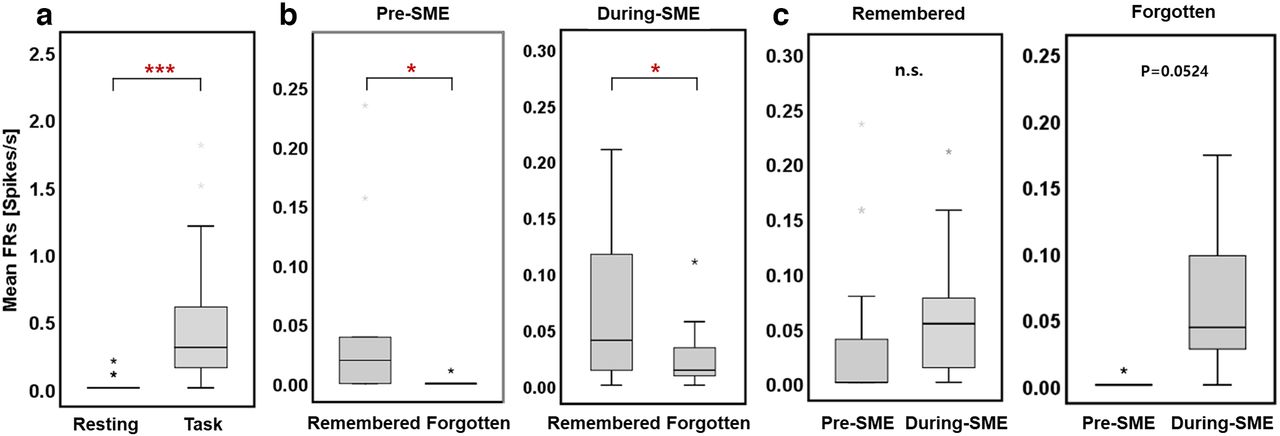
    Group-level comparison of spiking activity in the hippocampus during an item memory task. a, The mean firing rates across neurons (n = 11) during resting states versus memory encoding showed a significant increase in firing rates for memory encoding compared with resting states (Wilcoxon signed-rank test, ***p < 0.001). b, Group-level comparison of pre-SME and during-SME hippocampal mean firing rates. During the preceding stimulus, hippocampal firing rates were higher for subsequent remembered than forgotten words (left, Wilcoxon signed-rank test, **p < 0.01). However, the trend during-SME hippocampal activity was not significant (right, Wilcoxon signed-rank test, p = 0.058). c, Pre-SME and during-SME mean firing rates for an operation of words subsequently remembered (left) versus forgotten (right). The difference between pre-SME and during-SME was not significant in both remembered and forgotten words (Wilcoxon signed-rank test, p = 0.0518, p = 0.082, respectively).

  • Figure 5.
    • Download figure
    • Open in new tab
    • Download powerpoint
    Figure 5.

    Hippocampal neuronal responses to subsequently remembered and forgotten word items. Shown are representative examples of hippocampal neurons that significantly increase firing rates across trials. Preceding stimulus and during-stimulus raster plot of firing rates during encoding sessions. The red lines mark stimulus onset at 0; the offset is at 3 s (x-axis). Raster rows represent single trials, and each dot represents an action potential. Next, the preceding stimulus and during-stimulus raster plot of firing rates for the remembered and forgotten conditions during encoding (**p < 0.01, *p < 0.05, permutation-based p-value).

  • Figure 6.
    • Download figure
    • Open in new tab
    • Download powerpoint
    Figure 6.

    Hippocampal neuronal responses to subsequently remembered and forgotten word items. a, The mean firing rates between resting states and encoding. Mean firing rates for the encoding duration are significantly higher than the resting state. b, The hippocampal preceding stimulus neuronal activity (pre-SME) is significantly higher in subsequently remembered items compared with subsequently forgotten items (***p < 0.001, *p < 0.05, permutation-based p-value). This neuron was selective to only preceding stimulus with similar increases in activity in the during-stimulus condition. c, The hippocampal spiking activity indicated the difference between preceding stimulus and during-stimulus activity was negligible in both remembered and forgotten conditions (n.s., p = 0.0524, respectively, permutation-based p-value). n.s. indicates not significant.

  • Figure 7.
    • Download figure
    • Open in new tab
    • Download powerpoint
    Figure 7.

    Group-level comparison of spiking activity in the hippocampus during an associative memory task. a, Presubsequent memory effect (pre-SME) and (b) during-SME mean firing rates as an operation of words subsequently remembered versus forgotten words.

Tables

  • Figures
    • View popup
    Table 1

    List of patient demographics, pathology, and neuropsychological evaluation

    SubjectAgeSexEpilepsy diagnosisWAIS-IVWMS-R
    VCIPRIWMIPSIFSIQMQ
    Sub 130–40MRTLE1168698849566
    Sub 220–30MLTLE10010798559073
    Sub 360–70MRTLE114941129210377
    Sub 450–60MLTLE728478727056
    Average39.7 (17.3)--101 (17.6)93 (9.0)97 (12.1)76 (13.9)90 (12.2)68 (8.0)
    • Intelligence was measured with the Korean Wechsler Memory Scale (K-WMS) and memory with the Wechsler Memory Scale (WMS). RTLE, right temporal lobe epilepsy; LTLE, left temporal lobe epilepsy; VCI, verbal comprehension index; PRI, perceptual reasoning index; WMI, working memory index; PSI, processing speed index; FSIQ, full scale IQ; MQ, memory quotient.

Back to top

In this issue

eneuro: 10 (2)
eNeuro
Vol. 10, Issue 2
February 2023
  • Table of Contents
  • Index by author
  • Masthead (PDF)
Email

Thank you for sharing this eNeuro article.

NOTE: We request your email address only to inform the recipient that it was you who recommended this article, and that it is not junk mail. We do not retain these email addresses.

Enter multiple addresses on separate lines or separate them with commas.
Hippocampal Neuronal Activity Preceding Stimulus Predicts Later Memory Success
(Your Name) has forwarded a page to you from eNeuro
(Your Name) thought you would be interested in this article in eNeuro.
CAPTCHA
This question is for testing whether or not you are a human visitor and to prevent automated spam submissions.
Print
View Full Page PDF
Citation Tools
Hippocampal Neuronal Activity Preceding Stimulus Predicts Later Memory Success
Soyeon Jun, June Sic Kim, Chun Kee Chung
eNeuro 31 January 2023, 10 (2) ENEURO.0252-22.2023; DOI: 10.1523/ENEURO.0252-22.2023

Citation Manager Formats

  • BibTeX
  • Bookends
  • EasyBib
  • EndNote (tagged)
  • EndNote 8 (xml)
  • Medlars
  • Mendeley
  • Papers
  • RefWorks Tagged
  • Ref Manager
  • RIS
  • Zotero
Respond to this article
Share
Hippocampal Neuronal Activity Preceding Stimulus Predicts Later Memory Success
Soyeon Jun, June Sic Kim, Chun Kee Chung
eNeuro 31 January 2023, 10 (2) ENEURO.0252-22.2023; DOI: 10.1523/ENEURO.0252-22.2023
del.icio.us logo Digg logo Reddit logo Twitter logo Facebook logo Google logo Mendeley logo
  • Tweet Widget
  • Facebook Like
  • Google Plus One

Jump to section

  • Article
    • Abstract
    • Significance Statement
    • Introduction
    • Materials and Methods
    • Results
    • Discussion
    • Acknowledgments
    • Footnotes
    • References
    • Synthesis
  • Figures & Data
  • Info & Metrics
  • eLetters
  • PDF

Keywords

  • hippocampus
  • memory
  • neuronal activity
  • prediction
  • single-unit activity

Responses to this article

Respond to this article

Jump to comment:

No eLetters have been published for this article.

Related Articles

Cited By...

More in this TOC Section

Research Article: Confirmation

  • The Nature and Origin of Synaptic Inputs to Vestibulospinal Neurons in the Larval Zebrafish
  • Amyloid β-Peptide Effects on Glucose Regulation Are Dependent on Apolipoprotein E Genotype
  • Glycolytic System in Axons Supplement Decreased ATP Levels after Axotomy of the Peripheral Nerve
Show more Research Article: Confirmation

Cognition and Behavior

  • Neuronal representation of a working memory-based decision strategy in the motor and prefrontal cortico-basal ganglia loops
  • Strawberry additive increases nicotine vapor sampling and systemic exposure but does not enhance Pavlovian-based nicotine reward in mice
  • Attention Without Constraint: Alpha Lateralization in Uncued Willed Attention
Show more Cognition and Behavior

Subjects

  • Cognition and Behavior

  • Home
  • Alerts
  • Visit Society for Neuroscience on Facebook
  • Follow Society for Neuroscience on Twitter
  • Follow Society for Neuroscience on LinkedIn
  • Visit Society for Neuroscience on Youtube
  • Follow our RSS feeds

Content

  • Early Release
  • Current Issue
  • Latest Articles
  • Issue Archive
  • Blog
  • Browse by Topic

Information

  • For Authors
  • For the Media

About

  • About the Journal
  • Editorial Board
  • Privacy Policy
  • Contact
  • Feedback
(eNeuro logo)
(SfN logo)

Copyright © 2023 by the Society for Neuroscience.
eNeuro eISSN: 2373-2822

The ideas and opinions expressed in eNeuro do not necessarily reflect those of SfN or the eNeuro Editorial Board. Publication of an advertisement or other product mention in eNeuro should not be construed as an endorsement of the manufacturer’s claims. SfN does not assume any responsibility for any injury and/or damage to persons or property arising from or related to any use of any material contained in eNeuro.