

Article Figures & Data
Figures
- Figure 1.
Learning occludes repetitive synaptic stimulation-induced and kainate-induced AHP reduction. A, Schematic description of the four-arm maze. Protocols for trained and pseudotrained rats are similar: an electronic “start” command randomly opens two of eight valves (V), releasing a positive-cue odor (P) into one of the arms and a negative-cue odor (N) into another. Eight seconds later, the two corresponding guillotine doors (D) are lifted to allow the rat to enter the selected arms. On reaching the far end of an arm (90 cm long), the rat body interrupts an infrared beam (I; arrow) and a drop of drinking water is released from a water hose (W) into a small drinking well (trained rats: only if the arm contains the positive-cue odor, pseudotrained rats: random assignment of odors to rewards). B, Position of the intracellular recording electrode in layer II of the anterior piriform cortex in acute coronal brain slices and the stimulation electrode in layer Ib. C1, Time line of long-term AHP reduction induced by tetanic synaptic stimulation of trained (black) and control (red) rats. C2, Averaged typical traces of a neuron from a trained (black) and pseudotrained (red) rat before (light shade) and after (dark shade) synaptic stimulation. The AP peaks are clipped to facilitate comparison of the postburst AHP. C3, Direct comparison of the absolute AHP amplitudes before and 20 min after synaptic stimulation for control (red) and trained (black) rats. D1, Time line of long-term AHP reduction induced by kainate application to brain slices of trained (blue) and control (green) rats. D2, Averaged typical traces of a neuron from a trained (blue) and pseudotrained (green) rat before (light shade) and after (dark shade) kainate application. The AP peaks are clipped to facilitate comparison of the postburst AHP. D3, Direct comparison of the absolute AHP amplitudes before and 20 min after kainate application for pseudotrained (green) and trained (blue) rats. Values are represented as the mean ± SEM. **p < 0.01.
- Figure 2.
Activity-induced AHP reduction is mediated by intrinsic activity of second-messenger systems, and not by synaptic activation of kainate receptors. A, Averaged typical traces in three neurons taken from naive rats before (light shade) and after (dark shade) repetitive synaptic stimulation. The AP peaks are clipped to facilitate comparison of the postburst AHP. Repetitive synaptic stimulation-induced post-AHP reduction is abolished by prior application of the ERK inhibitor UO126 (top traces), prior application of the PKC activator OAG (middle traces), or by prior kainate application (bottom traces). B, Direct comparison of the absolute AHP amplitudes before and 20 min after repetitive synaptic stimulation in the presence UO126 (black), OAG (red), or kainate (blue). Each of the three treatments blocked the synaptic activity-induced AHP reduction. Note that the agonists kainate and OAG reduced the averaged AHP in naive neurons to the averaged value observed in neurons from trained rats, while the antagonist OU126 had no direct effect on the AHP. Values represent the mean ± SEM. C, Synaptic currents evoked by stimulating the intrinsic fibers pathway (layer Ib) do not contain a kainate receptor-mediated component. The application of GYKI53655 completely blocked the synaptic currents (top trace). The bottom trace shows the lack of response to a train of five stimuli at 200 Hz in the same cells after GYKI53655 application. D, Time line of the synaptic current amplitude before and after GYKI53655 application. Recordings were obtained in six pyramidal cells taken from three mice. Notably, the synaptic current amplitude is abolished in all recorded neurons. Values represent the mean ± SEM.
- Figure 3.
GluK2 activation is mandatory for AHP reduction and complex olfactory learning. A1, Averaged typical traces of a neuron from GluK2−/− mice (red) and wild-type littermates (black) before (light shade) and after (dark shade) kainate application. The AP peaks are clipped to facilitate the comparison of the postburst AHP. A2, Direct comparison of the absolute AHP amplitudes before and 20 min after kainate application for GluK2−/− mice (red) and wild-type littermates (black). B, While wild-type mice (black) show a gradual learning curve, resulting in acquisition of the complex OD task for most (22 of 23) of the subjects, GluK2−/− mice (red) performance mostly remains at the level of chance. Only 2 of 23 knock-out mice were able to learn the task. Average performance for each day was calculated for all 20 trials of the day. C, Swarmplot of the latency to find the cookie in the buried food task in wild types (black) and GluK2−/− mice (red). In this simple olfactory task, there is no difference between genotypes. Values represent the mean ± SEM. *p < 0.05.
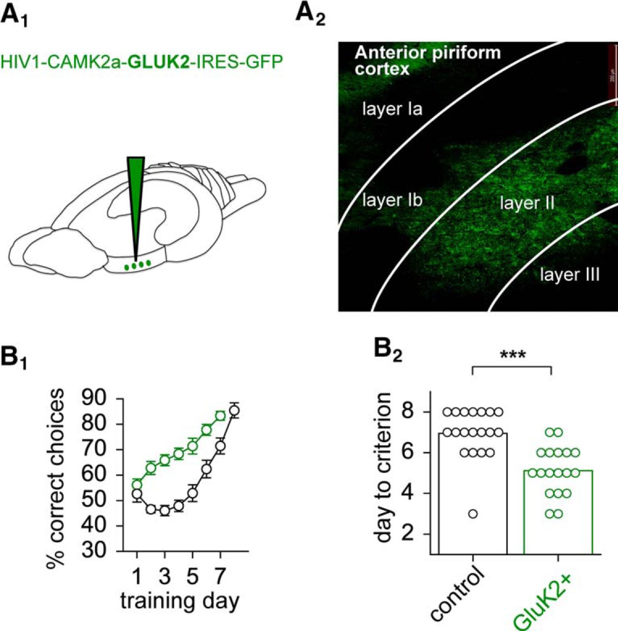
- Figure 4.
Enhanced complex olfactory learning is enhanced by GluK2 overexpression. A1, Schematic of virus injection in the rat piriform cortex. A2, Virus-infected pyramidal neurons in layer II indicated by GFP expression. Signal of GFP was enhanced using anti-GFP antibody. B1, Time line of performance as a function of training day for GluK2-overexpressing rats (green) and controls injected with GFP virus only (black). To allow a more detailed look at the progress in performance for each day, the averaged percentage of correct choices for all 20 trials of the day are plotted. B2, Swarmplot of the days needed to reach the criterion defined as a success rate of >80% in the last 10 trials. GluK2-injected rats (green) complete complex olfactory discrimination learning significantly faster than the control rats (black). Values represent the mean ± SEM. ***p < 0.001.


Tables
Naive-control Naive-GFP only Naive-GluK2 Trained-control Trained-GFP only Trained-GluK2 Pseudo-control Pseudo-GFP only Pseudo-GluK2
In this issue


