

Article Figures & Data
Figures
- Figure 1.
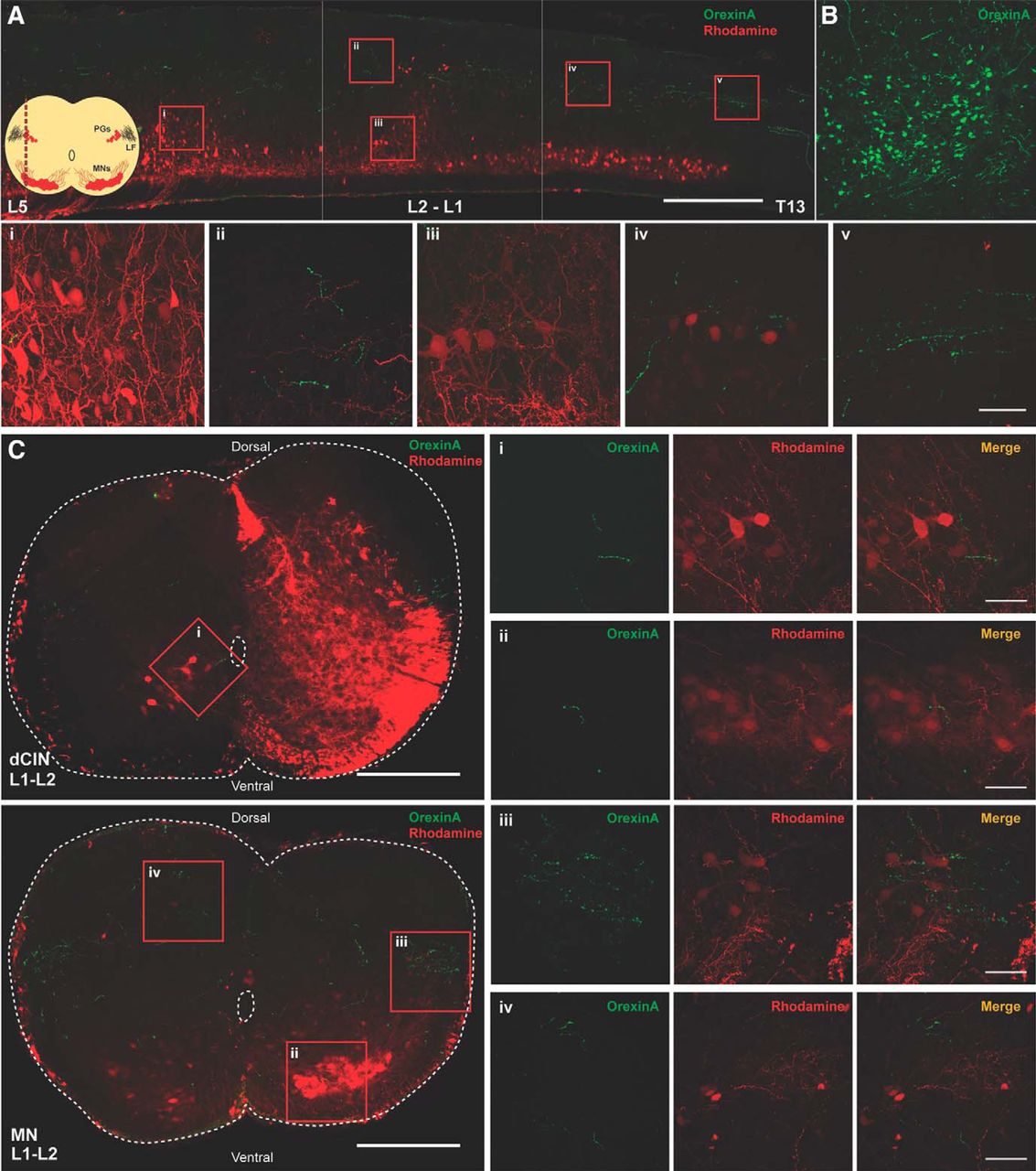
Orexinergic innervation of the developing (P3) spinal cord. A, Longitudinal section of spinal cord (30 μm) showing numerous orexinergic fibers projecting along the lateral funiculus throughout the lumbar spinal cord. Scale bar, 500 µm. Corner schematic shows the location of the longitudinal section (LF, lateral funiculus; PG, preganglionic motor neurons); 60× confocal images of select regions (scale bar, 50 µm): i, sparse orexinergic fibers around MNs in lumbar region; ii, dorsal fibers in the L1-L2 region; iii, rare fiber passing by L1-L2 MNs; iv, orexinergic fibers exiting the LF around the preganglionic MNs; v, orexinergic fibers in the LF, varicosities are evident. B, Positive control showing OXergic neurons in lateral hypothalamus. C, Transverse sections of L1-L2 spinal cord with rhodamine-labeled dCINs and rhodamine-labeled ventral MNs (scale bar, 250 µm); 60× confocal images of select regions (scale bar, 50 µm): i, orexinergic fibers apposing dCINs; ii, sparse fibers in MN pool; iii, orexinergic fibers exiting the LF around the preganglionic MNs; iv, dorsal fibers are prevalent.
- Figure 2.
Effect of OXA and OXB on spontaneous spinal cord motor activity. A, B, Representative neurogram of recorded motor spontaneous activity in absence of any drugs (pre-OX) and after bath application of OXA (A) and OXB (B; OXA and OXB, 300 nm, were added at the red dotted lines). C, D, Bar graphs representing the fold change of the tonic activity calculated as the AUC in different conditions: pre-OX (black); OXA (C) and OXB (D; 300 nM; red); washout of OXA or OXB (gray). E, F, Graphs representing the time course of the fold change in frequency of the spontaneous activity comparing pre-OX with the frequency observed at different times after OXA (E) and OXB (F) bath application. G, H, The fold change in amplitude of the spontaneous bursts during OXA and OXB bath application, respectively. N = 7/group for OXA and N = 10/group for OXB. RL, Right lumbar ventral roots; LL, left lumbar ventral roots (recorded at the second or fifth lumbar segmental level). Data are represented as the mean ± SD. *p < 0.05, **p < 0.01, ****p < 0.0001.
- Figure 3.
OXR antagonist abolished OXA or OXB-mediated excitation. A, B, Representative neurogram of recorded motor spontaneous activity when the OX1R and OX2R (dual receptor) antagonist TCS 1102 (10 μm) was applied to the isolated spinal cord preparation before the addition of 300 nm OXA (A) or OXB (B; indicated at the red dotted lines), respectively. Pre-OX + antagonist indicates period when antagonist was added to the aCSF. C, D, Bar graphs representing the fold change of the tonic activity: pre-OX + antagonist (black); OXA (C; 300 nm; red); OXB (D; 300 nm); washout (gray) of OXA (C) or OXB (D) in the presence of dual antagonist. E, F, Graphs representing the evolution of the fold change in frequency of the spontaneous activity comparing pre-OX, OXA (E), or OXB (F) observed at different times following the application of OXA or OXB in the presence of TCS 1102. G, H, The fold change in amplitude of the spontaneous bursts during OXA or OXB bath application, in the presence of TCS 1102. N = 4 for the dual receptor antagonist experiments, respectively. Data are represented as the mean ± SD.
- Figure 4.
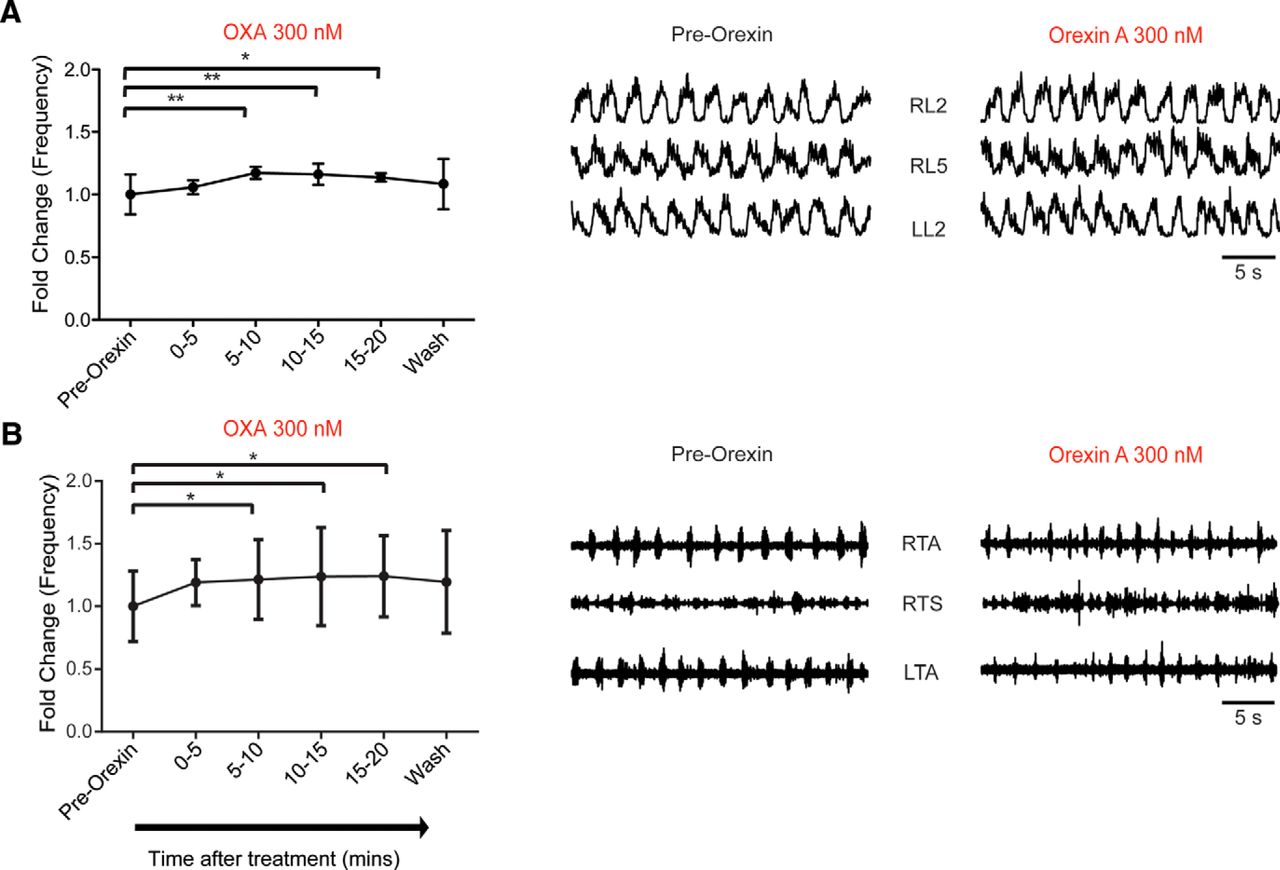
Orexin A modulates fictive locomotor rhythm frequency. A, Graph representing the time course of change in the frequency of fictive locomotor rhythm comparing the pre-OX frequency with the one observed at different times after the bath application of OXA (300 nm) obtained from an isolated spinal cord preparation (N = 7). Right, Representative neurograms showing the increase in frequency of the fictive locomotor rhythm induced by bath application of DA, NMDA, and 5-HT before and during OXA bath application. B, Graph representing the time course of the change in frequency of fictive the locomotor rhythm comparing pre-OX frequency with the one observed at different times after the application of OXA (300 nm) obtained from a spinal cord with leg-attached preparation (N = 5 preparations). Right, Representative EMG recordings showing the increased frequency of the fictive locomotor rhythm induced by bath application of DA, NMDA, and 5-HT before and during OXA bath application. RL, Right lumbar ventral roots; LL, left lumbar ventral roots (recorded at the second or fifth lumbar segmental level); RTA, right TA muscle; LTA, left TA muscle; RTS, right TS muscle. Data are represented as the mean ± SD. *p < 0.05, **p < 0.01.
- Figure 5.
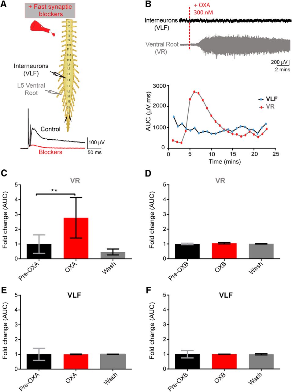
Orexins effects on motoneuron and interneuron populations. A, Top, Experimental setup; pulses delivered every 30 s were delivered to the L5 dorsal root through a suction electrode to elicit an evoked response recorded via suction electrodes placed on the right L5 ventral root (VR, recording of motoneuron pool) and onto the right L4 or L3 VLF to record activity from ventral horn interneurons projecting into the VLF. Bottom, Following the addition of fast neurotransmitter blockers, single pulses were delivered, and an evoked polysynaptic response was recorded from the VR and VLF (control, black trace) until no polysynaptic reflex was present (blockers, red trace). B, Top, Representative neurograms showing that the bath application of OXA 300 nm induced an increase in neurogram discharge recorded from the VR but not from the VLF. Bottom, Graphs showing the analysis of the change in tonic activity induced following bath application of OXA, a minute-by-minute analysis of the AUC (in microvolts per millisecond) of filtered, rectified, and reduced neurograms were obtained. C, E, Graphs representing the time course of the fold change in tonic activity from VR (C) and VLF (E) neurograms comparing the pre-OXA and post-OXA AUC (300 nm; N = 6 preparations). D, F, Graphs for pre-OXB and post-OXB for the VR (D) and VLF (F), respectively. Data are represented as the mean ± SD. **p < 0.01.
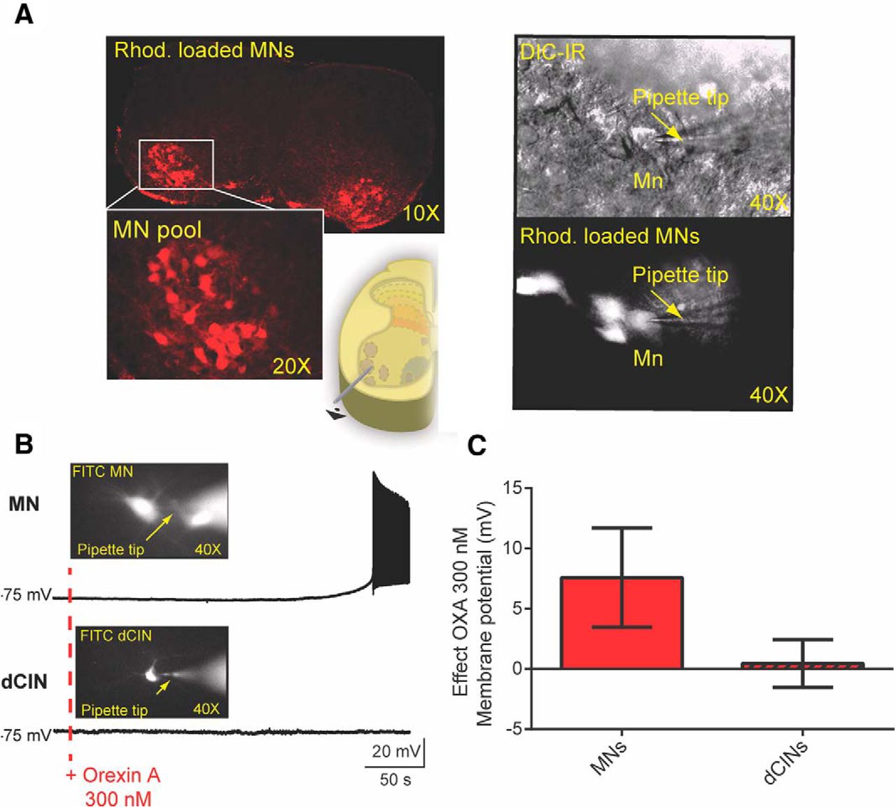
- Figure 6.
The effect of orexins on identified MNs and interneurons. A, Left, A 10× picture of a spinal cord showing MN pools previously retrogradely labeled by fluorescent Rhod crystals applied to the cut ends of ventral root L1-L2 axons. Bottom, A higher magnification of the MN pool (20×) and the schematic illustrating the ventrolateral position of the MN pools. Right, MN visually identified using IR-DIC optics in the ventral horn of the L1-L2 transverse slice (bottom right). B, OXA led to a depolarization of the MN recorded, but not the dCINs. The pictures show the patched cells that were visually identified via Rhod. Retrograde labeling, and the cells were also intracellularly labeled with FITC to identify post hoc which cells in the pool were recorded. C, Quantification of the effects of OXA 300 nm on MNs and dCINs (MNs, N = 22; dCINs, N = 14).
- Figure 7.
Orexin A modulates some intrinsic properties of identified motoneurons. A, Representative example of intracellular recordings showing an increase in the MN rheobase following OXA 300 nm (red traces). B, Bath application of OXA 300 nm blocked the delay of firing in an identified L1/2 MN. There is a shift toward the left for the spiking (see dashed line for the same MN and same current injected). C, Summary plot of changes in the first spike latency pre-OX and post-OX (MNs, N = 13). See Tables 1 and 2 for further details.
- Figure 8.
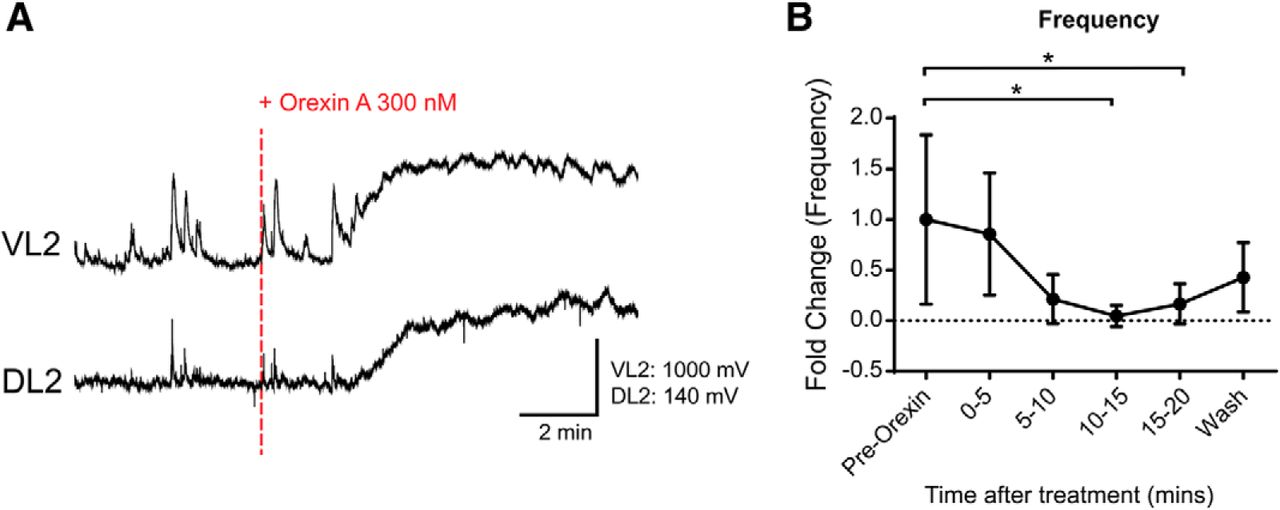
Orexin A decreases dorsal horn subthreshold potentials. A, Neurogram describing the potentials generated in ventral and dorsal L2 roots. Recordings have been filtered with a Butterworth 5 Hz low-pass filter. They depict 5 min preapplication and 10 min postapplication of 300 nm OXA to an intact isolated spinal cord. B, Frequency change in dorsal root subthreshold potentials taken in 5 min bins. Events were counted manually. Data are represented as the mean ± SD. *p < 0.05.
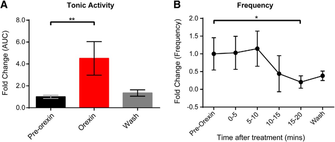
- Figure 9.
The effect of OXA on spontaneous activity in the dorsal horn-removed preparation. A, OXA (300 nm) increased tonic firing activity recorded from the L2 ventral roots in the isolated ventral spinal cord preparation with dorsal horn removed (N = 6 dorsal horn-removed spontaneous preparations; fold change in activity). B, Graph showing the decrease in spontaneous bursting activity recorded from the L2 ventral roots before and after the bath application of 300 nm OXA to a dorsal horn-removed preparation. Mean fold change in the frequency shown in 5 min bins. Data are represented as the mean ± SD. *p < 0.05, **p < 0.01.


Tables
- Table 1:
Summary of orexin A effects on some intrinsic properties recorded from motoneurons
MNs Parameters/conditions Pre-OX OXA 300 nm Washout Rin (MΩ) 80.60 ± 46.02 (13) 102.43 ± 38.41 (13)** 92.16 ± 43.04 (11)ns AP amplitude (mV) 72.48 ± 8.89 (13) 68.59 ± 10.61 (13)ns 69.08 ± 10.54 (12)ns AP threshold (mV) −51.08 ± 8.90 (13) −51.32 ± 8.87 (13)ns −52.34 ± 9.90 (12)ns Rheobase (pA) 478.6 ± 211.4 (13) 396.7 ± 181.7 (13)* 433.1 ± 233.3 (11)ns Average F/I slope (pA/Hz) 0.0855 ± 0.0692 (13) 0.0699 ± 0.0451 (13)* 0.0705 ± 0.0284 (10)ns Instantaneous F/I slope (pA/Hz) 0.1786 ± 0.206 (13) 0.1468 ± 0.165 (13)ns 0.1505 ± 0.209 (10)ns Values are the mean ± SD (number of motoneurons). Report of the statistical significance between control condition, pre-OX and OXA at 300 nm, and the control and washout of OXA for the following parameters: AP amplitude; action potential amplitude, AP threshold, action potential threshold, rheobase; average F/I slope, average frequency of spiking/intensity of current-injected slope; instantaneous F/I slope, instantaneous frequency/intensity slope. One-way repeated-measures ANOVA followed by Student–Newman–Keuls post hoc test was used to determine the significance between conditions.
nsp > 0.05; *p ≤ 0.05; **p ≤ 0.01.
dCINs Parameters/conditions Pre-OX OXA 300 nm Washout Rin (MΩ) 629.8 ± 527.2 (10) 626 ± 520.0 (10)ns 462.6 ± 217.0 (8)ns AP amplitude (mV) 73.69 ± 10.33 (10) 70.87 ± 9.25 (10)ns 70.82 ± 8.11 (9)ns AP threshold (mV) −57.74 ± 4.19 (10) −57.03 ± 3.2 (10)ns −57.91 ± 2.86 (10)ns Rheobase (pA) 52.11 ± 25.07 (10) 52.54 ± 31.09 (10)ns 51.58 ± 27.71 (9)ns Average F/I slope (pA/Hz) 0.3348 ± 0.16 (10) 0.2960 ± 0.181 (10)ns 0.2965 ± 0.127 (7)ns Instantaneous F/I slope (pA/Hz) 0.3996 ± 0.335 (10) 0.3896 ± 0.318 (10)ns 0.3512 ± 0.098 (7)ns Values are the mean ± SD (number of dCINs). Report of the statistical significance between control condition pre-OX and OXA at 300 nm, and the control and washout of OXA. ANOVA followed by Student–Newman–Keuls post hoc test was used to determine the significance between conditions.
nsp > 0.05.
In this issue


