

Article Figures & Data
Figures
- Figure 1.
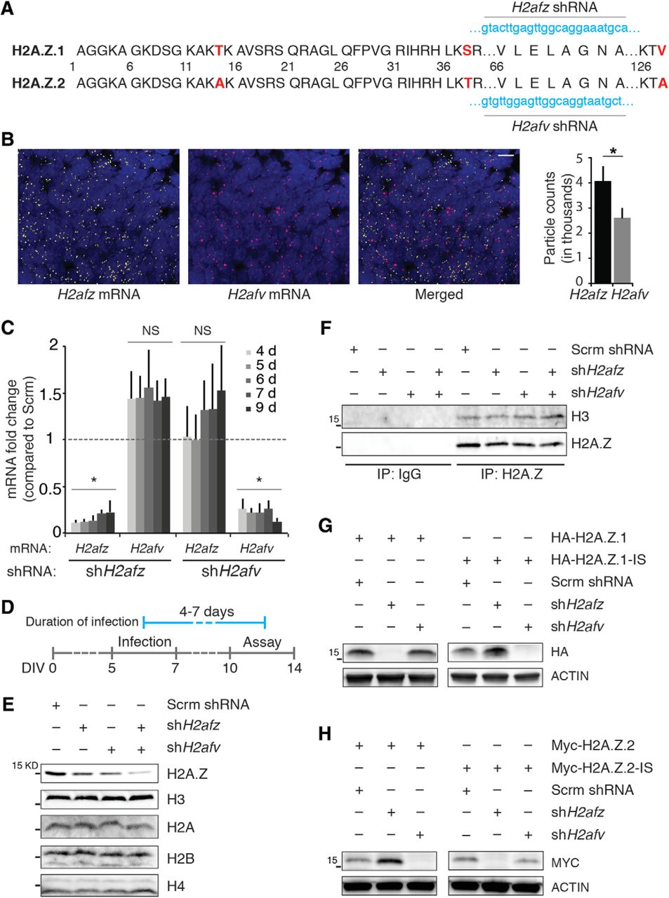
Validation of H2A.Z hypervariant-specific RNAi. A, H2A.Z.1 and H2A.Z.2 amino acid sequences are depicted where three different amino acid residues are indicated in red. The target sequences of shH2afz and shH2afv are noted in blue. B, left, Representative images of smFISH of H2afv and H2afz in E18 rat cortex. Right, Quantification of H2afz and H2afv molecules; particle count per 212 μm2. N = 3, *p < 0.05 (one-tailed unpaired t test). Scale bar, 10 μm. C, Cortical neurons in culture were infected with lentiviruses delivering either shH2afz or shH2afv and total RNA was collected after indicated number of days. Compared to control neurons, changes in H2afz and H2afv mRNA levels were determined by qPCR, normalized to Gapdh mRNA levels (internal control) and are depicted here as fold change. NS, not significant. *p < 0.05. N = 3–6. D, Timeline of infection and treatment for all assays performed in dissociated cortical neurons. Cells were infected with lentiviruses between 5 and 7 d after plating (gray line), and assays were performed between 4 and 7 d after infection (between DIV10 and DIV14; blue line). E, Total acid-extracted histone from neurons infected with lentiviruses delivering either shH2afz or shH2afv or both for 6–7 d, resolved by electrophoresis, and blotted for indicated histone. N = 3. Note the cumulative effect of depleting both hypervariants when probed with total H2A.Z antibody. F, IP of nucleosomal H2A.Z from nuclear extract of neurons infected with lentiviruses delivering either shH2afz or shH2afv or both for 6 d resolved by electrophoresis. N = 3. G, H, Neurons were coinfected with indicated constructs for 4–5 d and nuclear extracts were resolved by electrophoresis. IS, insensitive; shRNA target regions were swapped in these constructs. N = 3.
- Figure 2.
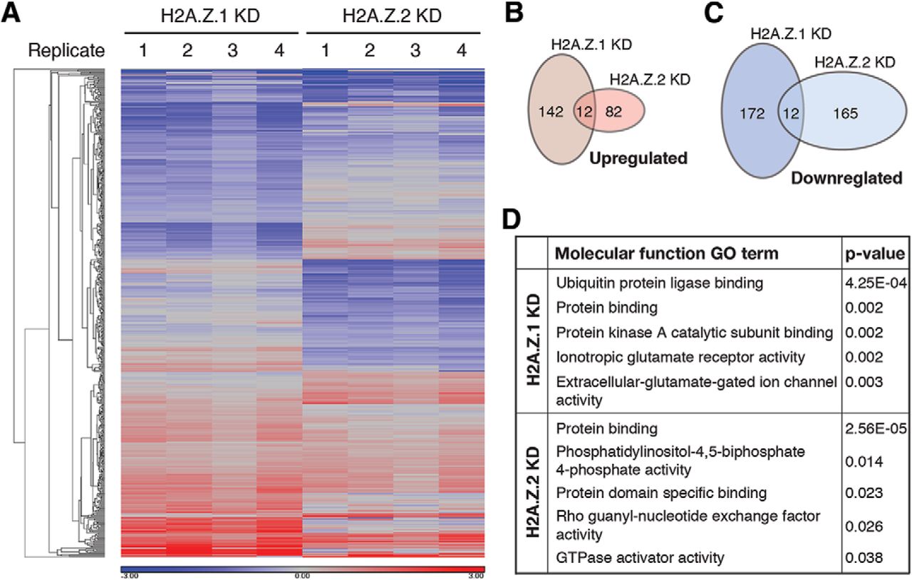
H2A.Z hypervariant depletion alters baseline expression of mostly nonidentical gene cohorts. A, Heat map showing changes in gene expression as detected by microarray analysis after depletion of H2A.Z.1 and H2A.Z.2 compared to control neurons. N = 4. Genes showing >1.25-fold change with p < 0.001 are shown here. B, C, Venn diagrams depicting total number of genes that are either upregulated (B) or downregulated (C) after depletion of H2A.Z.1 or H2A.Z.2. Note the nonoverlapping nature of such gene sets. D, Top five GO terms derived from molecular function gene ontology analysis (DAVID) for H2A.Z.1 and H2A.Z.2 target genes.
- Figure 3.
H2A.Z hypervariant-specific RNAi has synaptic effects. A, B, Amplitude of mEPSCs decrease after H2A.Z.1 or H2A.Z.2 depletion. Shown are example traces in A with quantification of mEPSC amplitude is in B (N = 3; n = 15 neurons for Sc, 10 for H2A.Z.1 KD, and 8 for H2A.Z.2 KD; *p < 0.05 and **p < 0.001). Neurons were infected for 5–7 d. C, mRNA levels of indicated genes, normalized to Gapdh, represented as fold change of their abundance in control neurons (broken line at 1 on y-axis). N = 3–5. *p < 0.05. D, Representative Western blottings of synaptic fraction from control or H2A.Z hypervariant-depleted neurons as indicated. Loading control (β-ACTIN) for individual blots are provided and molecular weights of the nearest protein ladder band is indicated. Arrow indicates quantified SHANK3 band (see text). Neurons were infected for 7 d. N = 3. Note: levels of synapsin, a presynaptic molecule, remain unaltered after hypervariant depletions.
- Figure 4.
H2A.Z hypervariant-specific RNAi has differential effects on activity-induced Arc transcription. A, Graphical representation of Arc mRNA level at 15 min after treatment with Bic + 4AP in indicated neuronal groups as detected by qPCR and normalized to Gapdh. N = 3–4 for each group; *p < 0.05. NS, not significant. B, C, Representation of Arc pre-mRNA level at different time points after treatment with Bic + 4AP (B) or TTX + PMA (C) in indicated groups of neurons (5–6 d postinfection) as detected by qPCR and normalized to Gapdh. N = 3–4 for each time point; *p < 0.05 and **p < 0.01. NS, not significant. Two sets of data in statistical comparison are represented by the position and the color of asterisk(s) or a NS. For example, in B, the green * on top of the 15 min H2A.Z.2 KD time point (red triangle) represents a statistical difference between the means of H2A.Z.2 and H2A.Z.1 KD group (green square) at the same time point. D, E, Representative Western blottings of whole-cell lysate from neurons infected as indicated for 5–6 d. Neurons were treated with Bic + 4AP (D) or TTX + PMA (E) for either 60 min to detect ARC, or for 15 min to detect phosphorylated ERK1/2. F, Representation of Arc pre-mRNA level at 15 min after treatment with Bic + 4AP in indicated neuronal groups as detected by qPCR and normalized to Gapdh. N = 3; **p < 0.01.
- Figure 5.
Distinct properties of HA-H2A.Z.1- and MYC-H2A.Z.2-containing nucleosomes. A, Representative image of immunocytochemical analysis of HA-H2A.Z.1 and MYC-H2A.Z.2 expression in unstimulated neurons. Approximately 70% neurons expressed both hypervariants. Scale bar, 20 μm. B, Co-IP assay from nuclear lysate preparation of neurons exogenously expressing a control vector or HA-H2A.Z.1 and MYC-H2A.Z.2. N = 3. C, Representative image of genomic DNA obtained from untreated or MNase-treated nuclei of neurons treated as described above. The band size of ∼150 bp indicates that MNase digestion predominantly produced mononucleosomes. N = 3. D, Co-IP assay from single nucleosome preparation of neurons exogenously expressing HA-H2A.Z.1 and MYC-H2A.Z.2. Antigen-antibody complex was resolved by electrophoresis and blotted for indicated histones. The 15-kDa protein ladder band is indicated in all panels. Arrow indicates unique band found only in anti-MYC precipitate. N = 3.
- Figure 6.
Both H2A.Z hypervariants are present near the Arc TSS and mediate its transcription. A, Representative Western blotting of whole-cell lysate from neurons infected as indicated for 5–6 d. Neurons were treated with TTX + PMA for 60 min to assess ARC expression. N = 3. B, top, Graphical map (not to scale) depicting relative position of ChIP primers near Arc TSS. Bottom, Quantification of H2A.Z, MYC-H2A.Z.2, and HA-H2A.Z.1 binding to the Arc TSS region in untreated neurons as determined by ChIP with antibodies against H2A.Z, MYC, and HA epitopes. N = 4. C, Quantification of H2A.Z, Ac-H2A.Z, MYC-H2A.Z.2, and HA-H2A.Z.1 binding to Arc TSS region in neurons treated with Bic + 4AP for indicated time periods. N = 4. Data are normalized as indicated and expressed as fold change compared to untreated samples (denoted by the broken line at 1 on y-axis).
- Figure 7.
H2A.Z.2 facilitates priming of the Arc promoter. A, Representative Western blotting of whole-cell lysate showing Ac-H2A.Z levels in neurons depleted of H2A.Z hypervariants as indicated. N = 3. B, Graphical map (not to scale) depicting relative position of ChIP primers near Arc TSS. C, Quantification of Ac-H2A.Z (normalized by total H2A.Z) binding to Arc TSS region in neurons treated as indicated, determined by ChIP. N = 4. Two sets of data in statistical comparison are represented by the position and the color of asterisk(s). D, ChIP data demonstrating binding of RPB1 to the Arc TSS region in control neurons or H2A.Z hypervariant-depleted neurons as indicated. N = 4. Note the enrichment of RPB1 at the TSS (primer 2) in control neurons (black) indicating promoter-proximal RNA Pol II pausing. E, ChIP data demonstrating binding of pSer5-RPB1 to the Arc TSS in control and hypervariant-depleted neurons. N = 3. *p < 0.05 and **p < 0.01.
- Figure 8.
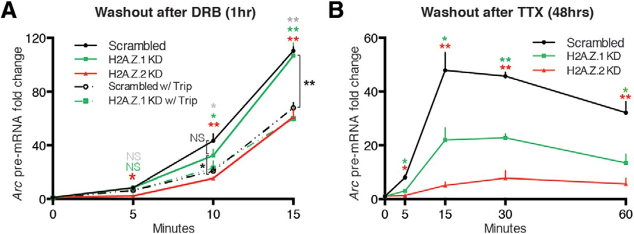
H2A.Z.1 plays context-dependent roles in Arc transcription. A, Graphical representation of the level of Arc pre-mRNA at different time points after 1-h treatment with DRB followed by its washout for indicated time periods in the presence or absence of Trip in indicated groups of neurons (infected for 5–6 d) as detected by qPCR and normalized to Rn18s (18s rRNA; DRB inhibition-insensitive, Pol III-dependent transcription). Gray, green and red asterisk(s) or NS represent statistical comparison between control (Scrambled) responses and responses in Scrambled w/Trip, H2A.Z.1 KD w/Trip and H2A.Z.2 KD, respectively. Connecting brackets delineate other comparisons, shown in black asterisk(s) or NS. B, Similar dataset as in A, except that control neurons or hypervariant-depleted neurons were treated with TTX for 48 h followed by its washout for indicated time periods. Two sets of data in statistical comparison are represented by the position and the color of asterisk(s) or NS. For both A, B, N = 4–5 for each time point; *p < 0.05 and **p < 0.01. NS, not significant.
- Figure 9.
H2A.Z.1 and H2A.Z.2 play context-dependent roles in activity-induced transcription of rapid IEGs. Graphical representation of pre-mRNA levels for noted IEGs at different time points after two treatment regimens. Left, Bic + 4AP treatment. Right, 48-h treatment with TTX followed by its washout for specified time periods in indicated groups of neurons as detected by qPCR and normalized to Gapdh. N = 4–7 for each time point; *p < 0.05 and **p < 0.01. NS, not significant. Two sets of data in statistical comparison are represented by the position and the color of asterisk(s).
- Figure 10.
Effect of H2A.Z hypervariant depletion on transcription of rapid IEGs and nonactivity-induced genes. A, Nanostring multiplexed gene expression data (heat maps) representing pre-mRNA levels in control and hypervariant-depleted neurons treated with Bic + 4AP for indicated times. B, Heat maps representing pre-mRNA levels in control and hypervariant-depleted neurons treated TTX for 48 h followed by washout and collected at indicated times. C, D, Violin plots depicting pre-mRNA abundance of all rapid IEGs from hypervariant-depleted neurons compared to that of control neurons treated with Bic + 4AP (C) or TTX for 48 h followed by washout (D) for indicated times. N = 3 for each treatment all time points. *p < 0.05 and **p < 0.01 (Wilcoxon paired nonparametric test).
- Figure 11.
Effect of H2A.Z hypervariant depletion on transcription of delayed IEGs. A, Nanostring multiplexed gene expression data (heat maps) representing pre-mRNA levels in control and hypervariant-depleted neurons treated with Bic + 4AP for indicated times. B, Heat map representing pre-mRNA levels in control and hypervariant-depleted neurons treated TTX for 48 h followed by washout and collected at indicated times. C, D, Violin plots depicting pre-mRNA abundance of all delayed IEGs from hypervariant-depleted neurons compared to that of control neurons treated with Bic + 4AP (C) or TTX for 48 h followed by washout (D) for indicated times. N = 3 for each treatment at all time points. *p < 0.05 and **p < 0.01 (Wilcoxon paired nonparametric test).


Tables
Target Forward primer Reverse primer Arc pre-mRNA GAATTTGCTATGCCAACTCACGGG AGTCATGGAGCCGAAGTCTGCTTT Btg2 pre-mRNA CTCTCTCTCTTGTTTCCTCCACAG TGTGGTTGATGCGGATACAGCGAT cFos pre-mRNA ACAGCCTTTCCTACTACCATTCCC CTGCACAAAGCCAAACTCACCTGT Cyr61 pre-mRNA ATGTATGAGTTTCAGCGTGTGGCG GTCTGCCTTCTGACTGAGCTGTAA Dusp1 pre-mRNA CTCTACGACCAGGTTAGTAGGAGT ACAGCCGCTTTCTCTATTCTCCCT Dusp6 pre-mRNA TCCTGTGCCTCTCACAAGCTGAAA AACTTACTGAAGCCACCTGCCAGA Fbxo33 pre-mRNA GCATCTACTTGGAGCTGGTGTTGT TCCACGCAAGCCTACCTGTTGTT Gadd45g pre-mRNA ACTCACGGCGCTTGTTCTTTCACA ATTCAGGACTTTGGCGGACTCGTA GAPDH pre-mRNA AACATGCACAGGGTACTTCGAGGA ACGACATACTCAGCACCAGCATCA Npas4 pre-mRNA GTTGCATCAACTCCAGAGCCAAGT ACATTTGGGCTGGACCTACCTTCA Nr4a3 pre-mRNA ATGGAGTGTCAACTGGCTTCTGAG GCCATAAGTCTGCGTGGCATAAGT GAPDH mRNA AGAGACAGCCGCATCTTCTTG GGTAACCAGGCGTCCGATAC H2Afz mRNA GAAGAAAGGACAACAGAAGACTGT CAGCTGTTAAGAGTATTTAGAGTCC H2Afv mRNA ACCCTATGCTCCCGTGTGTTAGAA AGGCAAAGATCAGCACCAACTCTG 18s rRNA CATTCGAACGTCTGCCCTAT GTTTCTCAGGCTCCCTCTCC ANP32e mRNA TCAGAAGTAGGAGAGGGAGAAG CCTTGGAGGGTCTAATCATCATC Symbol Scrambled expression shH2AZ.1 expression log2 fold change p value FDR Ace 4.140032802 5.231924578 1.091891777 2.67E—06 0.049404732 BF565662 3.628967203 5.376799174 1.747831971 1.69E—05 0.065026891 TC524990 8.870590012 10.29163907 1.421049062 1.88E—05 0.065026891 Ccdc23 14.47197211 15.09673068 0.624758569 2.34E—05 0.065026891 RGD1307943_predicted 5.328316737 7.637953753 2.309637017 3.41E—05 0.076745729 RGD1563203_predicted 10.11295573 10.66114527 0.548189542 3.52E—05 0.076745729 AW918709 11.15514358 12.49064688 1.335503299 4.75E—05 0.07711271 Ddx39 11.53226567 12.3812449 0.848979229 5.93E—05 0.07711271 CX570116 8.384790847 8.916443761 0.531652913 6.90E—05 0.07711271 BI293610 9.493734401 9.936922735 0.443188333 7.00E—05 0.07711271 ENSRNOT00000013636 10.29622089 11.25318643 0.956965537 7.45E—05 0.07711271 Slc13a5 8.198920861 9.995278642 1.796357781 7.61E—05 0.07711271 ENSRNOT00000028053 6.936499815 7.576197512 0.639697697 7.67E—05 0.07711271 Ddc 6.457510865 8.216773518 1.759262653 7.75E—05 0.07711271 Hdmcp 6.071940673 7.657559709 1.585619036 8.10E—05 0.07711271 CA339586 11.74191409 12.75911189 1.01719779 9.56E—05 0.07711271 Prlr 3.864580889 4.720040256 0.855459367 9.71E—05 0.07711271 TC533299 10.13831539 10.92955405 0.791238655 9.94E—05 0.07711271 DV722215 4.7082684 6.286053239 1.577784839 0.000100574 0.07711271 TC538999 5.251634416 6.808826043 1.557191627 0.000112895 0.07711271 Ube2i 11.90457203 12.34706984 0.442497806 0.00013949 0.07711271 BF542749 10.74023303 11.68091656 0.940683533 0.000154434 0.07711271 Ext2_predicted 10.8894966 11.22399898 0.334502379 0.000161949 0.07711271 RGD1311752_predicted 8.703882692 10.44148626 1.737603564 0.000169954 0.07711271 TC522315 2.79964542 4.101760357 1.302114937 0.000179121 0.07711271 AA942848 8.500936128 9.102749214 0.601813086 0.000182133 0.07711271 RGD1308031 5.736834927 6.676339719 0.939504792 0.000186685 0.07711271 Raph1_predicted 6.652550515 9.501294881 2.848744366 0.000196584 0.07711271 Col5a3 9.457522664 10.38121689 0.923694229 0.000206023 0.07711271 BF523017 10.85115842 11.3588647 0.507706279 0.000206202 0.07711271 CB547155 9.262431376 9.602202763 0.339771388 0.000218209 0.07711271 Pcsk3 9.563934918 10.34288495 0.778950036 0.000220488 0.07711271 TC545808 8.730436041 9.609236409 0.878800369 0.000220793 0.07711271 Mfap5_predicted 5.818035999 7.453695841 1.635659842 0.000225576 0.07711271 LOC684302 9.817000952 10.28928105 0.472280097 0.000228335 0.07711271 BE111891 1.08420639 1.983656252 0.899449863 0.000230407 0.07711271 Adam3 1.795552474 4.569825212 2.774272738 0.00024164 0.07711271 RGD1309534 8.024579741 8.473932865 0.449353124 0.00025109 0.07711271 XM_224155 1.757920019 4.374588989 2.61666897 0.000254712 0.07711271 Anks1_predicted 4.785255527 5.643215295 0.857959768 0.00025571 0.07711271 TC564875 9.056765795 9.929394592 0.872628797 0.000260742 0.07711271 Zfand2b 10.84791412 11.55774178 0.709827653 0.000270732 0.07711271 CA333998 11.83201426 12.28062694 0.448612678 0.000284527 0.07711271 Mfng 7.749259513 8.266369008 0.517109494 0.000296828 0.07711271 Bambi 8.812389606 9.914938916 1.10254931 0.000302915 0.07711271 XM_346066 1.076776976 2.82468633 1.747909353 0.000311005 0.07711271 Spg21 11.23012332 11.55721656 0.327093237 0.000318552 0.07711271 CO387496 4.453414129 6.536747702 2.083333573 0.000333125 0.07711271 TC554267 8.373468927 9.14689548 0.773426553 0.000346481 0.07711271 Dcn 5.529606864 8.465423531 2.935816667 0.000347064 0.07711271 RGD1309062 6.563975115 8.064163786 1.500188671 0.000352004 0.07711271 Gprk6 12.47356751 13.45420856 0.980641054 0.000358039 0.07711271 RGD1305243_predicted 7.220436673 7.549994374 0.329557701 0.000358398 0.07711271 DV716578 8.728614931 9.434993476 0.706378545 0.00036259 0.07711271 Lipl3_predicted 4.392258287 6.178702783 1.786444496 0.000363005 0.07711271 Mmp24 15.9433976 16.61068899 0.667291392 0.000369777 0.07711271 Tmem77 9.818583769 10.37533496 0.55675119 0.000374996 0.07711271 Ard1_predicted 11.53917153 12.08142809 0.542256562 0.000383345 0.07711271 RGD1566292_predicted 11.21332683 11.62680095 0.413474114 0.000388946 0.07711271 RGD1311331_predicted 7.875456813 8.872995293 0.997538481 0.000400441 0.07711271 BF556192 12.80155585 13.55394357 0.752387717 0.000416696 0.07711271 Ulk1_mapped 8.808173483 9.951782485 1.143609002 0.000418969 0.07711271 XM_341055 6.720993554 7.689316998 0.968323445 0.000434274 0.07711271 Pxn 7.025381997 7.696209005 0.670827008 0.000443884 0.07711271 Prdm7_predicted 8.455624612 9.496578258 1.040953646 0.000449472 0.07711271 Sdcbp2 3.853370711 4.661073334 0.807702623 0.000449476 0.07711271 Nkiras2_predicted 10.80772401 12.34366608 1.535942062 0.000453754 0.07711271 RGD1306939 5.039293136 5.763551483 0.724258347 0.000460339 0.07711271 BF549650 10.67675061 11.62149087 0.944740258 0.000464965 0.07711271 Tcte3_predicted 1.596662365 4.522208185 2.92554582 0.000467652 0.07711271 Rhebl1 12.07802518 12.52188936 0.443864183 0.000468091 0.07711271 RGD1565763_predicted 3.722812164 5.448624313 1.72581215 0.000488696 0.07711271 Setmar 7.220618123 8.453991365 1.233373242 0.000490118 0.07711271 Vwa1 6.169798444 6.711947958 0.542149513 0.000494053 0.07711271 Limk1 13.40359822 14.23252143 0.82892321 0.000495236 0.07711271 Adam24_predicted 1.398107927 3.625631203 2.227523276 0.00049853 0.07711271 DV720603 12.79953832 13.65543137 0.855893047 0.000504802 0.07711271 XM_215391 5.439407969 6.039017881 0.599609912 0.000510659 0.07711271 LOC498368 8.728806947 9.512459784 0.783652838 0.0005142 0.07711271 Zcwpw1_predicted 8.549635796 9.0135438 0.463908005 0.000519295 0.07711271 RGD1306839_predicted 13.69986612 14.17485686 0.474990743 0.000521826 0.07711271 Itga1 8.054333478 8.953630387 0.899296909 0.000524578 0.07711271 AI136665 2.376789645 3.389671123 1.012881477 0.000524619 0.07711271 Pcdh19_predicted 13.08120857 13.962638 0.881429428 0.000533667 0.07711271 BF564180 5.808806457 6.675428768 0.866622311 0.000536071 0.07711271 TC543571 1.274446722 2.996226578 1.721779856 0.000539336 0.07711271 Atp1b3 12.99375325 13.39955207 0.405798822 0.000540772 0.07711271 Kctd10 13.95844395 14.54894225 0.590498302 0.000541089 0.07711271 RGD1565709_predicted 2.770943739 4.053574585 1.282630846 0.000544688 0.07711271 MGC108896 9.927683288 10.78529474 0.857611447 0.00054487 0.07711271 Kctd12_predicted 5.830704506 6.729043191 0.898338685 0.0005464 0.07711271 TC544355 5.942384994 6.836536724 0.894151731 0.000547615 0.07711271 Reps1_predicted 12.54940511 12.97880545 0.429400346 0.000564254 0.077922785 Serpini2 3.851552344 5.63331535 1.781763007 0.000567829 0.077922785 RGD1559697_predicted 7.915361188 8.797601241 0.882240053 0.000592885 0.077922785 RGD1311517 9.050275883 9.381635441 0.331359557 0.000597652 0.077922785 Mobkl2b_predicted 6.180803886 6.909643322 0.728839436 0.00059883 0.077922785 Emp2 6.804079694 7.619840368 0.815760674 0.000601579 0.077922785 Ralgds 11.79767933 12.51812827 0.72044894 0.000605254 0.077922785 AY387076 1.468185811 3.89156566 2.423379849 0.00061619 0.077922785 AA799294 11.50558544 12.18395453 0.678369094 0.000618851 0.077922785 Brms1 11.25655293 12.15467322 0.898120294 0.000619669 0.077922785 Ankrd39_predicted 10.36845557 10.96848401 0.600028443 0.000637893 0.07819106 AI136427 6.037482625 7.335216483 1.297733859 0.000644122 0.07819106 Gjb3 3.57422167 5.651481798 2.077260129 0.000644829 0.07819106 AI501306 3.777011055 5.108158861 1.331147806 0.000653803 0.07819106 AW918392 11.51957268 12.3825602 0.862987521 0.000659521 0.07819106 Nmnat3 5.564449892 6.751676458 1.187226566 0.000661028 0.07819106 CB546810 5.801761173 6.857025975 1.055264802 0.000661036 0.07819106 XM_341150 7.490252385 7.995806521 0.505554136 0.000662575 0.07819106 ENSRNOT00000049215 5.487755572 6.491754516 1.003998945 0.000677502 0.07819106 Sat2_predicted 11.41485986 12.07821741 0.663357553 0.000679078 0.07819106 Sult5a1_predicted 4.028117727 5.018790107 0.99067238 0.000684007 0.07819106 Lifr 4.766105414 5.676759972 0.910654558 0.00068905 0.07819106 Sbk1 7.24921258 8.326023234 1.076810655 0.000691331 0.07819106 AW920693 5.251069432 6.325335641 1.074266209 0.000708009 0.078249597 Bag1_predicted 12.05780626 12.62124582 0.56343956 0.000709335 0.078249597 RGD1565253_predicted 6.764567091 7.373216758 0.608649667 0.000742443 0.07936277 Entpd1 4.234569182 5.684165794 1.449596612 0.000749336 0.07936277 RGD1564089_predicted 8.457456075 8.924711958 0.467255883 0.000750321 0.07936277 Apom 7.669673822 8.452252945 0.782579124 0.000753429 0.07936277 LOC682679 10.40772142 11.04564296 0.63792154 0.000755786 0.07936277 RGD1307465 4.52394686 5.029958008 0.506011148 0.000759693 0.079499818 RGD1560410_predicted 9.611168666 10.1887279 0.577559237 0.000766138 0.079628835 TC523645 7.803548588 8.242734021 0.439185432 0.000774025 0.080124927 TC539202 11.55544897 11.94668132 0.391232348 0.000783413 0.080601813 Centg3_predicted 12.84619462 13.9344075 1.088212888 0.000803447 0.081294469 RGD1311747_predicted 14.02434855 14.43900737 0.414658825 0.000825545 0.082438477 Shank1 10.5815021 11.58272927 1.001227168 0.000834894 0.082830708 Tacstd2 4.848974747 7.909738294 3.060763548 0.000838122 0.082831536 Tm6p1 11.90498256 12.32688801 0.421905453 0.000842078 0.082831536 Epb4.1l4a_predicted 7.708633228 8.084206681 0.375573454 0.000855828 0.082831536 TC539235 12.57769919 12.98429461 0.40659542 0.000857171 0.082831536 Slc14a2 4.278113831 5.499582517 1.221468686 0.000862335 0.082831536 XM_233669 11.02643522 11.3979456 0.371510374 0.000863122 0.082831536 Zcrb1 11.54763267 11.88828262 0.340649953 0.000871627 0.082831536 TC537987 10.44925387 11.35946001 0.910206148 0.000882803 0.083021035 Txnl5_predicted 12.08651224 12.77338822 0.686875981 0.000891118 0.083021035 Mc4r 4.776734293 6.357261766 1.580527472 0.000907649 0.083047388 Mapkapk2 12.24825701 13.41044477 1.162187759 0.000920767 0.083047388 Gpr113_predicted 2.71239918 4.350850097 1.638450917 0.000930512 0.083047388 RGD1563440_predicted 7.495317587 7.96665657 0.471338983 0.000939173 0.083047388 RGD1311484 7.4650719 8.873662257 1.408590356 0.000941116 0.083047388 Kremen1 3.748222464 5.539540629 1.791318165 0.000944467 0.083047388 Ppa2_predicted 14.6818083 15.0607799 0.378971597 0.000954418 0.083047388 Sirt7_predicted 11.76272884 12.11246965 0.349740806 0.000958533 0.083047388 TC562299 6.335779103 6.981390852 0.645611749 0.00096025 0.083047388 TC556681 11.48157966 12.18693567 0.70535601 0.000961791 0.083047388 Rasd2 9.911490969 11.62097176 1.709480787 0.000964146 0.083047388 Hsdl1 9.808903868 10.18720536 0.378301493 0.000966111 0.083047388 LOC500377 5.693389243 8.157926491 2.464537247 0.000966231 0.083047388 Dhrs4 7.792333232 8.478215325 0.685882094 0.000966303 0.083047388 TC562439 6.699930546 8.249882838 1.549952292 0.00097222 0.083047388 Scamp3 11.23002496 11.81910346 0.589078502 0.000984701 0.083582002 - Table 3.
List of DEGs after H2A.Z.1 and H2A.Z.2 KD-part B (downregulated after H2A.Z.1 KD)
symbol scrambled expression shh2az.1 expression log2 fold change p value fdr loc287522 6.259898423 3.781938662 —2.477959761 6.60e—06 0.049404732 rgd1566064_predicted 12.81190937 9.362864495 —3.449044872 7.18e—06 0.049404732 tomm34_predicted 9.80058324 8.124036057 —1.676547183 8.08e—06 0.049404732 ck363699 6.61546991 5.78793267 —0.827537241 1.87e—05 0.065026891 bf562307 9.734906301 8.443322223 —1.291584078 2.29e—05 0.065026891 tc553984 10.57601225 9.76833986 —0.807672386 2.34E—05 0.065026891 AI599250 7.713399206 7.087788209 —0.625610997 3.23E—05 0.076745729 BC098774 7.237548308 5.570550667 —1.666997641 4.77E—05 0.07711271 Grik4 9.223661505 8.523675197 —0.699986308 5.03E—05 0.07711271 Sdhd 14.37797406 13.56563435 —0.812339706 6.16E—05 0.07711271 Napg 10.37368659 9.853267401 —0.520419193 6.42E—05 0.07711271 Mrrf 7.127263566 6.065031172 —1.062232394 8.17E—05 0.07711271 BC091405 10.38125201 9.282285641 —1.09896637 8.74E—05 0.07711271 Tarbp2 12.45655726 12.07159614 —0.384961117 9.39E—05 0.07711271 RGD1309546_predicted 8.534002377 7.502899404 —1.031102973 0.000106301 0.07711271 Grin2c 8.899255961 6.97904754 —1.920208421 0.000106996 0.07711271 TC566987 6.659279818 5.827413166 —0.831866652 0.000108607 0.07711271 TC552897 5.933248175 4.801232308 —1.132015867 0.00010879 0.07711271 Prkar2a 6.163801451 5.313528282 —0.850273169 0.000111853 0.07711271 Glt1d1_predicted 5.121800999 4.103526035 —1.018274964 0.000112311 0.07711271 BF555161 10.59364955 9.470574079 —1.123075469 0.000117617 0.07711271 TC538830 12.53146816 11.24170839 —1.289759763 0.000121745 0.07711271 RGD1566054_predicted 9.115400025 8.101685773 —1.013714251 0.000122074 0.07711271 Lrp11_predicted 13.07487226 11.70424527 —1.370626989 0.000124681 0.07711271 TC536024 7.13278452 6.240473074 —0.892311445 0.000125993 0.07711271 Cited2 10.85269214 8.990111494 —1.862580645 0.000136342 0.07711271 BF397813 7.19066408 5.792890555 —1.397773525 0.000144061 0.07711271 AI112975 6.352713186 5.377218263 —0.975494923 0.000146482 0.07711271 RGD1566367_predicted 5.6358019 5.240674268 —0.395127632 0.00014795 0.07711271 Polr3h_predicted 11.44293632 10.23386016 —1.209076162 0.000150598 0.07711271 Pygb 12.45213506 11.79157662 —0.660558447 0.000154638 0.07711271 AW142634 8.922013872 7.829003261 —1.093010611 0.000156724 0.07711271 BF565628 10.29869369 9.349174788 —0.949518899 0.000157086 0.07711271 Gdap1_predicted 8.365334903 6.792027238 —1.573307664 0.000164033 0.07711271 Ryr2 8.079272174 7.27621553 —0.803056644 0.000164158 0.07711271 BF555945 7.644955812 5.93488388 —1.710071931 0.000166291 0.07711271 Chchd1_predicted 13.92529609 13.52993193 —0.395364161 0.000185585 0.07711271 CB718612 8.141380063 7.317726555 —0.823653508 0.00018998 0.07711271 BF401583 3.807342642 3.135350749 —0.671991893 0.000196042 0.07711271 DV718172 13.52733676 12.61872235 —0.908614414 0.000205766 0.07711271 AW921183 10.34179328 9.467173785 —0.874619492 0.000208601 0.07711271 Atp6v1a1_predicted 10.42042831 10.06973958 —0.350688726 0.000209702 0.07711271 Hexim2_predicted 9.914767395 9.287364476 —0.627402919 0.000237955 0.07711271 Pigh_predicted 6.450865425 5.643206561 —0.807658864 0.000246319 0.07711271 LOC307783 5.032882194 3.665875684 —1.367006509 0.000248578 0.07711271 LOC690163 8.287898848 6.848782046 —1.439116802 0.000252011 0.07711271 RGD1566086_predicted 6.787042716 6.316220869 —0.470821846 0.000252689 0.07711271 TC525615 11.81161754 10.5813283 —1.230289236 0.000256806 0.07711271 Pou3f1 10.95885519 9.374839483 —1.584015704 0.000261757 0.07711271 AW920545 8.612227937 8.006194471 —0.606033466 0.000263301 0.07711271 TC530561 6.361469677 4.977425946 —1.38404373 0.000266017 0.07711271 Vps45 11.18560958 10.64488764 —0.540721933 0.000266506 0.07711271 RGD1565289_predicted 13.51664684 12.46921634 —1.047430504 0.000283049 0.07711271 Klhl20_predicted 8.808515699 7.624582956 —1.183932742 0.000283307 0.07711271 Pdzk8_predicted 8.242171572 7.347607077 —0.894564495 0.000305242 0.07711271 TC560958 11.93185862 11.11468764 —0.817170979 0.000305894 0.07711271 Cops7a_predicted 13.1106929 12.4375094 —0.673183498 0.000306494 0.07711271 TC528997 9.156193399 8.115543858 —1.040649541 0.000308314 0.07711271 RGD1310351_predicted 10.85564614 9.902288125 —0.953358011 0.00031047 0.07711271 Pdcd8 12.75065713 12.03492925 —0.715727881 0.00031379 0.07711271 Ica1 12.17364623 11.6622667 —0.511379533 0.000314358 0.07711271 LOC680426 11.2010015 10.36982724 —0.831174259 0.000315713 0.07711271 DV719080 10.87559452 9.495677787 —1.379916733 0.000318635 0.07711271 TC545343 7.438383629 6.879531146 —0.558852483 0.000332478 0.07711271 Gsta3 12.17020876 10.20865792 —1.961550836 0.00033429 0.07711271 Mfn2 10.55727808 10.01586245 —0.541415627 0.000342094 0.07711271 AABR03068037 7.785306555 7.319303528 —0.466003028 0.000351693 0.07711271 BE113961 10.80779587 9.878175532 —0.92962034 0.00035311 0.07711271 TC519727 11.59791019 10.81753792 —0.780372272 0.000357927 0.07711271 TC528585 7.453045217 6.948322568 —0.504722649 0.000359377 0.07711271 RGD1311345 8.400406852 7.683659105 —0.716747748 0.000361038 0.07711271 TC543044 11.15897338 10.80731226 —0.351661122 0.00036687 0.07711271 TC538360 10.10529859 9.492938508 —0.612360085 0.000369532 0.07711271 RGD1306053 14.18807422 12.78633165 —1.401742574 0.00037825 0.07711271 Tmem55a 10.29421099 9.628109886 —0.666101101 0.000385931 0.07711271 CK478004 4.998539607 4.108978333 —0.889561275 0.000389178 0.07711271 Cog6 8.794988406 7.924249265 —0.870739141 0.000390394 0.07711271 TC544658 11.70378121 11.34008183 —0.363699382 0.000392004 0.07711271 AA957814 7.99801766 6.667451213 —1.330566447 0.000392693 0.07711271 AA851065 10.69658123 9.076743189 —1.619838043 0.000397605 0.07711271 CB545107 10.80488687 9.566124168 —1.2387627 0.000400668 0.07711271 BP503923 11.97203025 10.95266512 —1.019365134 0.000411009 0.07711271 Dnajc8 11.88375963 11.47580858 —0.407951048 0.000412706 0.07711271 Grin2b 7.819443318 6.894573198 —0.92487012 0.000417686 0.07711271 Slc16a11_predicted 10.74873215 9.354260934 —1.394471217 0.000419341 0.07711271 Birc6_predicted 7.745470898 7.220010389 —0.525460509 0.000419662 0.07711271 Mib1_predicted 8.245212972 7.371903929 —0.873309043 0.000423797 0.07711271 TC559433 11.13137332 9.782275241 —1.349098076 0.000423917 0.07711271 AW916157 11.73649748 10.69087852 —1.04561896 0.000426872 0.07711271 AW917472 11.34281752 10.58589712 —0.756920404 0.000435355 0.07711271 Tap1 9.061885637 8.182214145 —0.879671492 0.000440342 0.07711271 AW916613 2.654368574 1.390282852 —1.264085722 0.000444876 0.07711271 Mfsd1_predicted 6.848503926 5.756632868 —1.091871059 0.000445474 0.07711271 AA818474 4.391921753 3.594459458 —0.797462295 0.000446082 0.07711271 TC563567 8.989583387 8.005430887 —0.9841525 0.000455838 0.07711271 RGD1306356 10.99949907 10.38002044 —0.61947863 0.000463089 0.07711271 DV715341 8.050123212 7.619079779 —0.431043434 0.000473159 0.07711271 BF563211 6.051837901 5.373536893 —0.678301008 0.000476289 0.07711271 Rims1 7.088218226 6.595183902 —0.493034323 0.000480843 0.07711271 AI102821 12.8265747 11.10486978 —1.721704924 0.000496254 0.07711271 Umps 10.19219914 9.598751948 —0.593447192 0.000499689 0.07711271 AI175475 6.709628141 5.644117818 —1.065510323 0.00050039 0.07711271 Nek6 8.871652763 7.678760217 —1.192892546 0.000500809 0.07711271 LOC290577 8.796783148 8.098975545 —0.697807603 0.000512747 0.07711271 Epb4.1l1 12.87470645 11.8197883 —1.054918153 0.000513513 0.07711271 AW917204 10.32038964 8.851718495 —1.468671143 0.000518431 0.07711271 Aggf1 8.624603215 7.155915536 —1.468687679 0.00052289 0.07711271 TC524569 8.967339631 8.112362678 —0.854976953 0.000530873 0.07711271 Acsbg1 14.91888151 14.34065365 —0.578227868 0.000532623 0.07711271 TC532756 7.481185857 6.76026886 —0.720916997 0.000537663 0.07711271 AW918535 14.8762029 14.0293993 —0.846803594 0.000543389 0.07711271 TC547881 8.611169112 7.435283572 —1.17588554 0.000544447 0.07711271 TC525657 7.989864402 6.913160036 —1.076704366 0.000558529 0.077922785 Dlst 14.58436665 14.05417512 —0.530191529 0.000568429 0.077922785 BE117446 4.271387612 3.642009885 —0.629377726 0.000571062 0.077922785 TC556147 11.20330191 9.935224997 —1.268076913 0.000571171 0.077922785 Rbm18_predicted 8.265474859 7.532437984 —0.733036875 0.000573185 0.077922785 Chgb 16.35659504 15.63431894 —0.722276099 0.000576277 0.077922785 LOC682205 8.396143751 7.517812838 —0.878330913 0.000576944 0.077922785 Gpr173 6.652605716 5.860519811 —0.792085905 0.000590354 0.077922785 XM_216096 7.832298665 6.566407732 —1.265890933 0.000599328 0.077922785 Ppp6c 9.764572047 9.414556134 —0.350015913 0.000605339 0.077922785 XM_343871 10.90379578 10.24265728 —0.661138501 0.000606072 0.077922785 Fgd4 8.357984576 7.228528075 —1.129456501 0.000610696 0.077922785 CX569250 9.795475601 9.227937407 —0.567538194 0.00061442 0.077922785 Hap1 12.02268819 11.01938568 —1.003302507 0.000616681 0.077922785 AW915320 9.170075196 8.417656657 —0.752418539 0.000618451 0.077922785 Rragd_predicted 10.70444865 9.093806459 —1.610642189 0.000619331 0.077922785 TC541972 10.34640635 9.316275627 —1.030130724 0.00063095 0.07819106 AI230360 7.918189901 7.234716989 —0.683472912 0.000642562 0.07819106 Igf2r 13.17561599 12.47214529 —0.703470695 0.000648788 0.07819106 DV719617 10.27750625 9.107724317 —1.169781934 0.000651766 0.07819106 Mrpl18_predicted 12.51441646 11.58262484 —0.931791612 0.000662315 0.07819106 TC568517 7.18069051 6.439772542 —0.740917968 0.000674076 0.07819106 Adsl_predicted 12.53997034 11.84135281 —0.698617527 0.000679503 0.07819106 TC519890 15.20193502 13.84576879 —1.356166231 0.000682539 0.07819106 Spg20 8.308415518 7.58305852 —0.725356998 0.000686525 0.07819106 AI170696 6.942817153 6.027001338 —0.915815815 0.00069311 0.07819106 Ube2v2 9.661199706 8.572804044 —1.088395662 0.000693451 0.07819106 LOC498295 12.20451881 11.83959221 —0.364926599 0.000698494 0.078249597 TC560921 5.207732953 4.297097796 —0.910635157 0.000700069 0.078249597 BF555924 9.985597821 9.469135844 —0.516461977 0.000702983 0.078249597 Pfkm 14.51654843 14.05002374 —0.466524695 0.000708505 0.078249597 TC564512 8.333396639 7.32224411 —1.011152528 0.000721932 0.07899534 LOC500295 9.420651641 8.540467639 —0.880184003 0.000724825 0.07899534 Sf3b2_predicted 12.44354911 11.6085621 —0.83498701 0.000725793 0.07899534 BF523428 13.15045896 12.09237333 —1.058085628 0.000726436 0.07899534 Zfp297b 8.14767076 7.796674244 —0.350996515 0.000734886 0.07936277 AA998677 6.022499947 5.66326418 —0.359235767 0.000738839 0.07936277 Commd10 13.07110695 12.22251811 —0.848588842 0.000741977 0.07936277 Usp13_predicted 8.880457276 8.301686706 —0.57877057 0.000751682 0.07936277 TC539690 11.53252368 10.19001348 —1.342510194 0.000754935 0.07936277 AW142620 10.54231861 9.200592376 —1.341726234 0.000763691 0.079628835 Keap1 12.00883633 11.60287219 —0.405964134 0.000792061 0.081218196 Gsk3b 10.9550178 10.01148948 —0.943528324 0.000797188 0.081294469 RGD1310192_predicted 10.30348412 9.743106299 —0.560377818 0.000800586 0.081294469 AW919892 9.009219476 8.398195424 —0.611024052 0.00080164 0.081294469 CX570570 9.009092379 8.150761251 —0.858331128 0.00081345 0.081955526 CO403204 7.341185431 6.396430715 —0.944754716 0.000815344 0.081955526 DV727788 9.546149034 8.506229032 —1.039920002 0.000820046 0.082157822 Crebl2 8.237847284 7.059695143 —1.178152141 0.000832899 0.082830708 RGD1566078_predicted 15.4991444 14.43680942 —1.062334982 0.000843282 0.082831536 TC536252 6.791496764 6.081404474 —0.71009229 0.000848504 0.082831536 RGD1310383_predicted 9.675442727 8.875211356 —0.800231371 0.000854032 0.082831536 Ndrg2 16.92933608 16.23958962 —0.689746461 0.000855608 0.082831536 AA819653 6.401981606 5.54072971 —0.861251897 0.000864825 0.082831536 Nr1d2 4.102851341 2.907566815 —1.195284526 0.000870343 0.082831536 AI030552 6.242106917 4.813012178 —1.429094739 0.000872853 0.082831536 TC557120 11.58985733 11.08039255 —0.509464782 0.000878637 0.083021035 CB547657 7.11769739 5.41137132 —1.706326069 0.000885415 0.083021035 TC551191 7.699104736 6.244599038 —1.454505698 0.000887765 0.083021035 Aytl2_predicted 7.999183956 7.427312229 —0.571871727 0.000891151 0.083021035 Ddef2_predicted 7.639303041 7.13151159 —0.507791451 0.000898719 0.083047388 Ratsg2 11.68645688 11.30312412 —0.383332762 0.000913048 0.083047388 ENSRNOT00000043649 12.10622909 11.2676376 —0.838591496 0.00093425 0.083047388 Slc4a4 10.52371038 9.660948829 —0.862761553 0.000935657 0.083047388 Copb2 12.96384037 12.52074638 —0.443093992 0.000945107 0.083047388 Plekhc1 9.749091344 9.407128148 —0.341963196 0.000946928 0.083047388 RGD1561030_predicted 5.157115548 4.180877924 —0.976237624 0.000950336 0.083047388 TC525601 13.50942052 12.9991972 —0.510223326 0.000950464 0.083047388 Phf7 8.191447181 7.356775616 —0.834671565 0.000953909 0.083047388 BQ782721 4.908138931 4.310952068 —0.597186864 0.000963823 0.083047388 AA925274 5.579121622 5.090008894 —0.489112728 0.000969917 0.083047388 DV727304 11.16319859 10.59010722 —0.573091369 0.000975685 0.083047388 Symbol Scrambled expression shH2AZ.2 expression log2 fold change p value FDR Ahcyl1_predicted 11.30812916 15.70257541 4.394446257 1.55E—09 4.75E—05 Mcm6 10.83536001 11.40618674 0.570826728 8.24E—06 0.035135455 Dut 11.35492304 11.9535574 0.598634355 3.46E—05 0.066139598 Timp2 15.05728576 15.80164866 0.744362903 3.81E—05 0.067378588 RGD1311752_predicted 8.703882692 10.33537822 1.631495533 4.06E—05 0.067378588 Zcwpw1_predicted 8.549635796 9.169491306 0.619855511 4.50E—05 0.067378588 XM_345006 6.85771805 7.739541418 0.881823368 4.87E—05 0.067378588 TC549858 11.27224616 11.75359808 0.481351913 4.91E—05 0.067378588 Hrasls_predicted 1.352310448 2.473461424 1.121150976 5.02E—05 0.067378588 BF289520 5.430397248 6.059021678 0.62862443 7.69E—05 0.071367889 Pold3 10.06736193 10.48690247 0.419540544 7.71E—05 0.071367889 RGD1307357_predicted 11.2118082 11.5384726 0.326664395 7.97E—05 0.07161003 Gtf3c5_predicted 7.76009444 8.355130115 0.595035675 9.19E—05 0.073611113 Bcl7c_predicted 11.52854784 12.20876021 0.68021237 0.000109679 0.073611113 Pomc 8.615004399 9.436951714 0.821947315 0.000109978 0.073611113 RGD1565291_predicted 12.39574606 12.90176243 0.506016372 0.000117613 0.074873201 BF542749 10.74023303 12.36932877 1.629095739 0.000127234 0.077757776 Abcc2 8.819186435 9.789025783 0.969839348 0.000139329 0.080329614 LOC363188 5.75509275 8.424133782 2.669041033 0.000147266 0.083333563 Cecr5_predicted 10.95802065 11.33987144 0.381850792 0.000176782 0.08855644 AA955618 9.344852039 9.669319187 0.324467148 0.000209302 0.09181501 AI044097 5.588998691 6.267474587 0.678475896 0.000221456 0.092025775 RGD1563319_predicted 8.12622631 9.457387834 1.331161525 0.000236732 0.092741286 Tbc1d2b 6.869829194 8.039756988 1.169927794 0.000255978 0.095023281 ENSRNOT00000027501 9.948158576 10.60865914 0.660500565 0.000307587 0.095557268 RGD1304587 12.28837897 12.94341986 0.655040885 0.000314183 0.095557268 TC542265 9.893597595 10.95071148 1.05711388 0.000351139 0.095557268 BM384088 9.56848326 10.08580713 0.517323868 0.000375917 0.095557268 BG668164 9.868977302 10.4107215 0.541744198 0.000393414 0.095557268 Centg3_predicted 12.84619462 13.64912229 0.802927674 0.000397418 0.095557268 Rfc2 10.69628045 11.25875805 0.562477599 0.000401566 0.095557268 Slc26a7_predicted 4.035522477 6.194273087 2.15875061 0.000404596 0.095557268 Fn1 8.587931475 9.897009695 1.30907822 0.00041856 0.095557268 Fxyd6 15.52204053 16.22851939 0.706478863 0.000424885 0.095557268 Ezh1_predicted 13.35214855 13.9436327 0.59148415 0.000436061 0.095557268 DV719732 9.123092282 9.737560979 0.614468697 0.000443541 0.095557268 B4galt7 12.13569399 12.61112377 0.475429774 0.000458424 0.095557268 Lzts2 10.13285103 10.82559939 0.692748367 0.000469953 0.095557268 Src 9.695578825 10.36460234 0.669023515 0.000512904 0.095557268 BF556192 12.80155585 13.64456502 0.84300917 0.000526513 0.095557268 RGD1564308_predicted 8.53728578 9.316997749 0.77971197 0.00053388 0.095557268 AW915160 11.81695687 12.84151282 1.024555952 0.000534083 0.095557268 BF289433 4.810359467 5.442835512 0.632476045 0.000539006 0.095557268 LOC312502 9.813073542 10.28343955 0.470366013 0.000543607 0.095557268 Slc35b4_predicted 10.07187502 10.57533921 0.503464186 0.000547104 0.095557268 Tcn2 8.702856424 9.597660836 0.894804412 0.000548566 0.095557268 Rabl4_predicted 12.99354632 13.58209364 0.588547326 0.000558303 0.095557268 Astn1 7.130219184 7.783368618 0.653149434 0.000564477 0.095557268 Slitrk1_predicted 11.38978024 11.86215613 0.47237589 0.000565343 0.095557268 LOC678926 1.529149853 3.301916023 1.77276617 0.000568879 0.095557268 Sfmbt1 10.57247665 11.52524221 0.952765564 0.000576444 0.095557268 Nfya 7.339752471 8.230520214 0.890767743 0.000594435 0.095557268 CA503664 8.706063302 9.30753056 0.601467258 0.000612386 0.095557268 CO398332 2.088031423 3.465147675 1.377116252 0.000617647 0.095557268 Ankrd5_predicted 0.986066003 1.893556424 0.907490421 0.000618681 0.095557268 RGD1561903_predicted 8.073236386 9.261517734 1.188281348 0.000624827 0.095557268 Nav1_predicted 9.311359188 10.05118484 0.739825648 0.000627604 0.095557268 Lonrf1_predicted 6.665069586 8.231081607 1.566012021 0.000637379 0.095557268 LOC291840 10.46425437 10.98618406 0.521929682 0.00064038 0.095557268 BF555594 7.297901913 7.803911007 0.506009094 0.000642572 0.095557268 Prcp_predicted 6.945396355 7.628166927 0.682770572 0.000650274 0.095557268 Cggbp1_predicted 10.10446551 10.67233084 0.567865332 0.000651252 0.095557268 LOC686668 14.22879788 14.64664596 0.417848086 0.000653103 0.095557268 Shank1 10.5815021 11.85728804 1.275785942 0.000653256 0.095557268 BI293610 9.493734401 9.933127533 0.439393131 0.000667597 0.095557268 RGD1308637 10.63111621 10.99277702 0.361660808 0.000669694 0.095557268 LOC683646 14.6252591 15.00216313 0.376904029 0.000672344 0.095557268 TC562439 6.699930546 8.729363905 2.029433359 0.00068664 0.095625151 RGD1566063_predicted 10.42408783 10.82360645 0.399518627 0.000691139 0.095625151 XM_214416 9.178834112 9.6187083 0.439874188 0.000699314 0.095625151 RGD1308557_predicted 7.420320921 8.285465612 0.865144691 0.000733081 0.09725562 RGD1566292_predicted 11.21332683 11.71063736 0.49731053 0.000751031 0.097309775 RGD1305166_predicted 9.134297704 9.759187045 0.624889341 0.000751656 0.097309775 Nkiras2_predicted 10.80772401 11.77210764 0.964383627 0.000753238 0.097309775 Cybasc3 7.043310663 7.609593274 0.566282611 0.000754734 0.097309775 Mrpl36_predicted 11.89399742 12.33502107 0.441023645 0.000782062 0.098010329 Gprk2l 8.779110352 9.38417973 0.605069378 0.00080672 0.098999718 LOC686766 5.260876485 5.911787655 0.65091117 0.000811953 0.099243336 Phf22 7.538016027 8.324087108 0.786071082 0.000836521 0.101357181 RGD1306143_predicted 6.850673885 7.335015831 0.484341946 0.00084597 0.101533717 ENSRNOT00000048826 12.49598977 12.94226435 0.446274573 0.000847305 0.101533717 Znf579_predicted 11.20811184 11.67550922 0.467397383 0.000856332 0.10181686 Hsdl1 9.808903868 10.14043298 0.331529114 0.000883112 0.102944694 Gprk6 12.47356751 13.25494457 0.781377066 0.0008894 0.102944694 Hsd17b2 3.414854355 5.901570573 2.486716218 0.000943101 0.104035619 Rnf8 12.29803729 12.87630368 0.578266392 0.000952234 0.104035619 CA510534 11.34797575 12.80389432 1.455918574 0.000958972 0.104035619 Arl2 10.91134235 11.45372552 0.542383164 0.000971266 0.104035619 BF550209 12.37525589 12.8730042 0.497748308 0.000974014 0.104035619 BG665133 11.20679244 12.33422623 1.127433798 0.000979781 0.104035619 LOC691075 9.060232543 9.478672438 0.418439896 0.000980214 0.104035619 RGD1308535_predicted 9.816766494 10.28292832 0.46616183 0.000993396 0.104035619 Zswim5_predicted 5.470774971 6.301634314 0.830859343 0.000996038 0.104035619 LOC690354 10.40224677 10.7558671 0.353620329 0.000997294 0.104035619 - Table 5.
List of DEGs after H2A.Z.1 and H2A.Z.2 KD-part D (downregulated after H2A.Z.2 KD)
Symbol Scrambled expression shH2AZ.2 expression log2 fold change p value FDR Rnf14 16.57094576 12.9463825 —3.624563259 1.91E—08 0.000291288 RGD1359108 13.30079953 7.879686571 —5.421112956 4.85E—08 0.000494164 Inpp5f_predicted 4.824671522 3.588112893 —1.236558629 1.13E—06 0.008652283 Cfl2_predicted 10.28475755 8.941614741 —1.343142814 5.78E—06 0.033552658 Tex2 6.820637729 5.774872776 —1.045764953 6.59E—06 0.033552658 Lypla3 10.13489074 9.305455152 —0.829435583 9.20E—06 0.035135455 Sbds 10.29127274 9.876346931 —0.414925805 1.21E—05 0.041035544 Poldip2_predicted 11.21978549 10.11515746 —1.104628032 1.36E—05 0.041433838 RGD1564626_predicted 7.402821848 6.749656546 —0.653165302 1.94E—05 0.05398694 Chchd3_predicted 13.15554854 11.50732751 —1.648221032 2.44E—05 0.062079366 Wee1 7.228033394 5.956374529 —1.271658865 2.65E—05 0.06223942 U78138 8.370979606 6.934655598 —1.436324008 3.15E—05 0.066139598 XM_343572 12.87985279 12.26365661 —0.616196176 3.39E—05 0.066139598 AI231051 5.91510764 4.336231214 —1.578876425 5.07E—05 0.067378588 Gad2 7.493926937 5.159298815 —2.334628122 5.51E—05 0.06840792 Tmem55a 10.29421099 9.510629962 —0.783581025 5.80E—05 0.06840792 Lsm14a_predicted 8.399662619 7.415627465 —0.984035154 6.05E—05 0.068497749 LOC300429 6.310030902 5.753092672 —0.556938229 6.85E—05 0.071367889 Sdcbp2 3.853370711 3.05190314 —0.801467571 6.94E—05 0.071367889 LOC691853 9.783324367 8.799255704 —0.984068662 7.16E—05 0.071367889 XM_215270 10.17499295 9.701596351 —0.4733966 7.43E—05 0.071367889 TC538215 9.067975732 7.252392924 —1.815582808 8.86E—05 0.073611113 BG663025 6.859758488 5.74663244 —1.113126048 8.89E—05 0.073611113 TC553657 8.723092234 8.029121219 —0.693971015 9.42E—05 0.073611113 LOC684322 15.44820244 14.34666049 —1.101541943 9.84E—05 0.073611113 Usp32_predicted 10.25821098 9.517372161 —0.74083882 0.000100946 0.073611113 ENSRNOT00000008889 8.21062512 6.810763402 —1.399861718 0.000104197 0.073611113 Rb1 8.11762273 6.908923008 —1.208699722 0.000105314 0.073611113 AA849985 6.116135406 5.241886237 —0.874249169 0.000110254 0.073611113 AW917590 14.02219537 12.7498481 —1.272347268 0.000111886 0.073611113 CB547899 7.72064826 7.029262406 —0.691385855 0.000113222 0.073611113 U78132 7.279222721 5.846742231 —1.43248049 0.000121025 0.075472833 Mcf2l 9.397430132 8.657675863 —0.739754268 0.00013013 0.077968283 DV713600 13.15808683 11.11117444 —2.046912388 0.00013701 0.080329614 TC539285 9.30257925 8.414415448 —0.888163802 0.000151429 0.084131469 BF555161 10.59364955 9.409913061 —1.183736486 0.000167666 0.087621405 Cdk5rap1 10.22213249 9.608946578 —0.61318591 0.000168425 0.087621405 CO399145 7.567286017 6.640966202 —0.926319815 0.000169575 0.087621405 BF544403 7.874729806 6.432614592 —1.442115214 0.000172048 0.087621405 RGD1308147 9.865899439 8.842423951 —1.023475489 0.000183836 0.090604523 Arl10 7.253170158 6.41752538 —0.835644778 0.000195782 0.09181501 Ddn 11.23443915 9.493619755 —1.740819393 0.00019651 0.09181501 AW535924 2.716704964 1.208805749 —1.507899216 0.000200473 0.09181501 AW917511 7.921316801 7.098393914 —0.822922887 0.000207115 0.09181501 Atxn1 7.381331792 6.174416218 —1.206915574 0.000210514 0.09181501 Eaf2 5.106563436 4.649076352 —0.457487084 0.000211747 0.09181501 TC557676 7.472657949 6.19779289 —1.27486506 0.000212226 0.09181501 Ttc12 7.900994459 7.523648839 —0.37734562 0.000213335 0.09181501 TC560921 5.207732953 4.559434897 —0.648298056 0.000217244 0.092025775 AI112975 6.352713186 5.466730174 —0.885983012 0.000228285 0.092025775 Vil2 9.860779156 8.967669681 —0.893109474 0.000228882 0.092025775 AA875008 5.494818563 4.700887004 —0.793931559 0.000235511 0.092741286 TC528454 8.35589654 6.885831687 —1.470064854 0.000247959 0.09493033 Hspca 14.42409811 13.70998635 —0.714111765 0.00025058 0.09493033 AI059618 4.100124355 1.829444923 —2.270679431 0.00025164 0.09493033 Rnf11_predicted 10.2186121 9.615662304 —0.602949799 0.000258106 0.095023281 Pole3 12.55132532 11.31822759 —1.233097729 0.000262232 0.095273647 RGD1564451_predicted 9.713981828 8.589317234 —1.124664593 0.000265021 0.095273647 LOC683512 10.77887127 9.314774611 —1.464096657 0.000269989 0.095557268 Stx6 12.20386283 11.83254524 —0.371317584 0.000275093 0.095557268 TC539851 11.07018722 9.38957993 —1.680607294 0.0002773 0.095557268 RGD1308581_predicted 11.33094265 10.731295 —0.599647641 0.000301272 0.095557268 RGD1305052_predicted 12.11367655 10.94947524 —1.164201304 0.000303392 0.095557268 Pde4a 9.320418187 8.426565098 —0.893853089 0.000307772 0.095557268 RGD1565785_predicted 3.454975278 1.716774529 —1.738200749 0.000308741 0.095557268 TC551503 10.21507032 9.012702885 —1.202367432 0.0003089 0.095557268 Sdhd 14.37797406 13.64470284 —0.733271224 0.00031574 0.095557268 AW144312 10.73124208 8.983532052 —1.747710029 0.000316237 0.095557268 AW144351 9.197071628 7.899938415 —1.297133212 0.000317995 0.095557268 AI599930 7.678810398 7.029318808 —0.64949159 0.0003243 0.095557268 Ssu72 10.81988308 10.15157164 —0.66831144 0.000326611 0.095557268 MGC125215 13.54599663 12.92503148 —0.620965157 0.000330105 0.095557268 TC524569 8.967339631 8.237038714 —0.730300917 0.000331746 0.095557268 Inpp4a 8.581584747 7.638446802 —0.943137945 0.000332111 0.095557268 TC540740 12.032179 11.48000894 —0.552170063 0.000338226 0.095557268 TC550497 12.02106544 11.01957227 —1.001493172 0.00034439 0.095557268 XM_580045 8.850520618 7.850512112 —1.000008505 0.0003564 0.095557268 XM_236784 11.16551643 10.17544134 —0.990075092 0.000365511 0.095557268 AW915589 9.355873567 7.887143897 —1.46872967 0.000371323 0.095557268 Garnl1 6.899821493 6.075097502 —0.824723991 0.00038265 0.095557268 H33079 11.05742197 10.63726213 —0.420159837 0.000383244 0.095557268 LOC689577 10.30988195 9.693679501 —0.616202449 0.000385184 0.095557268 LOC683630 9.849902817 9.088044225 —0.761858591 0.000385627 0.095557268 Arpc5 14.5853404 13.66460753 —0.920732865 0.000387329 0.095557268 RGD1305797_predicted 5.300372467 4.356613034 —0.943759433 0.000389433 0.095557268 TC546991 7.224586672 5.935472523 —1.289114149 0.000393929 0.095557268 TC538360 10.10529859 8.83610561 —1.269192983 0.000408086 0.095557268 Gdi2 11.96894942 10.24844069 —1.720508726 0.00041582 0.095557268 BM391850 6.763729833 6.106887394 —0.656842439 0.000416848 0.095557268 Habp4_predicted 12.75635898 12.10459874 —0.651760235 0.000419683 0.095557268 TC541875 10.06553783 9.450612436 —0.614925399 0.000440675 0.095557268 DV718172 13.52733676 12.71122856 —0.816108202 0.000445019 0.095557268 Pgam5 8.963640697 8.159415155 —0.804225543 0.0004591 0.095557268 TC557253 11.21445231 9.296227403 —1.918224906 0.000461312 0.095557268 Gs3 9.481151768 8.642831162 —0.838320607 0.000463394 0.095557268 LOC681501 11.05409438 10.18583791 —0.868256472 0.000473823 0.095557268 BF281819 13.04857617 11.8092362 —1.239339965 0.000477166 0.095557268 CX569261 12.28234494 10.68729318 —1.595051767 0.000484223 0.095557268 CK596627 11.19585087 10.35777472 —0.838076159 0.000485026 0.095557268 Eif2c1_predicted 8.545178383 8.070863133 —0.47431525 0.000487557 0.095557268 DV726726 11.92676689 11.51677995 —0.409986941 0.000496554 0.095557268 Nefh 8.341691009 6.745856633 —1.595834376 0.000498525 0.095557268 Fahd1 9.969599036 9.383759066 —0.585839969 0.000500438 0.095557268 Tacc1 9.627935629 8.305618957 —1.322316672 0.000502046 0.095557268 Ppp3r1 9.456090327 8.882689108 —0.573401219 0.000504727 0.095557268 AW920896 8.970038953 8.300730935 —0.669308018 0.000505679 0.095557268 Atp6v1h 8.289085697 7.934631332 —0.354454365 0.000506174 0.095557268 LOC310395 10.4119975 9.308354087 —1.103643409 0.000510548 0.095557268 Dcbld2 6.201411422 5.130397482 —1.07101394 0.000512876 0.095557268 Hspa9a_predicted 11.06428449 9.911168244 —1.153116244 0.000529115 0.095557268 TC563075 7.096430274 5.632571768 —1.463858506 0.000532538 0.095557268 TC562837 7.37169182 5.1955973 —2.17609452 0.000536894 0.095557268 Mkks 9.065711002 8.556237021 —0.509473981 0.000544585 0.095557268 Mapk15 5.821049535 5.019106294 —0.801943241 0.000545662 0.095557268 Snx27 9.577525457 8.670999828 —0.906525629 0.000550679 0.095557268 TC523914 5.475100961 4.273483854 —1.201617107 0.000559079 0.095557268 AI029023 12.72038418 10.95540125 —1.764982934 0.000577539 0.095557268 Abcg3 5.995608197 4.88764231 —1.107965887 0.000580682 0.095557268 TC540837 7.653046158 6.293159397 —1.359886761 0.000583829 0.095557268 CB718612 8.141380063 7.502377182 —0.639002881 0.000586497 0.095557268 CB547822 7.48574771 7.056202285 —0.429545426 0.000586655 0.095557268 Cxxc6_predicted 5.461267196 3.727894207 —1.733372989 0.000596532 0.095557268 AW917678 8.008522785 7.106552387 —0.901970397 0.000598751 0.095557268 Dlgap2 6.739059513 6.048951702 —0.690107811 0.000612053 0.095557268 RGD1305269_predicted 10.48363487 9.989398981 —0.494235885 0.000618636 0.095557268 TC543111 5.988905601 5.175476656 —0.813428946 0.000624841 0.095557268 AW915587 12.81417625 12.0617611 —0.752415147 0.000629005 0.095557268 LOC302495 8.838373315 6.8520573 —1.986316015 0.000632419 0.095557268 Arhgef9 10.96945893 10.06784789 —0.901611047 0.000635486 0.095557268 Atp5b 15.39132483 15.05979828 —0.331526546 0.000641824 0.095557268 AI044115 4.38279618 3.418203792 —0.964592388 0.000651088 0.095557268 TC523083 6.820128764 6.144807662 —0.675321103 0.000654296 0.095557268 Rab11fip4_predicted 8.321833638 7.268975447 —1.052858191 0.000661426 0.095557268 TC523910 7.889138456 6.19649015 —1.692648306 0.000661665 0.095557268 BE108224 3.824896985 1.37170849 —2.453188495 0.000669059 0.095557268 LOC499900 9.327866735 8.709208418 —0.618658317 0.000670927 0.095557268 TC537999 6.001132395 4.678122045 —1.32301035 0.000676124 0.095625151 RGD1308745_predicted 4.568246264 3.179251688 —1.388994575 0.000687651 0.095625151 LOC688553 3.191286936 1.693084684 —1.498202253 0.000696309 0.095625151 Morn1 7.972102174 6.856300055 —1.115802119 0.000699611 0.095625151 TC530505 4.769710018 3.940413803 —0.829296214 0.000710803 0.096012504 BF396589 4.318891305 3.588142974 —0.730748331 0.000713252 0.096012504 CO554673 9.464355537 8.372142043 —1.092213494 0.000729299 0.09725562 TC543488 5.436948213 2.78354383 —2.653404383 0.000730873 0.09725562 TC532226 6.929504114 4.480025699 —2.449478415 0.000735218 0.09725562 TC566921 8.943896089 7.990664073 —0.953232016 0.000747355 0.097309775 AW914885 10.02209083 9.545298515 —0.476792314 0.000763893 0.097903454 BF556147 7.287577236 6.440436939 —0.847140297 0.000765747 0.097903454 Trim2 12.92583443 12.5169377 —0.408896732 0.000769469 0.097969457 Gpr158_predicted 6.695389757 5.535821733 —1.159568025 0.000782843 0.098010329 BG666117 12.11075957 11.62393319 —0.486826385 0.000784673 0.098010329 TC541992 7.227521264 6.312830078 —0.914691186 0.000785637 0.098010329 TC525657 7.989864402 7.075628128 —0.914236274 0.000787993 0.098010329 TC560235 8.131101996 7.601540913 —0.529561083 0.000804594 0.098999718 LOC685076 11.93684157 10.40645356 —1.53038801 0.000818584 0.099655287 AW143088 11.05925856 10.17888615 —0.88037241 0.000852562 0.101764579 TC537691 11.03421255 10.1832052 —0.851007351 0.000872934 0.102944694 Clcn2 10.18273395 9.654510577 —0.528223376 0.000877455 0.102944694 DV719080 10.87559452 9.843234019 —1.032360501 0.000877857 0.102944694 DY471780 7.546202473 6.688303944 —0.857898528 0.000885155 0.102944694 Ppm1e 8.969105775 7.850748852 —1.118356922 0.000904556 0.103853197 TC538748 11.28808193 10.56735898 —0.720722956 0.000907445 0.103853197 AI170363 4.98690796 4.240445668 —0.746462292 0.000917269 0.104035619 TC523584 8.251731137 7.876311518 —0.375419619 0.000921387 0.104035619 AI145492 4.960957943 3.550461441 —1.410496501 0.000927701 0.104035619 CF109839 7.043330761 6.236742364 —0.806588397 0.000929916 0.104035619 TC559053 8.399715145 7.440335331 —0.959379814 0.000932371 0.104035619 AB040488 4.509816182 3.935696141 —0.574120041 0.000936815 0.104035619 CB544681 7.403628364 6.79873552 —0.604892844 0.000949976 0.104035619 Arhgef11 8.620841844 8.014495747 —0.606346097 0.00096325 0.104035619 AA800571 8.989335176 8.145442592 —0.843892585 0.000970797 0.104035619 AA965181 6.11182251 4.75033957 —1.36148294 0.000977883 0.104035619 Ripk5 6.33665685 5.821877894 —0.514778956 0.000978231 0.104035619 Phka1 5.544744433 4.356297924 —1.188446509 0.000984497 0.104035619 TC524191 12.34462962 11.67308416 —0.671545452 0.00098735 0.104035619 BG663084 9.722716915 8.835969466 —0.886747449 0.000995922 0.104035619 BP503923 11.97203025 10.96018715 —1.011843108 0.00099756 0.104035619
In this issue


