

Article Figures & Data
Figures
- Figure 1.
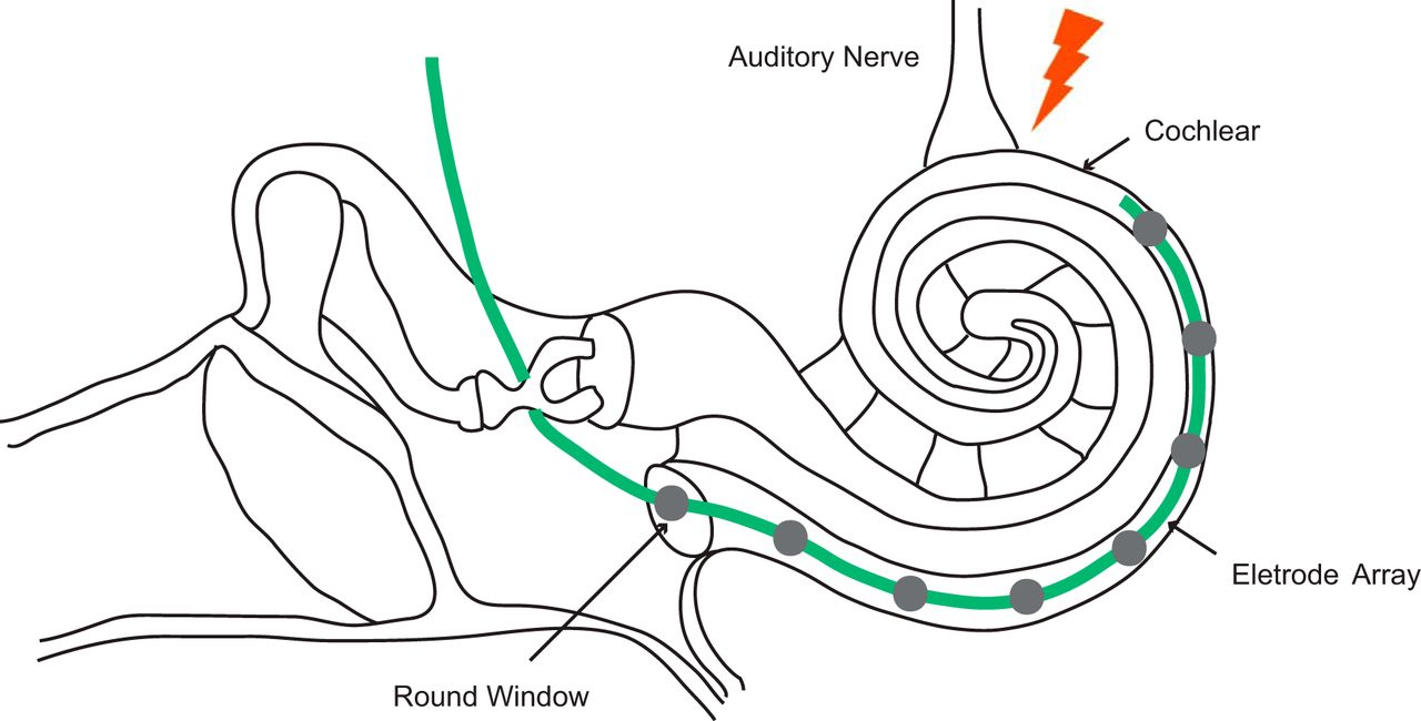
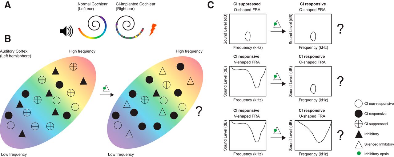
A, The setup used in Johnson et al., 2016. A CI implant with eight electrodes is implanted in the right ear, and the left ear is left acoustically intact. The perception of different frequencies is elicited by stimulating different electrodes along the tonotopic axis in the cochlea (color gradient). This strategy allows Johnson et al. to measure the response of single neurons in the left auditory cortex to both acoustic and CI stimulation in an awake marmoset, and thereby to examine the characteristics of neurons that respond, or fail to respond, to CI stimulation. Acoustic and CI stimuli were matched whenever possible. For CI stimulation, the response of a neuron is tested across different electrode positions and at multiple current levels, and analogously for acoustic stimulation, across a range of frequencies and at multiple sound levels. The CI electrode/frequency producing the significantly largest firing rate response is defined as the best electrode or best frequency of the neuron, for CI and acoustic stimulation, respectively. The receptive field of a neuron is described by electrode/frequency tuning curves across all current/sound levels. B, Hypothesis: interneurons, particularly PV interneurons, are important for effective cortical response to CI stimulation. Left, CI stimulation is surprisingly inefficient in activating A1 neurons (black circles) because many neurons are suppressed (crossed circle) by inhibitory interneurons (filled triangles). Right, decreasing inhibitory GABAergic interneuron activity using either optogenetics or pharmacology will increase the effectiveness of CI stimulation, and in particular, likely yield more O-shaped neuron activity either from previously suppressed (top) or evolved from V-shaped neurons (middle; see text). However, decreased GABAergic inhibition would likely come at the cost of broader V-shape tuning in already CI-responsive cells (bottom). Filled circle, CI responsive neuron; open circle, CI nonresponsive neuron; crossed circle, suppressed neuron; filled triangle, active inhibitory neuron; open triangle, inactivated inhibitory cell; green circle, inhibitory opsin or pharmacological blockage.

In this issue


